Download presentation
Presentation is loading. Please wait.

1
Chapter 19 Male Reproductive System
沈 阳 医 学 院 组织胚胎学教研室 丛敬

2
Ⅰ. General description 1. Consists of paired testes, genital ducts, accessory glands and penis. 2. Genital ducts epididymis ductus deferens ejaculatory ducts urethra. 3. Accessory glands seminal vesicles prostate bulbourethral glands

3
Ⅱ. Testes 1. General structure Capsule -- 2 layers:
the visceral layer of tunica vaginalis tunica albuginea Mediastinum testis Septum + testicular lobules Interstitial tissue

4
C. Testicular lobules --- highly coiled tubules contained in the lobules. a. Composition: Seminiferous tubules Short straight tubules Rete testis

5
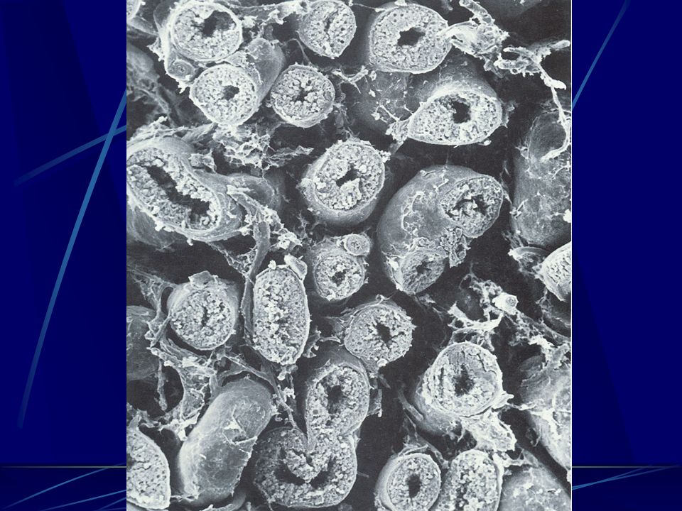
Testis section

7
b. Seminiferous tubules
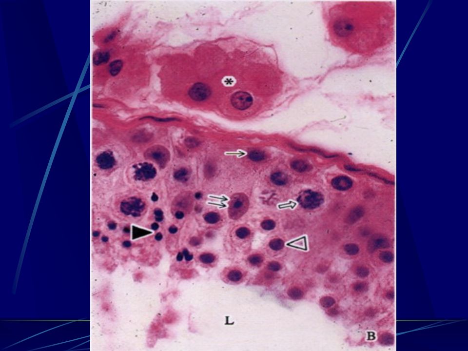
germinal epithelium: - a specialized stratified epi - consists of 2 types of cells: spermatogenic cells (male reproductive cells) Sertoli cells Lamina propria: basement membrane collagen layer myoid cells.
 Sertoli cells. Lamina propria: basement membrane. collagen layer. myoid cells.")
8
seminiferous tubules

9
Primary spermatocytes Secondary spermatocytes Spermatids Spermatozoa
2. Spermatogenic cells --include 5 successive cells. Spermatogonia Primary spermatocytes Secondary spermatocytes Spermatids Spermatozoa

10
A. Spermatogonia a. Adjacent to the BM, with round or ovoid nucleus.
b. Classified into type Ad, type Ap, and type B on the basis of nuclear staining. type A--nucleolus in the periphera of nuclear, Ad--nuclear dark ; Ap--nuclear pale type B ---nucleolus in the center of nuclear c. Type Ad (stem cells) →Type Ad → type Ad type Ap → type B Primary spermatocytes
 →Type Ad → type Ad. type Ap → type B. Primary spermatocytes.")
11
spermatogenesis

12
B. Primary spermatocytes
--- (stop in the prophase of first meiosis for 22 days ) a.The largest cells with nuclei in various stages of meiosis, easily visible, nuclear as woolen ball b. Contain diploid (2n) chromosomes (44+XY) and tetraploid (4n) DNA. c. Undergo the first meiosis (homologous chromosomes separate) producing the secondary spermatocytes.
 a.The largest cells with nuclei in various stages of meiosis, easily visible, nuclear as woolen ball. b. Contain diploid (2n) chromosomes (44+XY) and tetraploid (4n) DNA. c. Undergo the first. meiosis (homologous. chromosomes separate) producing the secondary. spermatocytes.")
13
C. Secondary spermatocytes
a. Smaller cells near the lumen, with round nucleus, and few in number. b. Contain haploid (1n) chromosomes (22+X or Y) and 2n DNA. c. Quickly undergo the second meiosis producing the spermatids. ※not easy to be seen for short-lived and quickly divide into second meiosis.
 chromosomes (22+X or Y) and 2n DNA. c. Quickly undergo the second. meiosis producing the spermatids. ※not easy to be seen for short-lived. and quickly divide into second. meiosis.")
14
D. Spermatids and spermiogenesis
a. The smallest, round or ovoid cells close to the lumen. b. Haploid cells (1n) in the number of chromosomes and in the amount of DNA. c. Do not divide, and transform from round cells into tadpole- like spermatozoa by spermiogenesis. d. spermiogenesis: spermatid spermatozoa
 in the. number of chromosomes and. in the amount of DNA. c. Do not divide, and transform. from round cells into tadpole- like spermatozoa by. spermiogenesis. d. spermiogenesis: spermatid spermatozoa.")
15
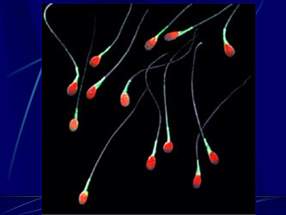
spermatozoon

16
d. Spermiogenesis includes following changes:
The nucleus becomes condensed and elongated. Golgi apparatus transforms into vesicle and then acrosome. Centrioles migrates to a position opposite acrosome and forms the flagellum (axoneme). Mi move and aggregate around proximal part of the flagellum forming a sheath. Much of cytoplasm is no longer required and cast off as residual bodies.
. Mi move and aggregate around proximal part of the flagellum forming a sheath. Much of cytoplasm is no longer required and cast off as residual bodies.")
18
E. Spermatozoon A mature spermatozoon has a head and a tail, about 60 m in length (head=5um,tail=55um) tadpole in shape.

19
E. Spermatozoon The head is pear-shaped, flattened and contains condensed nucleus (22+X or Y). The anterior 2/3 portion of the head is covered by the cap-like acrosome, which contains hydrolytic enzymes in it and is important for fertilization.
. The anterior 2/3 portion of the head is covered by the cap-like acrosome, which contains hydrolytic enzymes in it and is important for fertilization.")
20
E. Spermatozoon c. The tail is long and slender, containing a 9+2 microtubular axoneme (structure for motility) in its core. It is subdivided into 4 segments: neck middle Principal terminal piece
 in its core. It is subdivided into 4 segments: neck. middle. Principal. terminal piece.")
21
E. Spermatozoon ①The neck is very short and contains one centriole.
②The middle has a sheath of mitochondria which provides the energy for motility.

22
E. Spermatozoon ③The principal has a fibrous sheath providing support and protection. ④The terminal contains only the axoneme.

23
E. Spermatozoon d. The spermatozoa in the testes appear morphologically mature but functionally immature, that is, they are non-motile and do not fertilize the ovum.

24
Spermatogenesis about 64 days from spermatogonium to spermatozoon
The spermatogenesis is the process by which spermatogonia divide and differentiate into spermatozoon about 64 days from spermatogonium to spermatozoon 3 stages

25
Spermatogenesis

26
3. Sertoli cell (supporting cell)
pyramidal in shape, basal--close to the BM apical--close to the lumen lateral --project to lateral processes --tight junction

27
3. Sertoli cell (supporting cell)
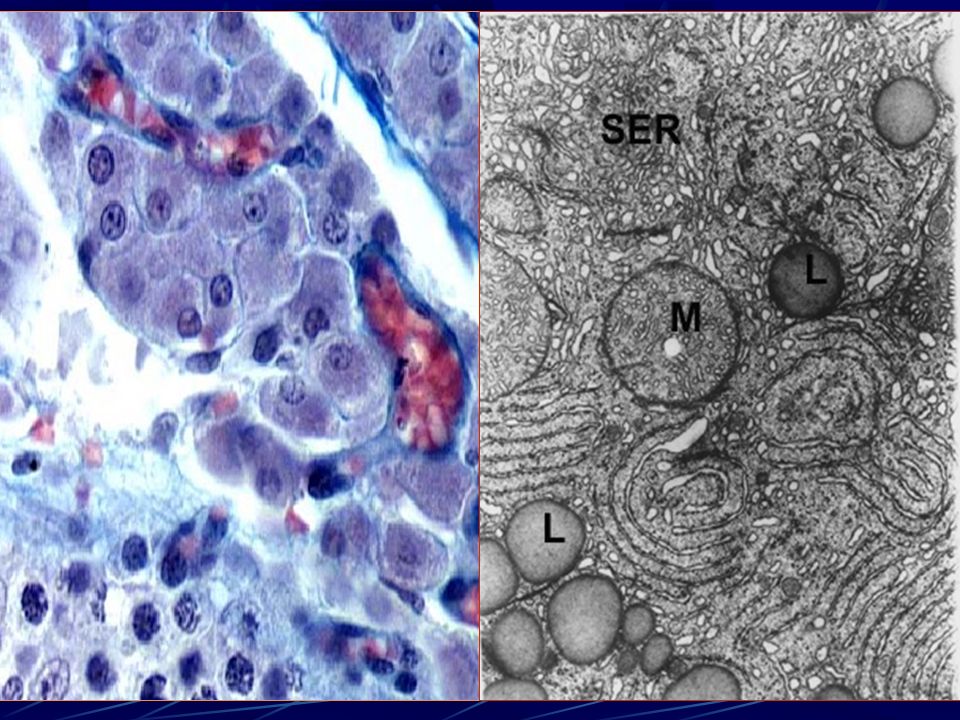
a. Irregular outline b. Elongated triangular and pale-stained nucleus with a prominent nucleolus. c. Abundant SER, as well as RER, Golgi apparatus, etc. d. Tight junctions between adjacent cells near their bases, which lie in a basal compartment and adluminal compartment

28
e. function support, protect, nourish, regulate and release germinal cell secret androgen-binding protein (ABP); bind to androgen, maintain the level of androgen concentration of lumen. And inhibin suppresses FSH synthesis and release.
; bind to androgen, maintain the level of androgen concentration of lumen. And inhibin suppresses FSH synthesis and release.")
29
e. function phagocytose degenerated cells and residual bodies from spermiogenesis. constitute the blood-testis barrier resistant to most factors: radiation, high temperature, infection, malnutrition, aging, etc.

30
blood-testis barrier constitute: endothelium and BM of capillary,
CT, and BM of seminiferous epithelium tight junction between the Sertoli cells.

31
blood-testis barrier function:
--- provide micro-environment for spermatogenesis --- avoid spermatozoon passing through the wall of seminiferous tubule and from autoimmune reaction

32
4. Leydig (interstitial) cells
Large, round or polygonal cells usually in groups, present in the interstitial tissue. Nucleus is large, pale-stained with 1-2 nucleoli. Cytoplasm is acidophilic and has abundant SER, lipid droplets, and Mi with tubular cristae. Secrete testosterone under the control of LH.

35
Control factors during spermatgenesis
hypothalamus hypophysis testis hormone regulate GnRH FSH LH Sertoli cell interstitial cell (leydig) ABP Inhibin androgen spermatogenesis
 ABP. Inhibin. androgen. spermatogenesis.")
36
5. straight tubules ( tubulus rectus )
position: near the mediastinum testis shape : straight and short canal structure: simple cuboidal cells without seminiferous epithelium (thinner wall)
")
37
6. rete testis position: within the mediastinum testis
shape: labyrinthine network (large and irregular lumen) structure: as straight tubules
 structure: as straight tubules.")
38
Ⅲ. Epididymis 1. Consist of : --head: the efferent ductules
(8-12 pieces). --body and tail : a highly coiled tube, ductus epididymis (only 1 pieces).
. --body and tail : a highly coiled tube, ductus. epididymis (only 1 pieces).")
39
2. Ductus efferents Ductus efferents have an epithelium composed of groups of nonciliated cuboidal cell alternating with ciliated cells. The nonciliated cells absorb much of the fluid secreted by the seminiferous tubules. The cilia beat towards the epididymis and assist in the transportation of the spermatozoa. The activity of ciliated cells and fluid absorption create a fluid flow that sweeps spermatozoa toward the epididymis.

40
3. ductus epididymis The ductus epididymis is lined with a pseudostratified columnar epithelium, containing 2 types of cells. Tall principal cells form a smooth lumen surface andhave numerous stereocilia. Short base cells may be germinative.

41
Ductus epididymis Ductus efferentes

42
epididymis duct Efferent duct

43
4. Has both absorptive and secretory functions
Absorbs most of the fluid that leaves the testis. Secretes carnitine, glycerylphosphorylcholine, sialic acid, etc. Spermatozoa become mature functionally, acquiring motility and fertilizability when they slowly pass the epididymis.

44
Ⅳ. Prostate 1. Compound glands around the urethra;

45
Ⅳ. Prostate covering: CT and SM parenchyma: 30-50 compound
tubuloaveolar gland. -- mucosal gland in the innermost zone(within urethral mucosa) -- submucosal gland in the middle zone (within submucosal layer) -- main gland in the peripheral zone (other portions)
 -- submucosal gland in the middle zone (within submucosal layer) -- main gland in the peripheral zone (other portions)")
46
2. simple epi ( cuboidal, columnar and pseudostratified )
3. within the cavity, there may be prostatic concretion, HE: round or oval acidophilic bodies showing concentric layers ※especially in older man.

Similar presentations
 produce androgen-testosterone ---gernital ducts: store.>")


 Lecturer of Comparative Anatomy and Embryology Zoology Department, Faculty of Science, MANSOURA UNIVERSITY EGYPT.>")


