Download presentation
Presentation is loading. Please wait.

1
Transport in Humans

2
Topics Overview of human transport system Components of Blood Structure of Heart Coronary Heart Disease Blood Vessels

3
OVERVIEW

4
Overview Recall in Sec 1, we studied respiration All living cells in our body (from our brain to our muscles to our organs) perform respiration to have the energy to function Recall that in order to respire, oxygen is required, and carbon dioxide is given out Where does all this oxygen come from? Where does all this carbon dioxide go?

5
Lungs

6
Overview Recall in Sec 1 we studied about the lungs in respiration The lungs is where gaseous exchange take place in our body We absorb oxygen from the atmosphere at the lungs We also give out carbon dioxide to the atmosphere at the lungs Gaseous exchange occurs by diffusion

7
Overview How does oxygen from the lungs reach the rest of the body (which needs it for respiration?) How does carbon dioxide from the rest of the body reach the lungs? Through the movement of our blood The system where our blood moves to and fro from our lungs to the rest of the body is known as the cardiovascular system Note: “cardio” is the root word for “heart”

8
Did you know? (not in syllabus) The cardiovascular system is not the only system of moving fluids in the body There is another system called the lymphatic system, which is an important part of our immune system
 The cardiovascular system is not the only system of moving fluids in the body There is another system called the lymphatic system, which is an important part of our immune system.")
9
COMPONENTS OF BLOOD

10
Components of Blood 1. Red Blood Cells 2. Plasma 3. White Blood Cells 4. Platelets

11
Red Blood Cells Red Blood Cells (also called erythrocytes) account for the red colour of our blood It is biconcave in shape and has no nucleus Contains the protein haemoglobin (which contains Iron), which is the protein which absorbs oxygen to the red blood cell
 account for the red colour of our blood It is biconcave in shape and has no nucleus Contains the protein haemoglobin (which contains Iron), which is the protein which absorbs oxygen to the red blood cell")
12
Red Blood Cells Is bright red when oxygenated, dark red when deoxygenated Produced in our bone marrow Anemia is the disease when you have too few red blood cells (or haemoglobin) in your blood
 in your blood")
13
Blood Plasma Plasma is the liquid portion of our blood It is actually pale yellow (“straw”) in colour It contains mostly water, with dissolved proteins, sugars, minerals and other substances (including carbon dioxide)
 in colour It contains mostly water, with dissolved proteins, sugars, minerals and other substances (including carbon dioxide)")
14
White Blood Cells Also known as leukocytes, white blood cells have a nucleus (unlike red blood cells) There are different types of white blood cells, but they all assist in the role of immunity Lymphocytes produce antibodies (which attack pathogens or flag them for other cells to kill them) Phagocytes attack pathogens by devouring them Leukemia is a disease where the body produces too much white blood cells
 There are different types of white blood cells, but they all assist in the role of immunity Lymphocytes produce antibodies (which attack pathogens or flag them for other cells to kill them) Phagocytes attack pathogens by devouring them Leukemia is a disease where the body produces too much white blood cells")
15
Platelets Also known as thrombocytes, platelets are mainly used for clotting the blood to stop bleeding (e.g. during a cut) Platelets also have no nucleus
 Platelets also have no nucleus.")
16
Components of Blood When you put blood in a centrifugue, the components of blood will separate as shown:

17
STRUCTURE OF THE HEART

18
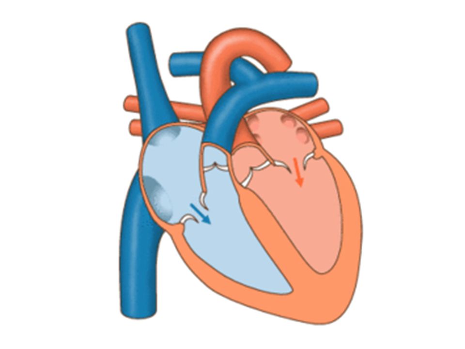
Structure of the Heart Recall the basic purpose of the heart: – bring oxygenated blood from lungs to rest of body – bring deoxygenated blood from rest of body to lungs A blood vessel which carries blood AWAY from the heart is called an artery A blood vessel which carries blood TOWARDS the heart is called a vein The pulmonary artery / vein connect the heart to the lungs

19
Structure of the Heart Note the left/right inversion Left side of the heart is slightly bigger than the right side Each side of the heart has 2 chambers – an atrium and a ventricle

20
Structure of the Heart Blood enters from outside the heart into the atrium Blood the flows from the atrium into the corresponding ventricle. A one-way valve prevents the blood from flowing back from the ventricle into the atrium The muscle in the ventricle wall contracts, pushing the blood out of the heart through the pulmonary artery (right) or aorta (left) Semi-lunar valves prevent the blood from flowing back into the ventricles
 or aorta (left) Semi-lunar valves prevent the blood from flowing back into the ventricles.")
22
BLOOD VESSELS

23
Blood Vessels 1) Arteries 2) Vein 3) Capillaries
 Arteries 2) Vein 3) Capillaries")
24
Arteries Blood which is pumped out of the heart (ventricles) goes straight into the arteries first That is why blood pressure in the arteries is the highest Arteries have the thickest layer of muscular wall to withstand this high pressure Pulmonary artery brings deoxygenated blood from heart to lungs Aorta brings oxygenated blood from heart to other parts of body
 goes straight into the arteries first That is why blood pressure in the arteries is the highest Arteries have the thickest layer of muscular wall to withstand this high pressure Pulmonary artery brings deoxygenated blood from heart to lungs Aorta brings oxygenated blood from heart to other parts of body")
25
Veins Veins are the blood vessels which bring back the blood to the heart Much lower pressure than arteries, so their walls need not be as thick Veins contact valves to prevent backflow of deoxygenated blood (note: pulmonary vein does not have valves because it carries oxygenated blood) You can see this using your own veins! (https://www.youtube.com/watch?v=A6isZ4dCT Mo)https://www.youtube.com/watch?v=A6isZ4dCT Mo
 v=A6isZ4dCT Mo.")
26
Capillaries After oxygenated blood has been pumped into the arteries, they bring it to the various parts of the body which needs the oxygenated blood (e.g. brain, muscles, etc.) When it reaches this destination, diffusion occurs between the blood and the destination tissue, – allowing for oxygen (and other nutrients) to be transferred from the bloodstream to tissue – carbon dioxide (and other waste products) to be transferred to the bloodstream from tissue Capillaries have very very thin walls (only one cell thick) to facilitate this diffusion.
 When it reaches this destination, diffusion occurs between the blood and the destination tissue, – allowing for oxygen (and other nutrients) to be transferred from the bloodstream to tissue – carbon dioxide (and other waste products) to be transferred to the bloodstream from tissue Capillaries have very very thin walls (only one cell thick) to facilitate this diffusion..")
27
Careful!! Do not use the term “cell wall” to refer to the thickness of the artery, vein or capillary. Recall what is a cell wall! Cells in humans don’t have cell walls!!! Use “artery wall” or “the wall of the vein”, etc.

28
CORONARY HEART DISEASE

29
Coronary Heart Disease The heart beats around 115 200 times per day. The muscles of the heart (particularly the ventricular muscles) do a lot of work Recall that muscles need oxygen (and blood!) in order to do their work The coronary arteries supply blood to the heart muscles However, if the arteries don’t manage to supply sufficient blood, the person suffers from coronary heart disease
 do a lot of work Recall that muscles need oxygen (and blood!) in order to do their work The coronary arteries supply blood to the heart muscles However, if the arteries don’t manage to supply sufficient blood, the person suffers from coronary heart disease.")
30
Coronary Heart Disease How does that happen? The coronary arteries may start to build up fatty deposits INSIDE the artery wall When that happens the lumen of the artery becomes smaller and smaller, constricting the artery Blood pressure will also increase when the artery is constricted

31
Coronary Heart Disease

32
Factors Causing Coronary Heart Disease High fat (and cholesterol) diet Smoking Stress Lack of Exercise
 diet Smoking Stress Lack of Exercise")
Similar presentations


 are veins? Y and Z.>")


