Download presentation
Presentation is loading. Please wait.

1
Membrane transport of small molecules and the electrical properties of membranes MB 207 – Molecular Cell Biology Part III: Internal Organization of the cell

2
Principles of membrane transport Barriers to the passage of most polar molecules. Allows the cell to maintain concentrations of solutes in its cytosol, differing from those in the extracellular fluid and in each of the intracellular compartments. Regulate movement of molecules into or out of cell. –Only very small uncharged molecules can readily diffuse i.e.. O 2, CO 2, water –Hydrophobic molecules pass through more readily than hydrophilic ones –Large uncharged polar molecules like glucose, and ions can not pass through: because the charge and high degree of hydration prevents them from entering the hydrocarbon phase of the bilayer. Relative permeability of bilayer to different classes of molecules: Rapid-Smaller & less association with water → Lipid bilayers are highly impermeable to charged molecules (ions), no matter how small.
, no matter how small..")
3
Membrane transport proteins To allow the passage of various polar molecules such as ions, sugars, amino acids, nucleotides and cell metabolites across cell membrane → membrane transport proteins Membrane transport proteins - responsible for transferring solutes across cell membrane. - multipass transmembrane proteins → enables specific hydrophilic solutes to cross the membrane without coming into direct contact with the hydrophobic interior of the lipid bilayer. - 2 major classes: carrier proteins and channel proteins i)Carrier proteins - also called transporter or permeases - bind the specific solute to be transported and undergo a series of comformational changes to transfer the bound solute across the membrane. ii)Channel proteins - form hydrophilic channels through the membrane that allow the passage of solutes without a major change in the conformation of the protein.
Carrier proteins - also called transporter or permeases - bind the specific solute to be transported and undergo a series of comformational changes to transfer the bound solute across the membrane. ii)Channel proteins - form hydrophilic channels through the membrane that allow the passage of solutes without a major change in the conformation of the protein..")
4
Carrier proteins and channel proteins (A)A carrier protein alternates between two conformations, so that the solute- binding site is sequentially accessible on one side of the bilayer and then on the other. (B) A channel protein forms a water-filled pore across the bilayer through which specific solutes can diffuse. Transport through channel protein occurs at a much faster rate than transport mediated by carrier proteins.
 A channel protein forms a water-filled pore across the bilayer through which specific solutes can diffuse. Transport through channel protein occurs at a much faster rate than transport mediated by carrier proteins..")
5
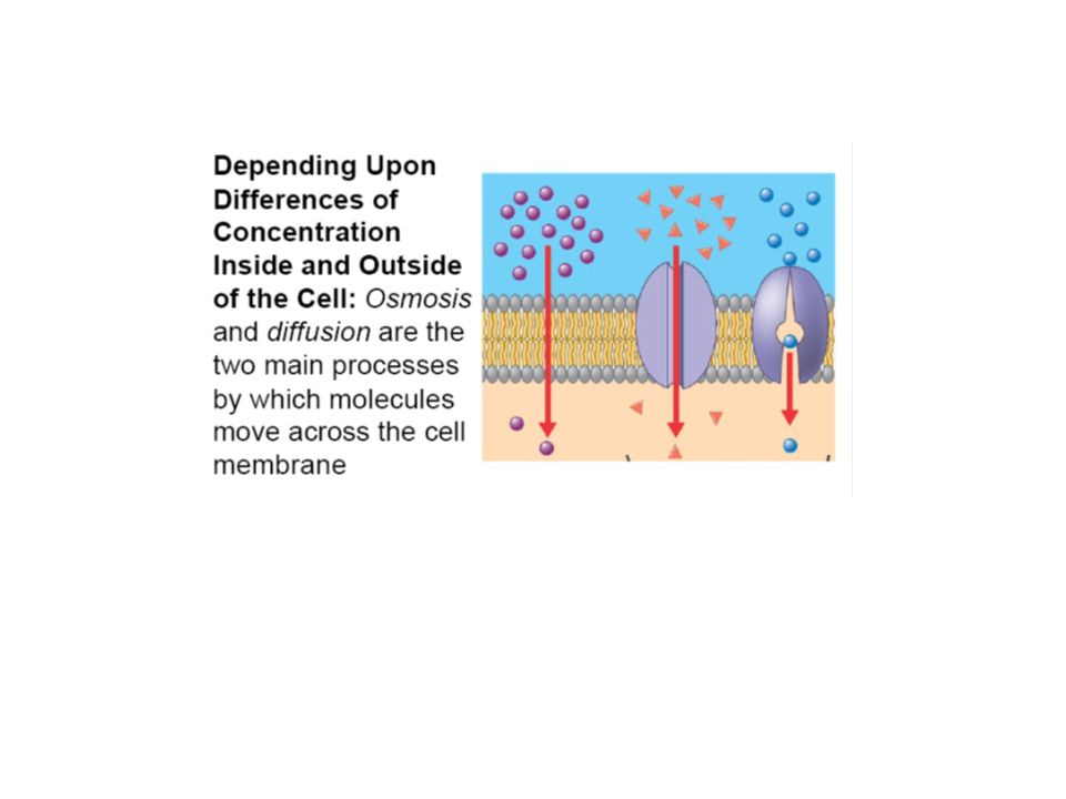
Traffic of substances across the plasma membrane Selective Bidirectional Depending upon differences of concentration inside and outside of the cell

8
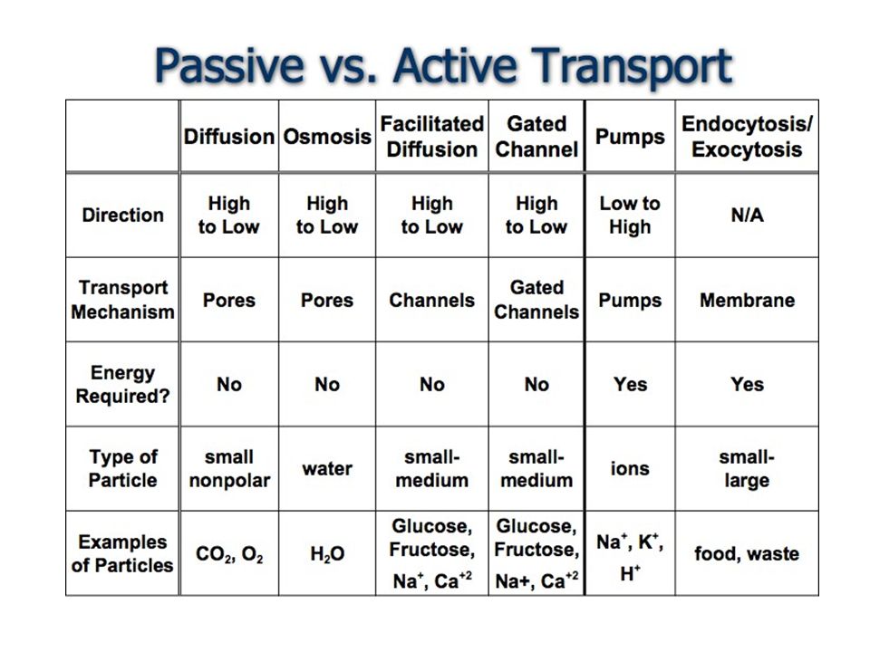
Passive and active transport Passive transport (facilitated diffusion): - moving solutes downhill of concentration gradient and requires no input of energy. Active transport: - moving solutes uphill of concentration gradient and requires input of energy.

10
Passive transport: Diffusion The movement of solutes across the membrane from an area of high concentration to an area of low concentration. → Membrane is permeable to molecules of dissolved solute. Equilibrium is reached when the solute concentration is the same in both areas.

11
Passive transport: Osmosis Osmosis occurs when the membrane between two areas is not permeable to the dissolved solute. Because solute cannot cross the membrane, water diffuses from areas of low concentration of solutes (more water) to areas of high concentration of solutes (less water). At equilibrium, the solute concentration will be equal on both sides of the membrane.
 to areas of high concentration of solutes (less water). At equilibrium, the solute concentration will be equal on both sides of the membrane..")
12
Passive transport: Facilitated Diffusion Facilitated diffusion is a protein- mediated passive (no energy required) diffusion of molecules across the cell membrane. Each transport protein transports just one type of molecule (selective).
..")
13
Active transport is a protein- mediated transport of molecules across the cell membrane against a concentration gradient (low to high solute concentration areas). It requires a boost of energy (ATP) to occur. As facilitated diffusion, is very selective. Glucose is actively transported through the plasma membrane of intestinal cells. Active transport
 to occur. As facilitated diffusion, is very selective. Glucose is actively transported through the plasma membrane of intestinal cells. Active transport.")
14
Small hydrophobic molecules that dissolve in lipid bilayers and increase their permeability to specific inorganic ions. Two classes of ionophores: a.Mobile ion carriers b.Channel formers Both are not coupled to energy sources, they permit the net movement of ions only down their electrochemical gradients Example: Valinomycin – mobile ion carrier, ring-shaped polymer that transports K + Gramicidin A - channel-forming ionophore, a dimeric compound of two linear peptides, which wind around each other to form a double helix Ionophores

15
Cells carry active transport in three main ways Solute is pumped uphill against its electrochemical gradient. a. Coupled carriers couple the uphill transport of one solute across the membrane to the downhill transport of another. b. ATP-driven pumps couple uphill transport to the hydrolysis of ATP. c. Light-driven pumps (bacterial cells), couple uphill transport to an input of energy from light
, couple uphill transport to an input of energy from light.")
16
Active transport can be driven by ion gradients Uniporters: carrier proteins that transport a single solute from one side of the membrane to the other. Uniporters: carrier proteins that transport a single solute from one side of the membrane to the other. Coupled: carrier proteins which transfer of one solute strictly depends on the transport of a second solute. Coupled: carrier proteins which transfer of one solute strictly depends on the transport of a second solute. - Couples transport involved either the simultaneous transfer of a second solute in the same direction (symporter) or (ii) in opposite direction (antiport).
 or (ii) in opposite direction (antiport)..")
17
Glucose carrier can be driven by a Na + gradient In the A state, the protein is open to the extracellular space whereas in the B state, it is open to the cytosol. Binding of Na + and glucose is cooperative, the binding of either ligand induces conformational change that greatly increases the protein’s affinity for the other ligand. Na + concentration is much higher in the extracellular space than in the cytosol, glucose is more likely to bind to the carrier in the A state. Therefore, both Na + and glucose enter the cell (via an A→B transition) much more often than they leave it (via an B→A transition). Na + -driven carrier proteins in the plasma membrane regulate Cytosolic pH - Most cells have one or more types of Na+-driven antiporters in their plasma membrane that help to maintain the cytosolic pH, about 7.2pH.
 much more often than they leave it (via an B→A transition). Na + -driven carrier proteins in the plasma membrane regulate Cytosolic pH - Most cells have one or more types of Na+-driven antiporters in their plasma membrane that help to maintain the cytosolic pH, about 7.2pH..")
18
Transcellular transport of glucose from lumen to extracellular fluid Na + -powered glucose symport –Na + gradient (higher outside) drive the active transport of glucose (lower outside) into the cell Uniport glucose transporter –Passive transport of glucose by the carrier protein down the concentration gradient Asymmetric distribution of carrier proteins in epithelial cells underlies the transcellular transport of solutes
 drive the active transport of glucose (lower outside) into the cell Uniport glucose transporter –Passive transport of glucose by the carrier protein down the concentration gradient Asymmetric distribution of carrier proteins in epithelial cells underlies the transcellular transport of solutes")
19
1.Glucose is pumped into the cell through the apical domain of the membrane by a Na + -powered glucose symport 2.Glucose passes out of the cell by passive transport mediated by a different glucose carrier protein in the basal and lateral membrane domains. 3.The Na + gradient driving the glucose symport is maintained by a Na + pump in the basal and lateral plasma membrane domains, which keep the internal concentration of Na + low. 4.Adjacent cells are connected by impermeable tight junctions, which have a dual function in the transport process illustrated: they prevent solutes from crossing the epithelium between cells, allowing a concentration gradient of glucose to be maintained across the cell sheet and they also serve as diffusion barriers within the plasma membrane, which help confine the various carrier protein to their respective membrane domains.

20
Example : Na + - K + pump or Na + pump [K + ] is 10-20 times higher inside the cells and reverse is true for [Na + ] [ ] differences are maintained by Na + -K + pump or Na 2+ pump (an antiporter which actively pumping Na + out of the cell and pumping K + in). Uses ATP to pump 3 Na + out and 2 K + in, against their electrochemical gradients Electrogenic, i.e. it drives a net current across the membrane Very important in regulating cell volume and pH –Most cells use 1/3 of the energy to feed this pump –Nerve cells use up to 2/3 of their energy to drive this pump as cells changes in Na+ and K+ concentrations due to firing of action potentials
![Example : Na + - K + pump or Na + pump [K + ] is times higher inside the cells and reverse is true for [Na + ] [ ] differences are maintained by Na + -K + pump or Na 2+ pump (an antiporter which actively pumping Na + out of the cell and pumping K + in).](http://images.slideplayer.com/25/7780657/slides/slide_20.jpg "Uses ATP to pump 3 Na + out and 2 K + in, against their electrochemical gradients Electrogenic, i.e. it drives a net current across the membrane Very important in regulating cell volume and pH –Most cells use 1/3 of the energy to feed this pump –Nerve cells use up to 2/3 of their energy to drive this pump as cells changes in Na+ and K+ concentrations due to firing of action potentials.")
21
A model of the pumping cycle of the Na + - K + pump P-type transport ATPase: Hydrolysis of ATP leads to the phosphorylation of the pump, which provides the energy necessary to turn over the Na + ions bound inside the cell towards the outside. The pump binds K + on the outside and through its dephosphorylation, it moves the K + ions into the cell. Na+ deptPhos- and K+ deptdephos- Induce conformation change

22
(1). The binding of Na + and (2) the subsequent phosphorylation by ATP of the cytoplasmic face of the pump induce the protein to undergo a conformational change. (3). Transfer the Na + across the membrane and releases it on the outside. (4). Then, the binding of K + on the extracellular surface and (5) the subsequent dephosphorylation return the protein to its original conformation, which (6) transfers the K + across the membrane and releases it into the cytosol. These changes in conformation are analogous to the A↔B transitions. Some Ca + and H + pumps (Ca 2+ pump or Ca 2+ ATPase) are also P-type transport ATPases. For example: sarcoplasmic reticulum membrane of skeletal muscle cells.
 the subsequent phosphorylation by ATP of the cytoplasmic face of the pump induce the protein to undergo a conformational change. (3). Transfer the Na + across the membrane and releases it on the outside. (4). Then, the binding of K + on the extracellular surface and (5) the subsequent dephosphorylation return the protein to its original conformation, which (6) transfers the K + across the membrane and releases it into the cytosol. These changes in conformation are analogous to the A↔B transitions. Some Ca + and H + pumps (Ca 2+ pump or Ca 2+ ATPase) are also P-type transport ATPases. For example: sarcoplasmic reticulum membrane of skeletal muscle cells..")
23
Osmosis is the movement of water from low osmotic pressure (dilute solution) to high osmotic pressure (concentrated solution). Cells swell in hypotonic and shrink in hypertonic solutions If the external solution balances the osmotic pressure of the cytoplasm it is said to be isotonic In the present of quabain, an inhibitor of the Na + - K + pump, the cells will burst. Na + - K + pump play important role in maintaining osmotic pressure of the cells by pumping out Na +. It controls the [ solute ] inside the cell, there by regulating the osmolarity (tonicity). Na + -K + pump: required to maintain Osmotic balance and stabilize cell volume
. Na + -K + pump: required to maintain Osmotic balance and stabilize cell volume.")
24
Channel Proteins form hydrophilic pores across membranes. have narrow, highly selective pores that can open and close. involved mainly in inorganic ion transport ie. Na +, K +, Ca 2+ or Cl - → ion channels more efficient compared to carried protein cannot coupled to an energy source to perform active transport, mediating passive transport.

25
Important properties of ion channels: i)Ion selectivity - permitting some inorganic ions to pass but not others. - the pore is narrow enough in places to force permeating ions into intimate contact with the walls of the channel so that only ions of appropriate size and charge can pass. ii)Are not continuously open - gated, which allows them to open briefly and then close again. - gate opens in response to a specific stimulus.
Are not continuously open - gated, which allows them to open briefly and then close again. - gate opens in response to a specific stimulus..")
26
Membrane potential arise when there is a difference in the electrical charge on the 2 sides of the membrane due to a slight excess of positive ions over negative ones on one side and a slight deficit on the other. All cells are typically have an excess of negative charge, cell has a negative resting membrane potential. Na + -K + pump helps maintain an osmotic balance across the membrane by keeping intracellular concentration of Na + low. → other cations have to be plentiful to balance the charge carried by the cell’s fixed anions. → balancing role is performed largely by K +, which is actively pumped into the cell by the Na + -K + pump and through K + leak channels in the plasma membrane. K + leak channels, is opened in unstimulated cells (resting) and mediate the resting membrane potential. → crucial role in maintaining the membrane potential across all plasma membranes. Voltage gated ions channel – generate action potentials (triggered by a depolarization of the plasma membrane) in electrically excitable cells
 and mediate the resting membrane potential. → crucial role in maintaining the membrane potential across all plasma membranes. Voltage gated ions channel – generate action potentials (triggered by a depolarization of the plasma membrane) in electrically excitable cells.")
27
Function of ion channels in generating action potential (AP)/ nerve impulse The AP is a membrane potential change caused by the flow of ions through ion channels in the membrane, depolarization of the plasma membrane. APs are rapidly propagated along the axons of the nervous system and over the surface of some muscle and glandular cells to generate electrical messages Voltage-gated cation channels are responsible for generating the APs Mechanism: –An AP is triggered by a shift in the local membrane potential to a less negative value, i.e. depolarization. –As cell depolarized, voltage-gated Na + channels open, allowing Na + to rush into the cell down its concentration gradient. –The Na + influx further depolarizes the membrane –As the [Na + ] inside the cell increases, Na + channels become inactivated –Opening of K + channels now account for the return of the membrane potential to a value more negative than the resting potential, i.e. hyperpolarization. –K + leak channels eventually change the membrane potential back to its normal resting value. –The AP travels forwards along the plasma membrane as a self-propagating wave of depolarization

28
Function of ion channels in generating action potential (AP)
")
29
Ligand-gated ion channel Are triggered by the binding of specific substances into to the channel protein ie. Neurotransmitter. Acetylcholine is the most common neurotransmitter for synapses between neuron outside CNS as well as for neuromuscular junctions. Example: Acetylcholine (Ach) receptor at the neuromuscular junction (excitatory). → The pore is lined by a ring of five transmembrane α helices. → In its closed conformation, the pore is thought to be occluded by the hydrophobic side chains by five leucines (one from each helix), which form a gate near the middle of the lipid bilayer. → The negatively charged side chains at either end of the pore ensure that only positively charged ion pass through the channel. Binding of Ach at the α-subunits lead conformational change that opens the gate leading to the influx of Na + which caused localized membrane depolarization Others: Glutamate-gated Ca 2+ channels & serotonin-gated cation channels
 receptor at the neuromuscular junction (excitatory). → The pore is lined by a ring of five transmembrane α helices. → In its closed conformation, the pore is thought to be occluded by the hydrophobic side chains by five leucines (one from each helix), which form a gate near the middle of the lipid bilayer. → The negatively charged side chains at either end of the pore ensure that only positively charged ion pass through the channel. Binding of Ach at the α-subunits lead conformational change that opens the gate leading to the influx of Na + which caused localized membrane depolarization Others: Glutamate-gated Ca 2+ channels & serotonin-gated cation channels.")
30
Neuromuscular transmission involves many different ion channels 1.The AP traveling down the axonal membrane reaches the terminal and causes opening of voltage-gated Ca 2+ channels that let Ca 2+ into the cell. 2.Raise in [Ca 2+ ] trigger the release of ACh by exocytosis. 3.ACh then binds the AChR which opens its cation channel leading to a change in the postsynaptic membrane potential 4.The AP propagates and eventually leads to release of Ca 2+ from the sarcoplasmic reticulum into the cytosol which then causes muscle fibers to contract. 5.Signaling is terminated by the enzyme acetylcholinesterase which hydrolyzes ACh The system of ion channels- stimulation of muscle contraction by a nerve impulse

31
Ligand-gated ion channel Example: -aminobutyric acid (GABA) receptor at postsynaptic membrane (inhibitory) Open Cl - channels that mediate a hyper- polarization of the postsynaptic membrane thus, suppressing the excitability of the postsynaptic cell Others: Glycine-gated Cl - channels
 receptor at postsynaptic membrane (inhibitory) Open Cl - channels that mediate a hyper- polarization of the postsynaptic membrane thus, suppressing the excitability of the postsynaptic cell Others: Glycine-gated Cl - channels")
32
Long term potentiation (LTP) in the mammalian hippocampus depends on Ca 2+ entry through NMDA-receptor channels LTP occurs on any occasion when a presynaptic cell fires (once or more) at a time when postsynaptic membrane is strongly depolarized. Mediated by NMDA receptors – selectively activated by artificial glutamate analog, N-methyl-D-aspartate. NMDA-receptor channels opens only when: i)Glutamate must be bound to the receptor. ii)Membrane must be strongly depolarized. → NMDA receptors are normally activated when conventional glutamate- gated ion channels are activated as well and depolarize the membrane.
Glutamate must be bound to the receptor. ii)Membrane must be strongly depolarized. → NMDA receptors are normally activated when conventional glutamate- gated ion channels are activated as well and depolarize the membrane..")
Similar presentations


 Plasma membranes are selectively permeable some molecules pass through membrane; some don’t Types of Membrane Transport.>")


 Membranes. Membrane transport Membranes are selectively permeable barriers Hydrophobic uncharged small molecules can freely diffuse.>")


 Nervous system functions Structure of a neuron Sensory, motor, inter- neurons Membrane potential Sodium.>")
