Download presentation
Presentation is loading. Please wait.

1
Chapter 11 The Cardiovascular System

2
The Cardiovascular System
Closed system of the heart and blood vessels Heart pumps blood Blood vessels allow blood to circulate to all parts of the body Blood is the transport vehicle Carries oxygen, nutrients, cell wastes, & hormones, to and from the cells to maintain homeostasis Function of the cardiovascular system To deliver oxygen and nutrients and to remove carbon dioxide and other waste products

3
The Heart Location About the size of your fist Mediastinum
Middle cavity of the thorax, between the lungs Pointed apex directed toward left hip The great vessels of the heart emerge from the base which points toward the right shoulder and lies beneath the second rib About the size of your fist Less than 1 lb. Apex- Approximately at the level of the 5th intercostal space where one would place a stethoscope to count the heart rate for an apical pulse

4
The Heart

5
The Heart: Coverings Pericardium – a double serous membrane
Visceral pericardium Hugs the external surface of the heart & is part of the heart wall Parietal pericardium Outside layer Serous fluid fills the space between the layers of pericardium Pericarditis- Inflammation of the pericardium results in an decrease in the amt of serous fluid. This causes the pericardial layers to bind and stick to each other, forming painful adhesions that interfere with heart movements.

6
The Heart: Heart Wall Three layers Epicardium Myocardium Endocardium
Outside layer This layer is the visceral pericardium Connective tissue layer Myocardium Middle layer Mostly cardiac muscle – Allows contraction Endocardium Inner layer Endothelium

7
External Heart Anatomy

8
The Heart: Chambers Right & left sides act as separate pumps
Four chambers Atria Receiving chambers Right atrium Left atrium Ventricles Discharging chambers Right ventricle Left ventricle

9
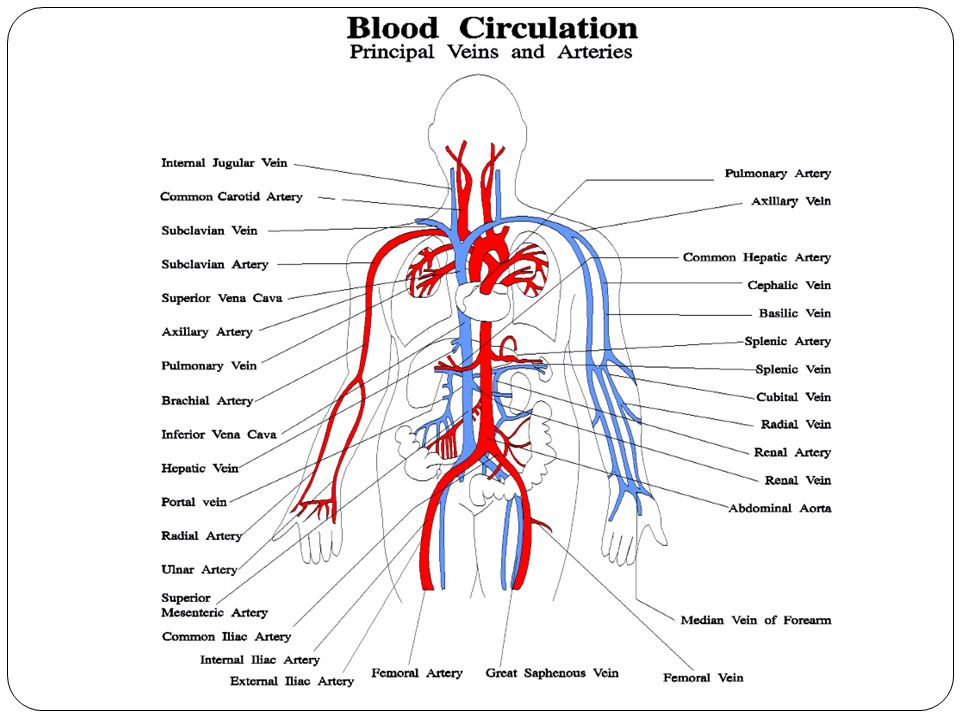
Blood Circulation

11
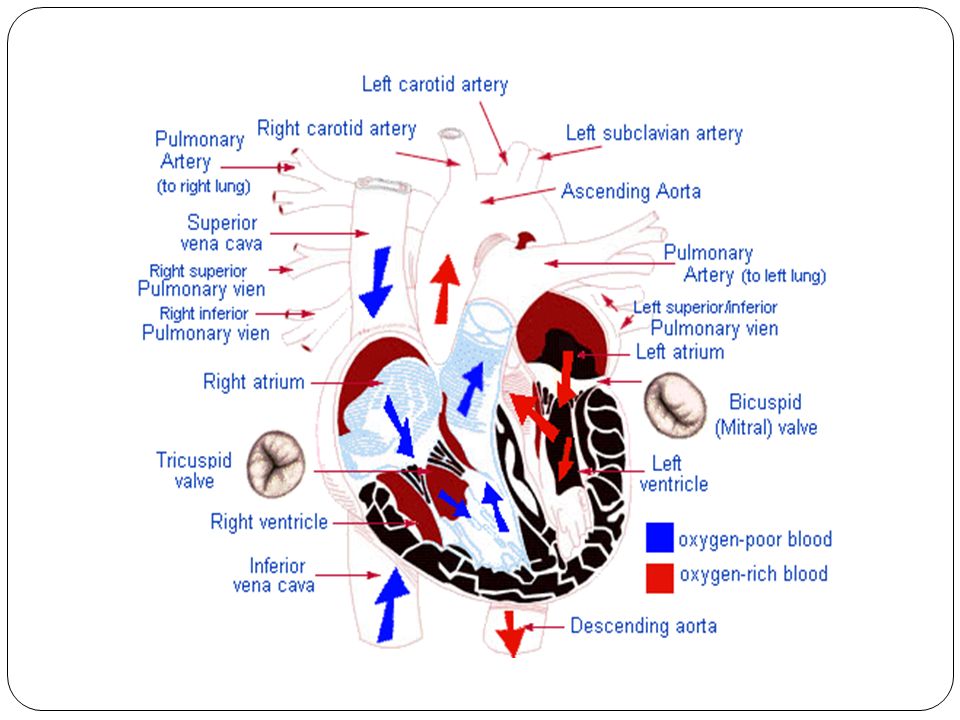
The Heart: Associated Great Vessels
Aorta Leaves left ventricle Pulmonary arteries Leave right ventricle Vena cava Enters right atrium Pulmonary veins (four) Enter left atrium
 Enter left atrium.")
12
The Heart: Valves Allows blood to flow in only one direction
Four valves Atrioventricular valves – between atria and ventricles Bicuspid (mitral) valve (left) Tricuspid valve (right) Semilunar valves between ventricle and artery Pulmonary semilunar valve Aortic semilunar valve
 valve (left) Tricuspid valve (right) Semilunar valves between ventricle and artery. Pulmonary semilunar valve. Aortic semilunar valve.")
13
The Heart: Valves Valves open as blood is pumped through
Held in place by chordae tendineae (“heart strings”) Close to prevent backflow Aortic Valve Replacement Surgery
 Close to prevent backflow. Aortic Valve Replacement Surgery.")
14
Operation of Heart Valves

15
Coronary Circulation Blood in the heart chambers does not nourish the myocardium The heart has its own nourishing circulatory system Right and left coronary arteries & their major branches Are compressed when the ventricles are contracting and fill when the heart is relaxed Cardiac veins Drain the myocardium Blood empties into the right atrium via the coronary sinus

16
Angina & Myocardial Infarction website

17
Bypass Surgery Website Coronary Bypass Surgery

18
Balloon Angioplasty Coronary Angioplasty & Stenting

19
Heart View

21
The Heart: Conduction System
Autonomic Nervous system Nerves = brakes & accelerators to decrease or increase heart rate Accelerators = Sympathetic Nervous system Brakes = Parasympathetic Nervous system Intrinsic conduction system- Nodal system Heart muscle cells contract, without nerve impulses, in a regular, continuous fashion Built into the heart tissue & sets its basic rhythm Causes heart muscle depolarization in only 1 direction (From the atria to the ventricles) Enforces a contraction rate of 75 beats/minute Heart beats as a coordinated unit
 Enforces a contraction rate of 75 beats/minute. Heart beats as a coordinated unit.")
22
Intrinsic Conduction System
Special type of tissue Sets the pace Sinoatrial (SA) node Located within the right atrium Pacemaker Starts each heartbeat Atrioventricular (AV) node Located at the junction of the right & left atria and ventricles Atrioventricular bundle (Bundle of His) Bundle branches (right and left) Purkinje fibers Spread within the muscle of the ventricle walls
 node. Located within the right atrium. Pacemaker Starts each heartbeat. Atrioventricular (AV) node. Located at the junction of the right & left atria and ventricles. Atrioventricular bundle (Bundle of His) Bundle branches (right and left) Purkinje fibers. Spread within the muscle of the ventricle walls.")
23
Heart Contractions

24
Electrocardiograms (EKG/ECG)
Three formations P wave: Small & signals the depolarization of the atria immediately before they contract QRS complex: Complicated shape Depolarization of the ventricles T wave: Repolarization of the ventricles

25
Electrocardiograms (EKG/ECG)
")
26
Abnormal EKG

27
Pathology of the Heart Abnormal ECG: Heart Block:
Damage to AV node Ventricles are partially or totally released from the control of the SA node Result = slower heart beat Other conditions can damage the SA node resulting in a slower heart rate Surgically installation of an artificial pacemaker Fibrillation Results from a lack of blood flow to the heart (ischemia) Rapid uncoordinated heartbeat that makes the heart useless as a pump Major cause of death from heart attacks in adults Tachycardia (If prolonged, can lead to fibrillation) +100 beats/min Bradycardia Less than 60 beats/min
 Rapid uncoordinated heartbeat that makes the heart useless as a pump Major cause of death from heart attacks in adults. Tachycardia (If prolonged, can lead to fibrillation) +100 beats/min. Bradycardia. Less than 60 beats/min.")
28
The Heart: Cardiac Cycle
Events of 1 complete heartbeat Both atria & ventricles contract and then relax Atria contract simultaneously and then relax ventricles then contract simultaneously and then relax Systole Contraction of the ventricles Diastole Relaxation of the ventricles

29
Filling of Heart Chambers – the Cardiac Cycle

30
The Heart: Cardiac Output
Cardiac output (CO) Amount of blood pumped out by each side of the heart (each ventricle) in one minute CO = (heart rate [HR]) x (stroke volume [SV]) Varies with the demands of the body Stroke volume (SV) Volume of blood pumped out by a ventricle with each heartbeat
 Amount of blood pumped out by each side of the heart (each ventricle) in one minute. CO = (heart rate [HR]) x (stroke volume [SV]) Varies with the demands of the body. Stroke volume (SV) Volume of blood pumped out by a ventricle with each heartbeat.")
31
The Heart: Regulation of Heart Rate
Stroke volume usually remains relatively constant Starling’s law of the heart – the more that the cardiac muscle is stretched, the stronger the contraction Changing heart rate is the most common way to change cardiac output

32
Regulation of Heart Rate
Increased heart rate Sympathetic nervous system stimulation Activated in times of “Fight or Flight” Hormones Epinephrine Thyroxine Exercise Fever Increases the metabolic rate of heart cells

33
The Heart: Regulation of Heart Rate
Decreased heart rate Parasympathetic nervous system stimulation Congestive heart failure Heart is worn out and pumps weakly Digoxin Works to provide a slow, steady, but stronger beat

34
Cardiac Output Regulation

35
Congestive Heart Failure (CHF)
Decline in pumping efficiency of the heart Leading to inadequate circulation Progressive condition Causes: Coronary atherosclerosis, high blood pressure and a history of multiple myocardial infarctions Left side fails Pulmonary congestion suffocation Right side fails Peripheral congestion and edema

36
Blood Vessels: The Vascular System
Taking blood from the heart to the tissues and back Arteries Arterioles Capillaries Venules Veins

37
Blood Vessels: Anatomy
Three layers (tunics) Tunic intima Lines the lumen or interior of the vessels Endothelium slick surface, decreases friction as blood flows through Tunic media Middle coat Smooth muscle Controlled by sympathetic nervous system Changes the diameter of the vessels Constriction blood pressure increases Dilation blood pressure decreases Tunic externa Mostly fibrous connective tissue Outermost tunic
 Tunic intima. Lines the lumen or interior of the vessels. Endothelium slick surface, decreases friction as blood flows through. Tunic media. Middle coat. Smooth muscle. Controlled by sympathetic nervous system. Changes the diameter of the vessels. Constriction blood pressure increases. Dilation blood pressure decreases. Tunic externa. Mostly fibrous connective tissue. Outermost tunic.")
38
The Vascular System

39
Differences Between Blood Vessel Types
Walls of arteries are the thickest Lumens of veins are larger than arteries Skeletal muscle “milks” blood in veins toward the heart Walls of capillaries are only one cell layer thick to allow for exchanges between blood and tissue

40
Movement of Blood Through Vessels
Most arterial blood is pumped by the heart Veins use the milking action of skeletal muscles to help move blood

41
Varicose Veins website

42
Capillary Beds Capillary beds consist of two types of vessels
Vascular shunt – directly connects an arteriole to a venule

43
Capillary Beds True capillaries – exchange vessels
Oxygen and nutrients cross to cells Carbon dioxide and metabolic waste products cross into blood

44
Diffusion at Capillary Beds

45
Vital Signs Arterial pulse Blood pressure Respiratory Rate
Body Temperature

46
Pulse Pulse – pressure wave of blood
Monitored at “pressure points” where pulse is easily palpated

47
Blood Pressure Measurements by health professionals are made on the pressure in large arteries Systolic – pressure at the peak of ventricular contraction Diastolic – pressure when ventricles relax Pressure in blood vessels decreases as the distance away from the heart increases

48
Blood Pressure Changes

49
Measuring Arterial Blood Pressure

50
Blood Pressure: Effects of Factors
Neural factors Autonomic nervous system adjustments (sympathetic division) Renal factors Regulation by altering blood volume Renin – hormonal control
 Renal factors. Regulation by altering blood volume. Renin – hormonal control.")
51
Blood Pressure: Effects of Factors
Temperature Heat has a vasodilation effect Cold has a vasoconstricting effect Chemicals Various substances can cause increases or decreases Diet

52
Variations in Blood Pressure
Human normal range is variable Normal 140–110 mm Hg systolic 80–75 mm Hg diastolic Hypotension Low systolic (below 110 mm Hg) Often associated with illness Hypertension High systolic (above 140 mm Hg) High diastolic (above 90) Can be dangerous if it is chronic
 Often associated with illness. Hypertension. High systolic (above 140 mm Hg) High diastolic (above 90) Can be dangerous if it is chronic.")
54
Varicose Veins website

55
Blood Distribution

Similar presentations


 Transport O 2, nutrients, hormones, cell wastes, etc…>")


