Download presentation
Presentation is loading. Please wait.

1
IN THE NAME OF GOD Chapter 165 EXTRA-AXIAL NEOPLASM OF THE POSTERIOR FOSSA Department Of Oto-rhino-Laryngology Of Isfahan Medical Science

2
DDX OF SKULL BASE NEOPLASMS POSTERIOR FOSSA: 1) Common Cerebellopontine Lesions: Acoustic neuroma- Meningioma - Epidermoid- Nonacoustic neuroma -Paraganglioma -Arachnoid cyst- Hemangiom 2) Petrous Apex Lesions : Cholesterol granuloma -Epidermoid - Asymmetric pneumatization -Retained mucus or mucocele - Petrous carotid artery aneurysm 3) Uncommon Cerebellopontine Lesions : Metastatic tumor -Lipoma –Dermoid-Teratoma - Chordoma - Chondrosarcoma- Giant cell tumor
 Common Cerebellopontine Lesions: Acoustic neuroma- Meningioma - Epidermoid- Nonacoustic neuroma -Paraganglioma -Arachnoid cyst- Hemangiom 2) Petrous Apex Lesions : Cholesterol granuloma -Epidermoid - Asymmetric pneumatization -Retained mucus or mucocele - Petrous carotid artery aneurysm 3) Uncommon Cerebellopontine Lesions : Metastatic tumor -Lipoma –Dermoid-Teratoma - Chordoma - Chondrosarcoma- Giant cell tumor")
3
4) Intra-Axial Tumors : Hemangioblastoma- Medulloblastoma - Astrocytoma - Glioma - Fourth ventricle tumor ANs most common tumors more than 90% Secondary tumors paragangliomas 10% of CPA neoplasms
 Intra-Axial Tumors : Hemangioblastoma- Medulloblastoma - Astrocytoma - Glioma - Fourth ventricle tumor ANs most common tumors more than 90% Secondary tumors paragangliomas 10% of CPA neoplasms")
4
Acoustic Neuroma : benign schwannoma vestibular eighth nerve. 8% -10% of all intracranial tumors. slowly growing- circumscribed-grossly displacing neural structures-firm and dense to soft with large cystic spaces. pressure neurovascular structures,-auditory and vestibular symptoms earlier in IAC than CPA. >95% unilateral -nonhereditary lesions

5
(NF) type 1 (von Recklinghausen's disease) : AD - gene : 17, arising both intracranially - extracranially Schwann cells of any nerve; <5% ANs bilateral AN is not a part of the syndrome.
 type 1 (von Recklinghausen s disease) : AD - gene : 17, arising both intracranially - extracranially Schwann cells of any nerve; <5% ANs bilateral AN is not a part of the syndrome.")
6
NF type 2 : bilateral AN in > 96% of patients. Schwannomas of the other cranial nerves, meningiomas and epandymomas are more in NF 2. gene : 22 ANs in NF 2 are onset early in life(often < 21 y) AN s < 30y close evaluation of the contralateral ear. technically more challenging to remove because of tendency to adhere to nearby structures.
 AN s < 30y close evaluation of the contralateral ear. technically more challenging to remove because of tendency to adhere to nearby structures..")
7
Malignant schwannomas rarely occur,often in: 1) NF 2) solitary schwannomas 3) pigmented schwannoma growth rate : variable, slow growing(0.2 cm/y) ANs that are not treated are potentially lethal Gradual enlargement leading to indentation of brainstem, ICP,death during of 5-15y
 NF 2) solitary schwannomas 3) pigmented schwannoma growth rate : variable, slow growing(0.2 cm/y) ANs that are not treated are potentially lethal Gradual enlargement leading to indentation of brainstem, ICP,death during of 5-15y")
8
Growth of ANs in three phases: 1) IAC growth : 7-8 nerve compression and Displacement 7,8 nerve and (AICA) 2) cisternal portion : Fourth ventricle shift often (2 to 3 cm ) -total ventricle obstruction - hydrocephalus - Trigeminal compression occurs at about the 3-cm 3) brainstem compression : cerebellar tonsil herniation -death
 IAC growth : 7-8 nerve compression and Displacement 7,8 nerve and (AICA) 2) cisternal portion : Fourth ventricle shift often (2 to 3 cm ) -total ventricle obstruction - hydrocephalus - Trigeminal compression occurs at about the 3-cm 3) brainstem compression : cerebellar tonsil herniation -death")
9
Signs and Symptoms: Most: (SNHL progressive unilateral)-tinnitus, dysequilibrium, facial hypesthesia loss SDS large tumors : facial sensation or corneal reflex (compression 5 (CN)) -compress the fourth ventricle and brainstem, ataxia, Increased ICP (headaches, nausea) Rotatory vertigo is far less common
-tinnitus, dysequilibrium, facial hypesthesia loss SDS large tumors : facial sensation or corneal reflex (compression 5 (CN)) -compress the fourth ventricle and brainstem, ataxia, Increased ICP (headaches, nausea) Rotatory vertigo is far less common")
10
>20% of AN with SSNHL may totally recover only 1% SSNHL have AN > 5% of AN have normal hearing any patient with asymmetric or sudden hearing loss (even after total recovery) should be R/O retrocochlear lesion.
 should be R/O retrocochlear lesion.")
11
Diagnostic Studies 1) Audiometry 2) (MRI) (MRI) – audiometry are not sensitive to detect small ANs 3) ABR: most sensitive even small tumors AN (95% -100%) 4) Stacked ABR :modified ABR -improved sensitivity for intracanalicular ANs
 Audiometry 2) (MRI) (MRI) – audiometry are not sensitive to detect small ANs 3) ABR: most sensitive even small tumors AN (95% -100%) 4) Stacked ABR :modified ABR -improved sensitivity for intracanalicular ANs")
12
IMAGING DDX OF CPA LESIONS: 1) Extra-axial : Most common (AN) -Common (Meningioma- Epidermoid) 2) Extradural : Common (Paraganglioma : glomus jugulare, vagale) 3) Intra-axial : (Rare) Astrocytoma, ependymoma, papilloma, hemangioblastoma, metastasis
 Extra-axial : Most common (AN) -Common (Meningioma- Epidermoid) 2) Extradural : Common (Paraganglioma : glomus jugulare, vagale) 3) Intra-axial : (Rare) Astrocytoma, ependymoma, papilloma, hemangioblastoma, metastasis")
13
Acoustic Neuroma: *Centered *on IAC- enlarge IAC -Spherical or ovoid- acute bone-tumor angle- in CT scan : Mostly isodense- often inhomogeneous – in MRI T1,T2 : Isointense or hypointense

17
MRI : lesions as small as 3 mm contrast CT can fail to detect ANs smaller than 1.5 cm CT primary imaging modality in patients ; 1) who cannot undergo MRI because of medical reasons (e.g., cardiac pacemaker or cochlear implant) 2) unmanageable phobic reactions Oxygen cisternography : used to diagnose small, mainly intracanalicular lesions
 who cannot undergo MRI because of medical reasons (e.g., cardiac pacemaker or cochlear implant) 2) unmanageable phobic reactions Oxygen cisternography : used to diagnose small, mainly intracanalicular lesions")
18
MENINGIOMAS 18% of intracranial tumors and 3% of CPA tumors arachnoid villae are the cells of origin benign but locally aggressive tumors globular mass firmly adherent to dura mater psammoma bodies thin investing capsule bone without destruction hyperostotic in 25%

19
The signs and symptoms ; similar ANs Small tumors: HL, tinnitus, imbalance Larger tumors : other cranial nerve involvement and hydrocephalus Diagnostic Studies Audiovestibular Testing 75% abnormal ABR Imaging: Unlike ANs, meningiomas are usually eccentric to the porous. 60% extend to the middle fossa

20
CT, 2/3 meningiomas ; hyperintense - homogeneous and calcified Hyperostosis (characteristic of meningiomas ) Eccentric IAC -Hemispherical,rarely plaquelike obtuse bone tumor angle in MRI T1 : Isointense or hypointense in T2 :Variable
 Eccentric IAC -Hemispherical,rarely plaquelike obtuse bone tumor angle in MRI T1 : Isointense or hypointense in T2 :Variable")
22
PRIMARY CHOLESTEATOMAS: originates from epithelial rests within the temporal bone or CPA. slow growing, and symptoms often do not become apparent until the second to fourth decade of life. expand, compression of surrounding structures variable shapes with irregular surfaces. They may burrow into crevices on the surface of the brain or dumbbell into the middle fossa.

23
Diagnostic Studies : Auditory Testing, SDS; is poorer than degree of pure-tone loss. ABR abnormal CT and MRI Epidermoids CPA should be DDX from arachnoid cysts- arachnoid cysts have smoother surfaces than primary cholesteatomas. epidermoids petrous apex should be DDX from the much more common cholesterol granulomas. primary cholesteatoma requires excision, whereas drainage is sufficient for cholesterol granulomas and arachnoid cysts

24
CT: less dense than brain + no enhancement, irregular margins-Anterolateral or posterolateral to brainstem- erosion- dumbbell into middle fossa or contralateral CPA –hypodense-peripheral calcium MRI: inhomogeneous and hypointense (on T1) homogeneous and isointense or hyperintense on T2. special MRI (diffusion-weighted images) (DWI) may differentiate
 (DWI) may differentiate.")
26
Facial nerve neuroma: Signs and Symptoms depend portion of the nerve affected. Peripheral involvement; parotid mass, ME involvement (CHL )- IAC or CPA involvement (SNHL) Unlike hemangiomas of the facial nerve, schwannomas do not produce facial weakness until the tumors are very large
- IAC or CPA involvement (SNHL) Unlike hemangiomas of the facial nerve, schwannomas do not produce facial weakness until the tumors are very large.")
27
Diagnostic Studies Auditory Testing : Impedance testing may reflect motor fiber impairment on ipsilateral reflex testing or CN VIII involvement on contralateral reflex testing. ABR : tumors arising in the IAC shows abnormalities Electroneurography (ENOG) may be reduced in facial nerve neuromas even when no facial weakness or tic is present,whereas ENOG remains normal in AN until the tumor becomes very large.
 may be reduced in facial nerve neuromas even when no facial weakness or tic is present,whereas ENOG remains normal in AN until the tumor becomes very large..")
28
Imaging: Intratemporal facial nerve lesions( bone destruction ) facial nerve neuromas and Ans usually impossible to distinguish with CT. Anterosuperior erosion of the IAC or erosion of the labyrinthine facial nerve canal if present may be the only diagnostic clue More distal tumors enlarge the geniculate ganglion and fallopian canal. Patients CPA neuromas should be warned preoperatively of a 1% risk of facial nerve neuroma,if preoperative ENOG is abnormal on the tumor side.

30
OTHER CRANIAL NERVE NEUROMAS ANs 95% of intracranial schwannomas 1) trigeminal neuromas are the next most common; arise intradurally, from the nerve root in the CPA and Meckel's cave, and extradurally, from the gasserian ganglion in the middle cranial fossa. Typically these lesions enlarge Meckel's cave and produce hypesthesia of the face

31
The tumor is nearly of CSF intensity because of the predominance of the intratumoral cystic components

32
2) Neuromas of CNs IX, X, and XI : smooth enlargement jugular foramen and hypesthesia and weakness of the palate, vocal cord,shoulder 3) Hypoglossal neuromas ; motor hemiatrophy of the tongue and enlargement hypoglossal canal on radiography.
 Neuromas of CNs IX, X, and XI : smooth enlargement jugular foramen and hypesthesia and weakness of the palate, vocal cord,shoulder 3) Hypoglossal neuromas ; motor hemiatrophy of the tongue and enlargement hypoglossal canal on radiography.")
34
Glomus Tumors first symptom : pulsatile tinnitus, after CHL develops. Involvement of the nerves of the jugular foramen and the hypoglossal nerve (progressive neurologic deficits) CT with bone review: irregular destruction of the jugular foramen. biopsy is not indicated in these lesions. If surgical resection requires manipulation of the artery, preoperative assessment of the adequacy of collateral flow via the circle of Willis is necessary- preoperative embolization when surgical resection is planned.
 CT with bone review: irregular destruction of the jugular foramen. biopsy is not indicated in these lesions. If surgical resection requires manipulation of the artery, preoperative assessment of the adequacy of collateral flow via the circle of Willis is necessary- preoperative embolization when surgical resection is planned..")
35
MRI : salt and pepper mixture limitations of MRI in evaluating paragangliomas: 1) bone changes not visualized 2) distinguishing tumor intensity from bone marrow is difficult MRI : information about infralabyrinthine and intracranial tumor extensions
 bone changes not visualized 2) distinguishing tumor intensity from bone marrow is difficult MRI : information about infralabyrinthine and intracranial tumor extensions")
38
Arachnoid Cysts : thin-walled sacs: contain yellow, entrapped CSF. theory : congenital developmental anomalies Symptoms : mass effect on surrounding structures similar ANs(retrocochlear pattern) similar to epidermoids (Enlargement of IAC ) CT : smooth-surface lesion, approximates CSF MRI : nonenhancing +isointensity or hypointensity T1 and hyperintensity T2 management of these lesions: 1) not total resection.surgical drainage via retrosigmoid 2) diuretic therapy
 similar to epidermoids (Enlargement of IAC ) CT : smooth-surface lesion, approximates CSF MRI : nonenhancing +isointensity or hypointensity T1 and hyperintensity T2 management of these lesions: 1) not total resection.surgical drainage via retrosigmoid 2) diuretic therapy.")
40
Hemangiomas; symptoms : compression of adjacent structures. **Capillary hemangiomas: arise of the geniculate ganglion. progressive facial weakness. pulsatile tinnitus. CT : smooth enlargement of the geniculate ganglion and enlargement of the labyrinthine portion of the fallopian canal by a soft-tissue mass. Enhancing, honeycomb bone, irregular -intratumoral bone spicules

41
Intratemporal vascular tumor in the region of the geniculate ganglion

42
***Cavernous hemangiomas: in the IAC symptoms typical of an AN. symptoms more rapidly than an AN CT and MRI; slightly more hyperintense than AN.

43
PETROUS APEX LESIONS ; Cholesterol Granulomas 1) occlusion + 2) Hemorrhage into the air cells results 3) foreign body reaction+ 4) granuloma formation. expansile lesion with extension into CPA and signs and symptoms of CN VIII dysfunction. CT: punched-out lesion - isodense mass -does not enhance-rim enhancement MRI, T1 and T2 : hyperintense Cholesterol granulomas ; more common than epidermoids.

44
CT, epidermoids ( rim enhancement) MRI: hyperintense on T1 and T2; whereas epidermoids are hyperintense only on T2. Total excision of cholesterol granulomas: unnecessary. Drainage

45
Asymmetric Petrous Apex Pneumatization should be distinguished from true neoplasms. lack of bone destruction or expansion on CT, absence of contrast enhancement with gadolinium hypointensity on T2-weighted distinguish this finding from a neoplasm

46
Petrous Carotid Artery Aneurysms rare, appear as expansile, well-defined masses preoperative identification is critical Carotid aneurysms may be confused with the radiologic appearance of chondrosarcomas.

47
Giant Cell Tumors extremely rare. originate from undifferentiated cells of the supporting connective tissue consist of multinucleated giant cells in a background of spindle-shaped stromal cells. Patients with this unusual condition have retrocochlear signs and symptoms. CT : diffuse lesion of the temporal bone compressing the contents of the IAC.

49
Metastatic Tumors ; metastasize to the CPA from ; lung, breast, prostate, oropharynx, and cutaneous melanomas suggest especially in a patient with a history of another malignancy rapid progression of symptoms and associated neurologic signs in addition to hearing loss and dizziness. lytic lesions in the petrous apex. other cranial neuropathies brainstem dysfunction

50
Chordomas arise in remnants of the embryonic notochord. 1/2 arise in the sacrococcygeal regions, 1/3 at the skull base in the region of the clivus or less commonly, the upper cervical vertebrae extensive bone destruction and progressive cranial nerve palsies. not unusual. frontoorbital headache and vision complaints (e.g., limited visual fields, diplopia, loss of acuity) are more common, occasionally the initial symptoms (extension into the CPA) CT: bone destruction, masses are homogeneous with moderate enhancement and a greater density than bone MRI : isointense T1 image and hyperintense T2 image
 are more common, occasionally the initial symptoms (extension into the CPA) CT: bone destruction, masses are homogeneous with moderate enhancement and a greater density than bone MRI : isointense T1 image and hyperintense T2 image.")
52
Chondrosarcomas ; clinically indistinguishable from chordomas except that they are centered more laterally. CT ; bone destruction and invasiveness. MRI ; hyperintense on T2 in the area of bone destruction in the skull base

53
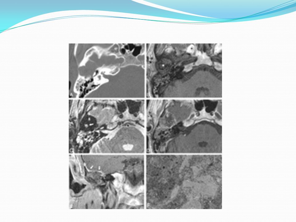
The mass is markedly but inhomogeneously enhancing, with components containing small, poorly enhancing or nonenhancing foci in Meckel's cave (arrow in A) and in petrous apex (arrows in B), as well as involvement of the longus colli muscle and jugular fossa (arrows in C).
 and in petrous apex (arrows in B), as well as involvement of the longus colli muscle and jugular fossa (arrows in C).")
54
Lipomas ; thinly encapsulated -soft, multilobular masses of typical adult adipose tissue. Lipomas within the IAC : symptoms typical of an AN. on CT : less dense than neuromas MRI is diagnostic; hyperintense on T1, nonenhancing with gadolinium, and hypointense on T2.

56
Dermoid Tumors ; skin-lined cystic tumor containing dermal and adnexal structures. The lining of the cyst : mature, stratified squamous epithelium. slowly expanding (symptoms similar to a primary cholesteatoma) CT ; non homogeneous cystic mass that contains calcium but less dense than brain
 CT ; non homogeneous cystic mass that contains calcium but less dense than brain.")
58
Teratomas ; arise from multipotential cells various tissues, representing more than one germ layer. Contain: ectodermal, mesodermal, endodermal Carcinomatous or sarcomatous : 10% -35% When malignant degeneration occurs, symptoms progress rapidly otherwise symptoms progress slowly like benign CPA tumors. CT : non homogeneous lesion of less density than brain without enhancing

59
INTRA-AXIAL TUMORS 1) arise from the brainstem (gliomas) 2) cerebellum :(medulloblastomas from the vermis or astrocytomas from the peduncles) 3) the fourth ventricle (choroid plexus papillomas and ependymomas). highly unusual In children, brainstem gliomas ; most common source of CPA neoplasms. Intra-axial tumors are usually isointense on T1 MRI and hyperintense on T2 images

60
Hemangioblastomas ; tumors of blood vessel origin in the cerebellum. They may also occur in the cerebral hemispheres and in association with a similar retinal tumor and may be multicentric. Histologically: benign, may produce major neurologic dysfunction by compression of the brainstem. Rapidly progressing signs and symptoms of cerebellar dysfunction Hearing and balance are likely to remain normal.

61
Imaging studies : intrinsic to the cerebellum that may extend into the CPA.

62
Medulloblastomas ; arise from the cells of the external granular layer of the cerebellar folia. exophytic masses on the cerebellum with extension into the CPA. Symptoms : destruction of cerebellar tissue and mass effect of the tumor on the CPA's adjacent structures. rapid development of symptoms + hearing loss and dizziness neurologic findings include facial weakness, dysmetria of speech and hand motion, perverted nystagmus,abnormal peripheral reflexes.

63
Temporal bone imaging : normal, CT and MRI show a lesion intrinsic to the cerebellum.

64
Brainstem Gliomas; Exophytic gliomas may arise on the surface of the pons and grow into the CPA. intrinsic to the brainstem. signs and symptoms similar to those of ANs, predominantly long tract signs. Patients with exophytic lesions may be diagnosed preoperatively to have ANs. Long tract signs in association with characteristic brainstem distortion on imaging make preoperative diagnosis possible in the patients with intrinsic lesions.

66
TUMORS OF THE FOURTH VENTRICLE Malignant Choroid Plexus Papillomas and Ependymomas arise from the fourth ventricle cause CPA symptoms by growing through the foramen of Luschka. early signs of CN VIII dysfunction. Ependymomas may calcify MRI: isointense with brain on T1 -mildly hyperintense to brain on T2 images

67
CPA ependymoma. A and B, Pre- and post-gadolinium images. The mass protruding into the right CPA from the foramen of Luschka shows clear demarcation and strong enhancement. C, T2-weighted image. The tumor is hyperintense and does not extend into the IAC

68
In malignant ; CT shows a mass with enhancing characteristics of a schwannoma Malignant ependymomas require multimodality therapy: 1) role of surgery is biopsy 2) brainstem decompression 3) management of hydrocephalus.
 role of surgery is biopsy 2) brainstem decompression 3) management of hydrocephalus.")
69
SELECTION OF SURGICAL APPROACH The principal goal : tumor removal with minimal postoperative morbidity. surgical approach should be tailored to patient's pathology and functional status. In cases without serviceable hearing, the translabyrinthine; 1) provides wide exposure 2) maximal facial nerve safety 3) minimal cerebellar retraction 4) low incidence of severe postoperative headache.
 provides wide exposure 2) maximal facial nerve safety 3) minimal cerebellar retraction 4) low incidence of severe postoperative headache..")
70
In meningiomas and tumors that do not affect the IAC and have not affected hearing: extended middle fossa approach is used if posterior and middle fossa exposure is necessary. retrolabyrinthine approach is used for limited lesions of the CPA only retrosigmoid approach : for more extensive lesions of the posterior fossa. Extensive anteromedial exposure is needed for lesions of the clivus.

71
Translabyrinthine: 1) Large, medium, or small cerebellopontine angle tumor 2) Wide exposure 3) facial nerve identified, immediate repair 4) limited cerebellar retraction 4) Total hearing loss Retrosigmoid (suboccipital): 1) Cerebellopontine angle tumors without extensive IAC 2) Hearing preservation possible 3) wide exposure 4) cerebellar retraction : hydrocephalus, intradural drilling may result in severe headaches
 Large, medium, or small cerebellopontine angle tumor 2) Wide exposure 3) facial nerve identified, immediate repair 4) limited cerebellar retraction 4) Total hearing loss Retrosigmoid (suboccipital): 1) Cerebellopontine angle tumors without extensive IAC 2) Hearing preservation possible 3) wide exposure 4) cerebellar retraction : hydrocephalus, intradural drilling may result in severe headaches")
72
Retrolabyrinthine: 1) CPA lesions without IAC 2) biopsy of cerebellopontine angle lesions 3) Hearing preservation possible 4) bone removal is extradural, no cerebellar retraction 5) Limited exposure Transcochlear: 1) Extensive lesions of petrous apex and clivus 2) Wide exposure of skull base with access to clivus, vertebral, and basilar arteries and full exposure of petrous carotid artery 3) Temporary facial nerve paralysis 4) total hearing loss
 CPA lesions without IAC 2) biopsy of cerebellopontine angle lesions 3) Hearing preservation possible 4) bone removal is extradural, no cerebellar retraction 5) Limited exposure Transcochlear: 1) Extensive lesions of petrous apex and clivus 2) Wide exposure of skull base with access to clivus, vertebral, and basilar arteries and full exposure of petrous carotid artery 3) Temporary facial nerve paralysis 4) total hearing loss")
73
Transotic: Same as transcochlear Middle fossa: 1) Intracanalicular tumors with minimal CPA involvement 2) good hearing Hearing preservation possible 3) Small tumors only 4) temporal lobe retraction Extended middle fossa : 1) Petroclival lesions involving posterior and middle fossa with good hearing 2) Hearing preservation possible 3) Extensive temporal lobe retraction
 Intracanalicular tumors with minimal CPA involvement 2) good hearing Hearing preservation possible 3) Small tumors only 4) temporal lobe retraction Extended middle fossa : 1) Petroclival lesions involving posterior and middle fossa with good hearing 2) Hearing preservation possible 3) Extensive temporal lobe retraction")
74
small tumors and good hearing have three options for surgical : 1) translabyrinthine approach that destroys hearing 2) the middle fossa approach in young patients with small, mainly intracanalicular tumors 3) the retrosigmoid approach in small tumors of the CPA that do not extend to the fundus of the IAC. Hearing preservation is usually not feasible in such lesions and the transcochlear or transotic approach may be used.

75
PATIENT MANAGEMENT AND SURGICAL COMPLICATIONS Preoperative Management 1) Preoperative antibiotics are not routinely used in uncomplicated cases. 2) Osmotic agents and diuretics for brain relaxation are usually not used in the translabyrinthine approach; however, the retrolabyrinthine, retrosigmoid, middle fossa, and extended middle fossa approaches require such techniques to minimize the need for retraction.
 Osmotic agents and diuretics for brain relaxation are usually not used in the translabyrinthine approach; however, the retrolabyrinthine, retrosigmoid, middle fossa, and extended middle fossa approaches require such techniques to minimize the need for retraction..")
76
Intraoperative Monitoring 1) Facial nerve monitoring is used routinely in all cases of posterior fossa 2) ABR monitoring is not routinely used.
 Facial nerve monitoring is used routinely in all cases of posterior fossa 2) ABR monitoring is not routinely used.")
77
Postoperative Care 1) The compression mastoid dressing until po 4th 2) neurologic intensive care unit for 1 to 2 days and then transferred to the general ward. 3) Limited activity is begun on the first postoperative morning and ambulation usually begins po 3-4 th
 Limited activity is begun on the first postoperative morning and ambulation usually begins po 3-4 th.")
78
Complications 1) Anteroinferior Cerebellar Artery changes in vital signs Atkinson's syndrome infarction of the lateral tegmental pons 2) Meningitis The mean time of onset of meningitis in patients with AN is 8 days postoperatively. Aggressive medical management

79
3) Cerebrospinal Fluid Leaks In translabyrinthine ; through the wound is rare(<10%)-usually responds : mastoid dressing. obliteration of the eustachian tube In rare case of persistent leakage, lumbar CSF drainage for 3 days In refractory cases, abdominal fat Hydroxyapatite cement

80
………..csf leak In the retrosigmoid : mastoidectomy is also obliterated with fat. In some institutions,fluid leakage through the eustachian tube. Aggressive bone wax over mastoid air cells is necessary to avoid leaks persistent leaks: fat obliteration of the mastoid and waxing of air cells are usually successful

81
4) Facial Nerve ideally managed ; immediate repair -interposition graft direct repair is impossible or intact nerve does not resume function within 1 year; facial hypoglossal anastomosis Temporalis muscle transfer
 Facial Nerve ideally managed ; immediate repair -interposition graft direct repair is impossible or intact nerve does not resume function within 1 year; facial hypoglossal anastomosis Temporalis muscle transfer")
82
5) Ophthalmologic facial paralysis and corneal insensitivity, in large tumors. managed prolonged facial paralysis (6 months) ; a brow lift and eyelid spring. management partial facial weakness: 1- lubricating drops 2- ointments 3-contact lenses 4- moisture chamber protection, and nightly taping of the affected eye is undertaken 5- gold weight implantation to the upper lid
 ; a brow lift and eyelid spring. management partial facial weakness: 1- lubricating drops 2- ointments 3-contact lenses 4- moisture chamber protection, and nightly taping of the affected eye is undertaken 5- gold weight implantation to the upper lid.")
Similar presentations


 Ass. Professor of Clinical Oncology Kasr El-Aini School of Medicine.>")


Primary Tumours: Benign Glomus tumour Malignant Carcinoma,sarcoma 2)Secondary Tumours: a) From adjacent areas like.>")


 10.1 A 10.1 B 10.1 C Precontrast sagittal T1 wtd. MRI of.>")

 H. Louis Harkey Department of Neurosurgery University of Mississippi Jackson, MS.>")


