Download presentation
Presentation is loading. Please wait.

1
IN THE NAME OF GOD THE COMPASSIONATE THE MERCIFUL

2
Histology Lecture 10 Nervous Tissue

3
Nervous Tissue Cells of nervous tissue are classified into two categories: neurons and neuroglial cells Neurons form an Integrated communication network They are responsible for receiving, processing, transmitting and motor functions of nervous system Perhaps about one trillion neurons exist in human nervous tissue Neurons are the functional units of nervous tissue There are more other cells which is called neuroglial cells that supporting and protecting neurons Vasculature are other important component of nervous tissue

4
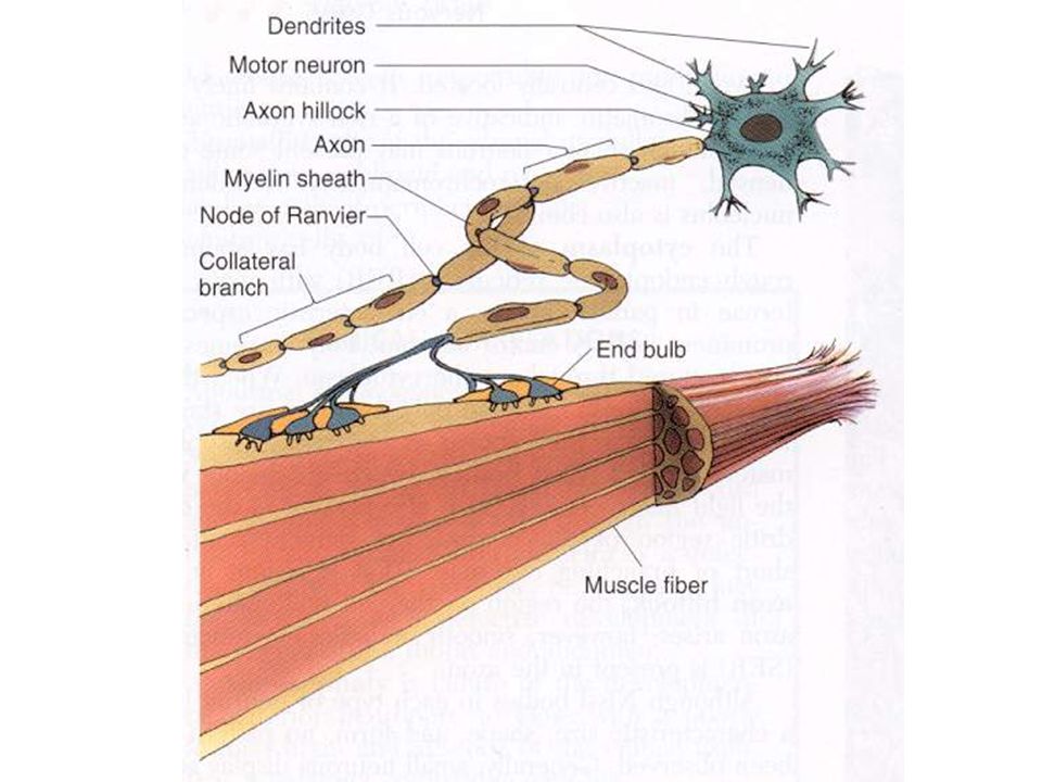
Neuron Structure Cell body/perikaryon (5-150 mm diameter) An axon (myelinated or unmyelinated) with terminals and synaptic junctions, up to 100 cm in length Dendrites specialized for receiving inputs
 An axon (myelinated or unmyelinated) with terminals and synaptic junctions, up to 100 cm in length Dendrites specialized for receiving inputs")
6
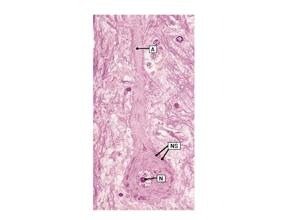
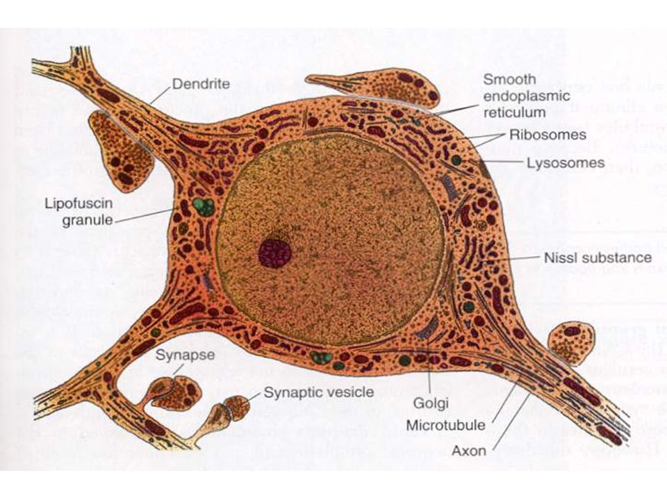
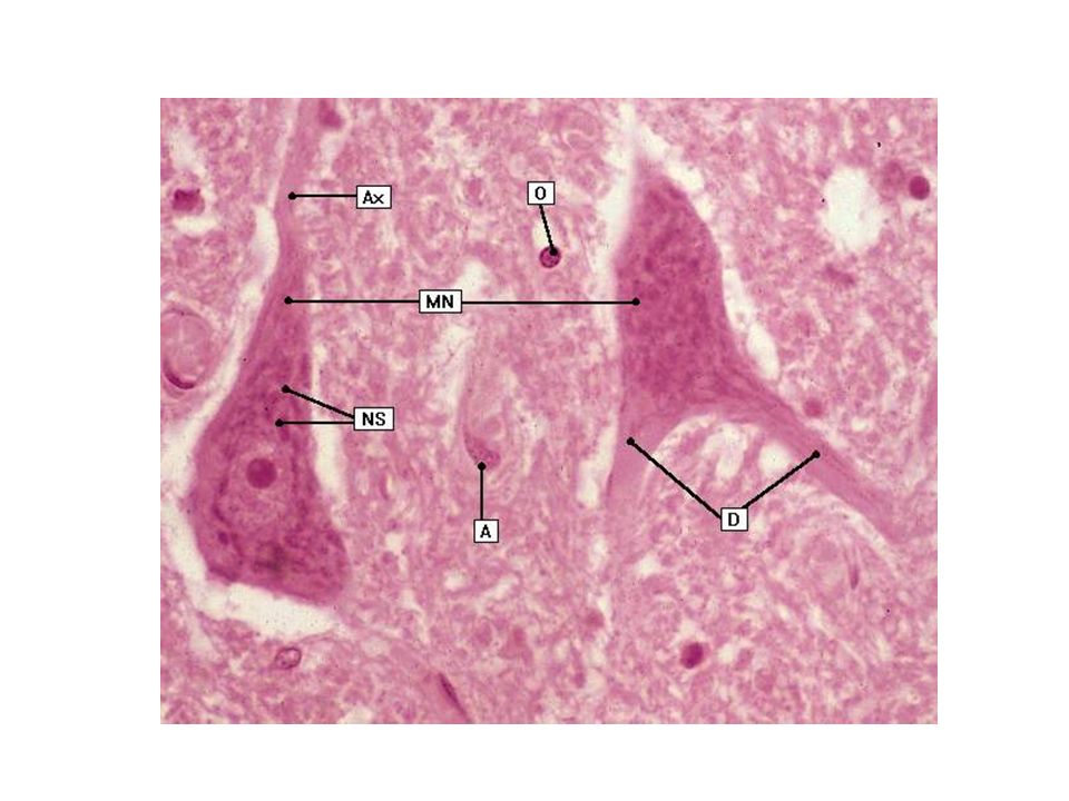
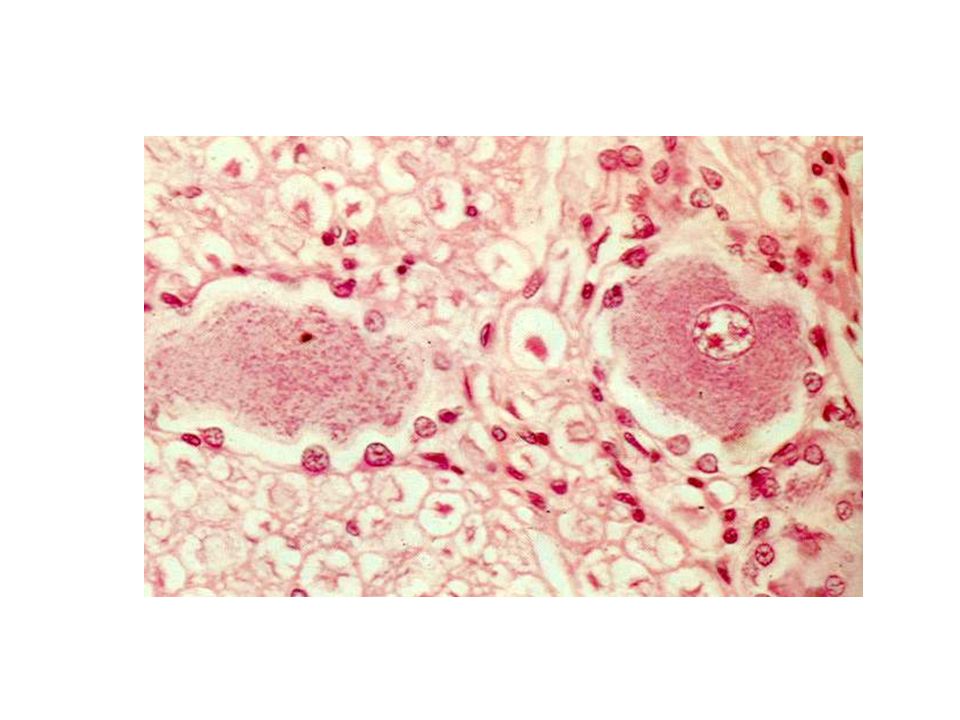
Perikaryon (Cell Body or soma) Nucleus: large, euchromatic, prominent nucleolus, usually centrally located Nissl bodies: clumps of RER absent at axon hillock Abundant amount of SER forming hypolemmal cisternae directly beneath cell membrane hypolemmal cisternae sequester Ca ions and contain proteins Prominent juxtanuclear Golgi apparatus and numerous mitochondria A centriol associated with a basal body
 Nucleus: large, euchromatic, prominent nucleolus, usually centrally located Nissl bodies: clumps of RER absent at axon hillock Abundant amount of SER forming hypolemmal cisternae directly beneath cell membrane hypolemmal cisternae sequester Ca ions and contain proteins Prominent juxtanuclear Golgi apparatus and numerous mitochondria A centriol associated with a basal body")
8
Inclusions: melanin (rare), lipofuscin and lipid droplets Melanin in certain region(Substantia nigra), and it may be by-product of the synthesis of dopamine & noradrenaline Lipofuscin more in older adults, crowd organelles and nucleus to one side and may affecting neuron function Lipid droplets may be energy reserve or the result of metabolism Cytoskeleton: Microtubules (24 nm), neurofilaments(10 nm, intermediate filaments), microfilaments (6 nm) Neurofibrils possibly are clumped bundles of neurofilaments Perikaryon
, lipofuscin and lipid droplets Melanin in certain region(Substantia nigra), and it may be by-product of the synthesis of dopamine & noradrenaline Lipofuscin more in older adults, crowd organelles and nucleus to one side and may affecting neuron function Lipid droplets may be energy reserve or the result of metabolism Cytoskeleton: Microtubules (24 nm), neurofilaments(10 nm, intermediate filaments), microfilaments (6 nm) Neurofibrils possibly are clumped bundles of neurofilaments Perikaryon")
10
Dendrites May be highly branched Receptive surface for synaptic junctions Tens of thousands of synapses on large dendrites Dendritic spines located on surface of some dendrites Spines diminish with age and poor nutrition

11
Axons 1 axon projects from cell body at axon hillock Some axons are up to 100 cm in large animals Axon hillock a pyramid shaped region of the soma that is devoid of RER Initial segment is a portion of axon from its origin to the beginning of myelin sheath, referred as Spike trigger zone At spike trigger zone summation of excitatory and inhibitory impulses occurred Collateral branches, Terminal arbor Myelinated or Unmyelinated

12
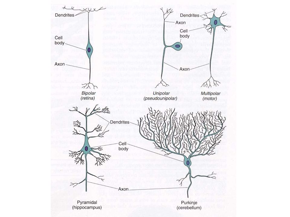
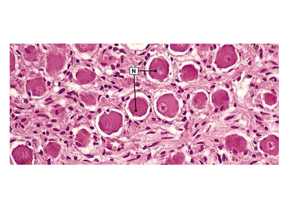
Functional Categories of Neurons Motor(efferent) neurons: conduct impulses to muscles, neurons, glands Sensory(afferent) neurons: receive sensation Interneurons: local circuit neurons Morphological Categories of Neurons Multipolar (the most common type of neuron) Bipolar ( located in vestibular and cochlear ganglion) Unipolar (located in dorsal root ganglia)
 neurons: conduct impulses to muscles, neurons, glands Sensory(afferent) neurons: receive sensation Interneurons: local circuit neurons Morphological Categories of Neurons Multipolar (the most common type of neuron) Bipolar ( located in vestibular and cochlear ganglion) Unipolar (located in dorsal root ganglia)")
16
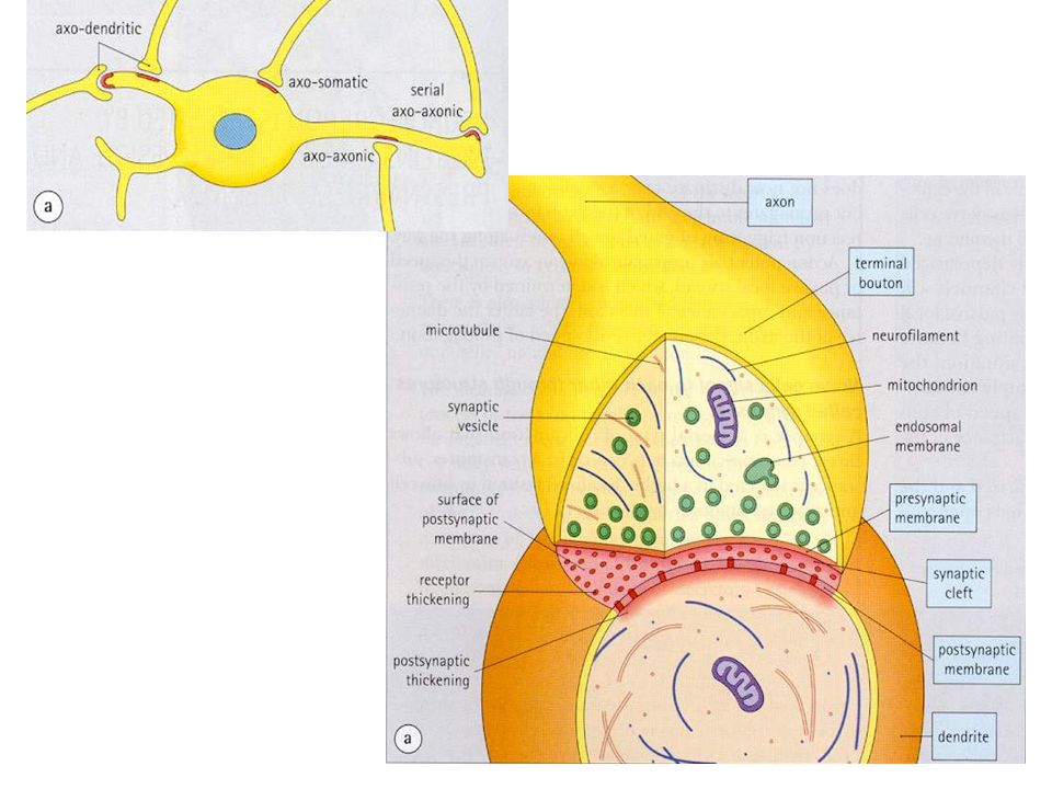
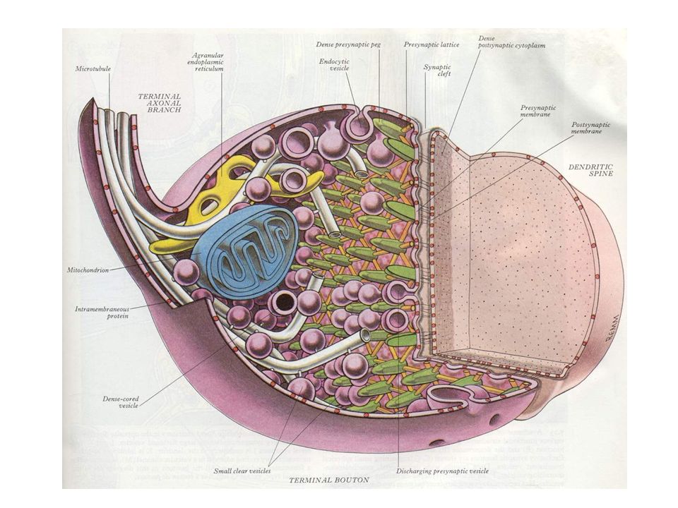
Synaptic Junctions Synapses are the sites where impulses are transmitted from a presynaptic cell to a postsynaptic cell Axon terminal form bouton terminal Presynaptic membrane contains mitochondria, and an abundance of synaptic vesicles with neurotransmitter, Presynaptic dense projections are associated with synaptic vesicles form active sites of synapse Other vesicles are reserve pool and adhere to actin Postsynaptic membrane contain receptors, and some dense materials Synaptic cleft 20-30 nm width, occupied by fine filaments

18
Synaptic Junctions Glial cells increase synaptic efficacy Asymmetric synapses are excitatory Form by a thick postsynaptic density and a 30 nm synaptic cleft Symmetric synapses are inhibitory Form by a thin postsynaptic density and a 20 nm synaptic cleft Need special staining to see by light microscopy

20
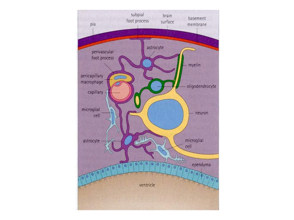
Features of Neuroglia Non-neural supporting cells of several types: astroglia, oligodendroglia, microglia and ependyma of CNS and Schwann cell, satellite cell of PNS Only nuclei visible by light microscopy without special staining There are several glial cells for each neuron Neuroglial cells have important functions

21
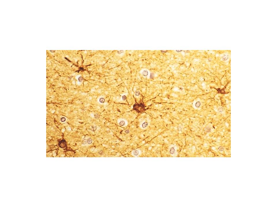
Astrocytes Largest, sperical/oval nucleus with small amount of heterochromatin, intermediate filament composed of glial fibrillar acidic protein Provide structural and metabolic support to neurons and act as scavengers of ions and neurotransmitters released into the extracellular space Fibrous type in white matter and protoplasmic type in gray matter Processes end as pedicels that surround vessels and form a layer at CNS surface (pia-glial membrane) Produce glial scar at injury Activity to maintain homeostasis May assist in maintaining Blood-Brain Barrier
 Produce glial scar at injury Activity to maintain homeostasis May assist in maintaining Blood-Brain Barrier")
24
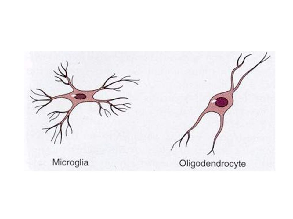
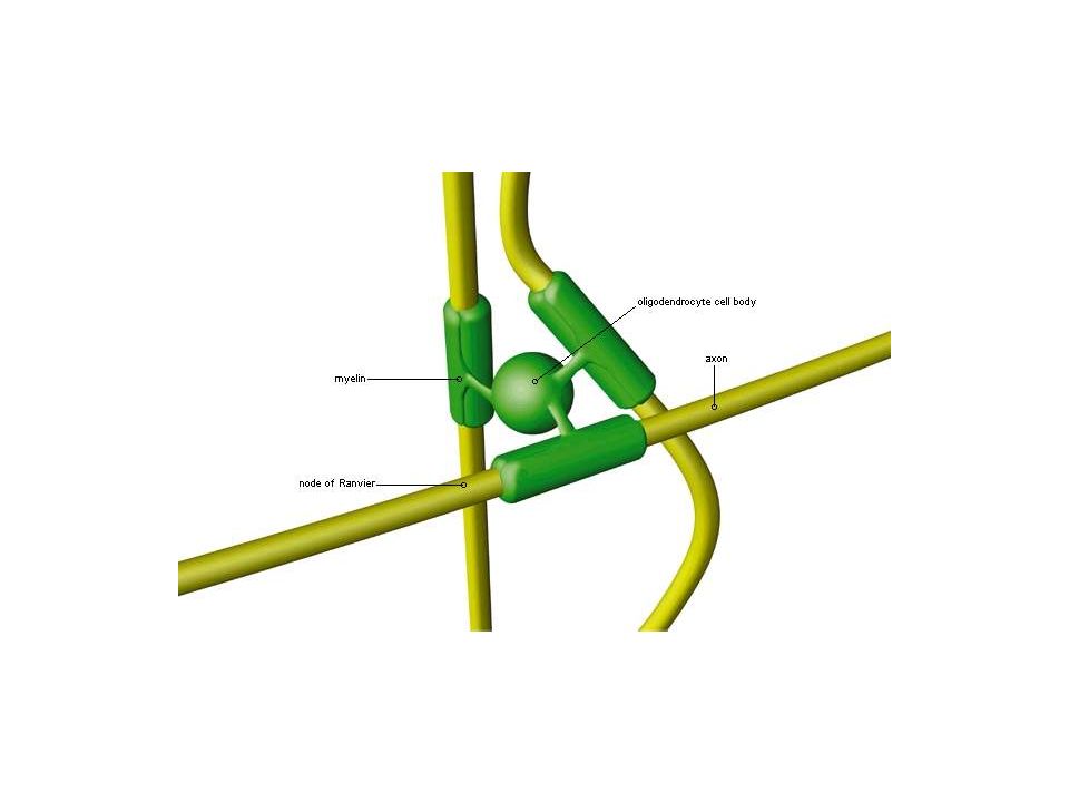
Oligodendrocytes Smaller than astrocytes; darker, round nucleus, abundant RER, well developed golgi apparatus, Interfascicular oligodendrocytes forming myelin of axon in CNS Function in electrical insulation Satellite oligodendrocytes are near cell bodies of large neurons, their function unclear Damaged in multiple sclerosis

28
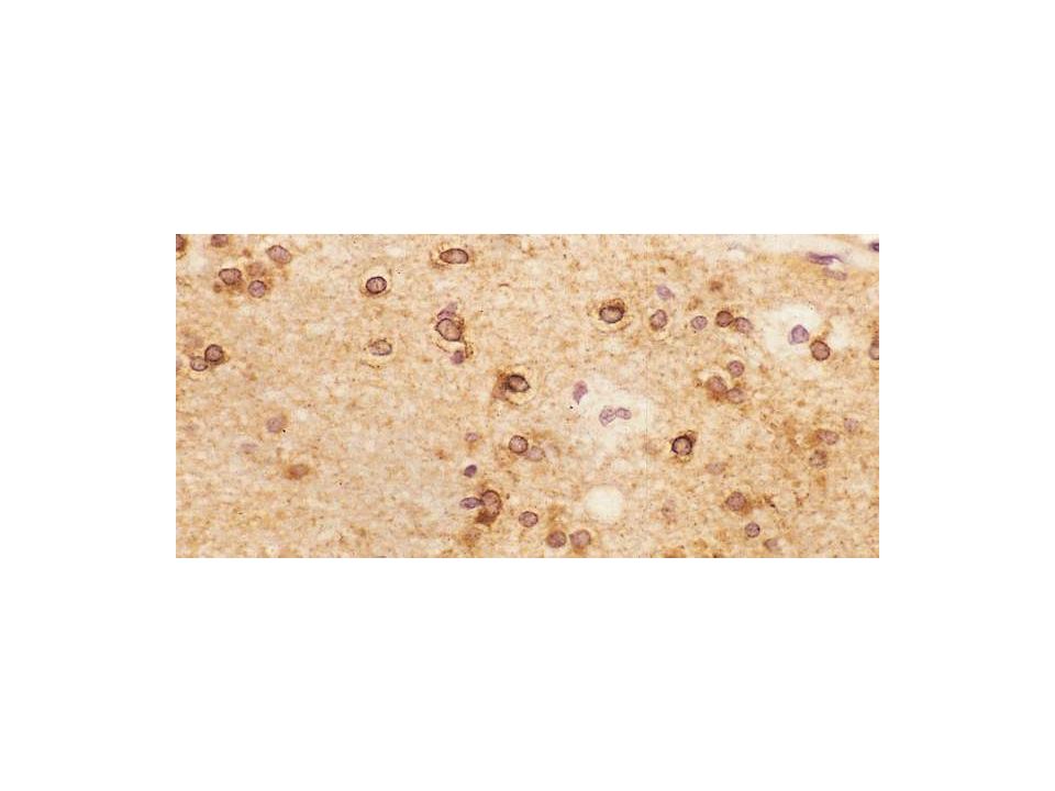
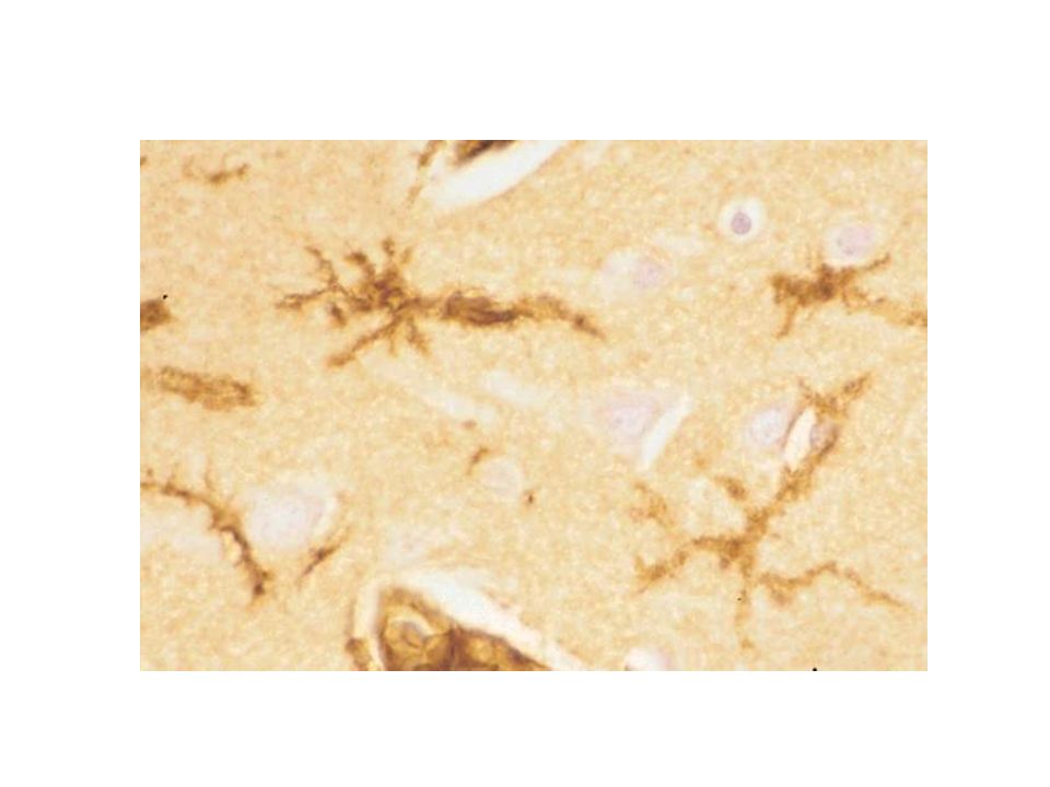
Microglial Cells Smallest neuroglial cell, with heterochromatin nucleus and short processes phagocytes and clearing debris and damaged structures in the CNS When activated act as antigen presenting cell Originate in the bone marrow

30
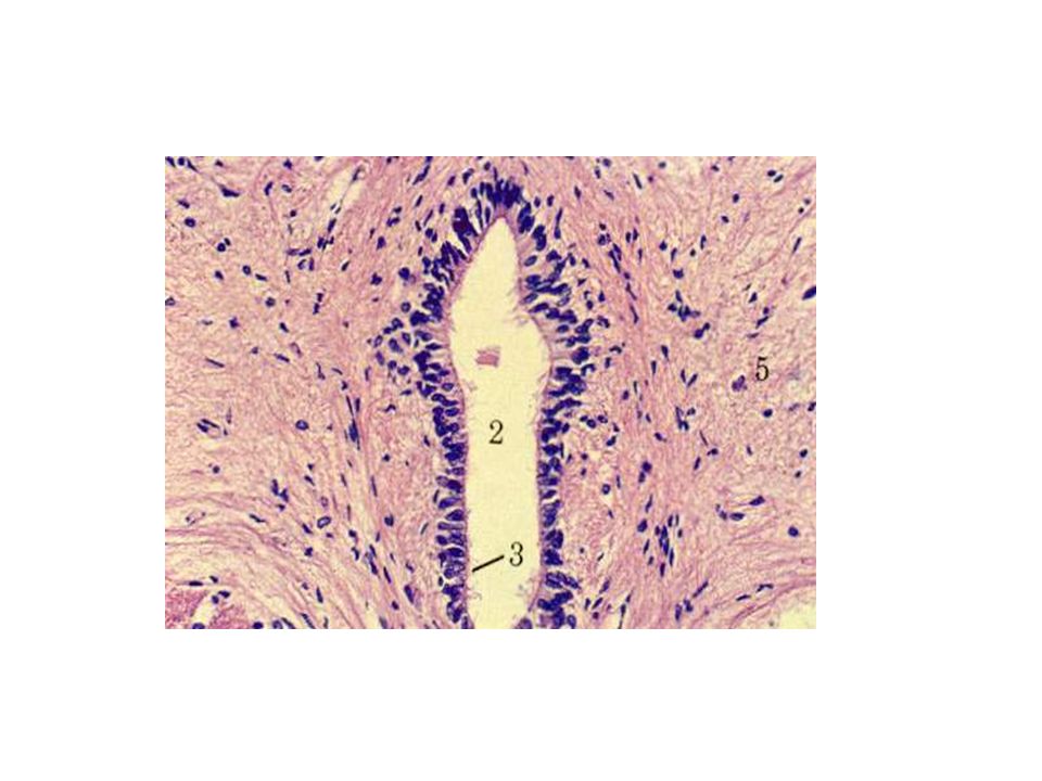
Ependymal Cells Line ventricles of CNS and cental canal of spinal cord Cuboidal or low columnar shape Some are ciliated, facilitate movement of CSF Participate in formation of Choroid plexus

33
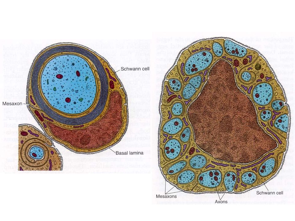
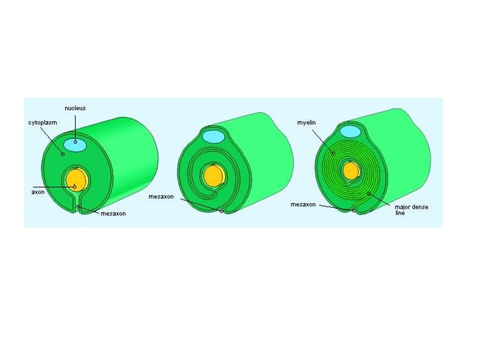
Schwann cells form both myelinated and unmyelinated covering over axons of the PNS Interruptions in myelin sheath is called nodes of Ranvier This cell has basal lamina that covers axon at nodes of Ranvier Schmidt-Lanterman clefts in myelin are schwann cell cytoplasm trapped within the lamellae of myelin A Schwann cell can myelinate just one internode of a single axon The length of an internodal segment is 200 to 1000 micrometer Schwann cells

36
Satellite cells Satellite cells are small support cells Resembling Schwann cells which surround the neuron cell bodies in ganglia

39
HR Mahmoudzadeh Sagheb Z Heidari MH Noori Mugahi Department of Histology

Similar presentations


 and gathers information.>")

 Integration:>")

>")
 1.Microglial cells –Scattered throughout CNS –Support neurons.>")

