Download presentation
Presentation is loading. Please wait.

1
M/29 C.C: Right hip pain

3
T1 Gd-enhanced FS T1 T2 Gd-enhanced FS T1

4
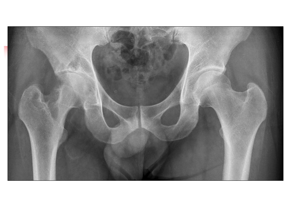
Answer: Tuberculous arthritis Findings: - Anteroposterior view of the hip shows periarticular osteoprosis, bony erosions, subchondral cysts with internal radiopaque density and capsular distension. Joint space narrowing of superior direction is also noted. - Axial MR images of right hip demonstrate bony erosions and joint capsular distension by the materials with central low and peripheral intermediate signal intensities on T2-weighted image and only peripheral enhancement on enhanced fat-suppressed T1-weighted image. The peripheral rims along the capsule has irregular thickness. Periarticular abscess adjacent to obturator externus muscle is seen. - Coronal MR images reveals subchondral lesions at both acetabulum and femoral head. The lesions shows low signal intensities with dark signal foci in central part and intermediate to high signal intensity in peripheral part, suggesting tuberculomas.

5
Diagnosis: Tuberculous arthritis, right hip. Differencial diagnosis: Pyogenic arthritis, Rheumatoid arthritis

6
Discussion: On plain radiograph, tuberculous arthritis usually shows Phemister’s triad; juxta- articular osteoporosis, peripherally located osseous erosions and progressive joint space narrowing. The MR findings of tuberculous arthritis are subchondral erosion, synovial proliferation and periarticular soft tissue abscess formation. The presence of a tuberculoma in intraarticular space or periarticular soft tissue is a specific finding in tuberculous arthritis, and it can differenciate them from pyogenic arthritis. Tuberculoma is composed of a central caseous necrosis and peripheral capsule with fibroblasts, epithelioid cells, macrophages and lymphocystes. Tuberculoma on MR scans demonstrates heterogeneously intermediate or variable signal intensities and peripheral rim enhancement on T1-weighted image and central low signal intensity with a few dark signal foci and peripheral intermediate signal intenty on T2-weighted image. The dark signal foci on T2-weighted image may be due to free radicals with irregular distribution and immobile saturated fatty acids, leading to T2- shortening. On the other hand, abscess of tuberculous arthritis shows various signal intensities and enhancing peripheral wall with smooth inner margin on T1-weighted image and high signal intensity on T2-weighted image.

7
References 1.Jan HW, Kim JW, Cho KH. MR findings of tuberculous arthritis: significance of tuberculoma. J Korean Radiol Soc 2001;44:237-241 2.Kim TK, Chang KH, Kim CJ, Goo JM, Kook MC, Han MH. Intracranial tuberculoma: Comparison of MR with pathologic findings. AJNR Am J Neuroradiol 1995;16:1903-1908 3.Suh JS, Lee JD, Cho JH, Kim MJ, Han DY, Cho NH. MR imaging of tuberculous arthritis: clinical and experimental studies. J Magn Reson Imaging. 199;6(1):185-189
:")
Similar presentations


:1848-1864 August 1, 2005 ©2005 by The Journal of Bone.>")


>")


