Download presentation
Presentation is loading. Please wait.

1
Diabetes Mellitus Type II

2
Beta Cell Failure in DM T2 signaling pathways implicated in β -cell failure

3
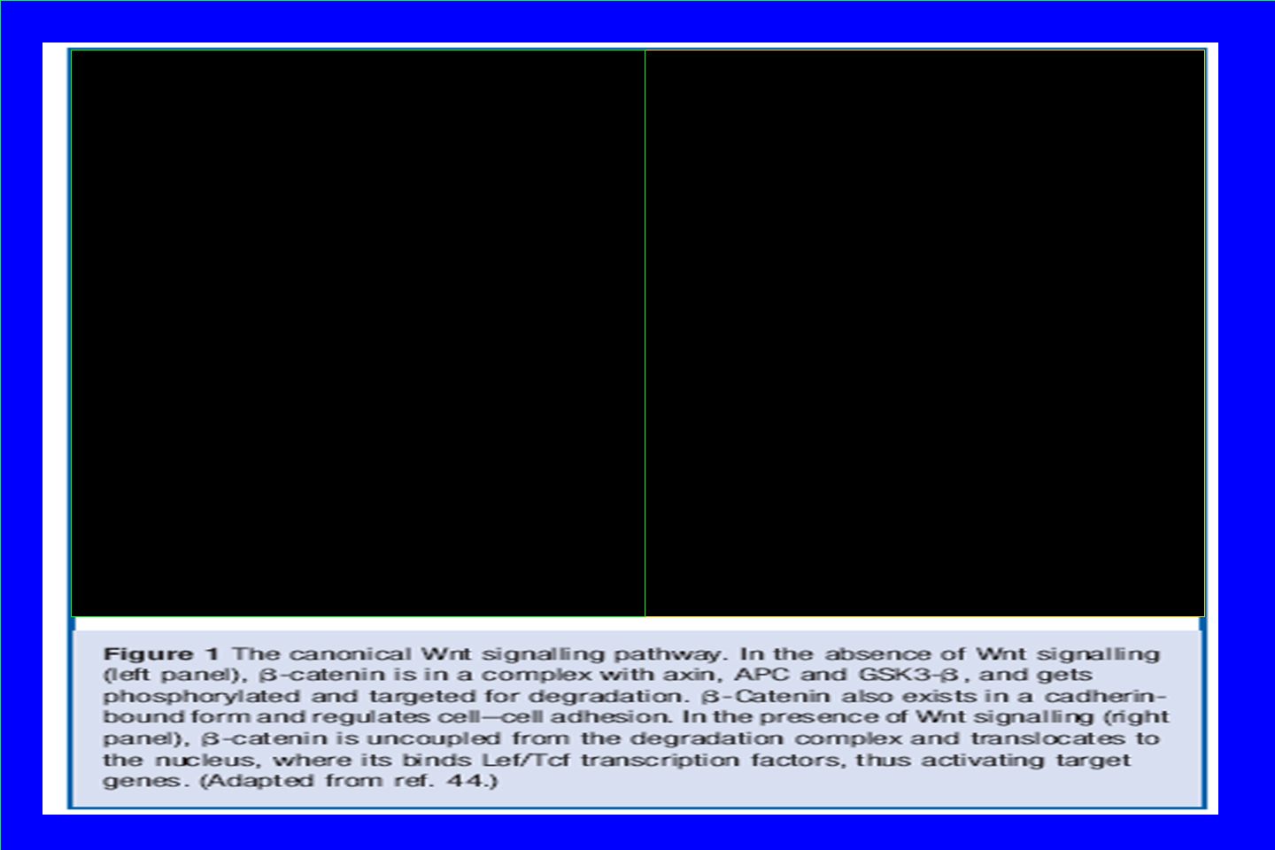
Wnt Signaling Pathway Controls organismal growth and differentiation

6
Wnt signalling Pathway and DM T2 1. Homozygous mutation of LRP5 in mice leads to defective glucose-stimulated insulin secretion from isolated islets in vitro. 2. Components of the Wnt pathway are present in the adult pancreas, and in particular multiple members of the frizzled family of Wnt receptors have been identified in the islet. 3. TCF7/L2 gene polymorphism affects B-cell function

7
While most studies have found little evidence that Wnt signaling is involved in endocrine differentiation or in the adult islet, there is some evidence for an effect of Wnt signaling on β-cell replication.

9
Main Objective To study the role of Wnt signaling in human diabetes Systematically examined components of the pathway in the pancreas of normal & DMT2 Mouse Studies

10
METHODS TISSUE PREPARATION 1. Paraffin-embedded from human pancreases: 5 non-diabetic & 9 DM T2 2. Freshly isolated & cultured pancreases: 3 non-diabetic (Isolated human islets) 3. Human Fetal Pancreases in 18-24 gestational weeks 4. Murine Pancreaes (Balb/c or C57/bl mice) 5. NEPCs WESTERN BLOT 1. Whole-cell extracts IMMUNO-HISTOCHEMISTRY 1. Paraffin samples 2. Frozen samples
 3. Human Fetal Pancreases in gestational weeks 4. Murine Pancreaes (Balb/c or C57/bl mice) 5. NEPCs WESTERN BLOT 1. Whole-cell extracts IMMUNO-HISTOCHEMISTRY 1. Paraffin samples 2. Frozen samples.")
11
I.Objective: To determine if TCF7L2 is upregulated in islets of type II diabetic patients TCF7L2 is a protein acting as a transcription factor. Several SNPs of the gene are associated with Type 2 DM

12
TCF/L2TCF3 TCF7L2 is upregulated in islets of type II diabetic patients

13
II. Objective: To determine if Wnt2b is upregulated in type II diabetes To test the hypothesis that the induction of TCF factors, being a both effector as well as a downstream target of Wnt Signaling, is a result of a more global activation of Wnt signaling

14
B Catenin Wnt2b In islets Wnt 2b is upregulated in Type 2 Diabetes

15
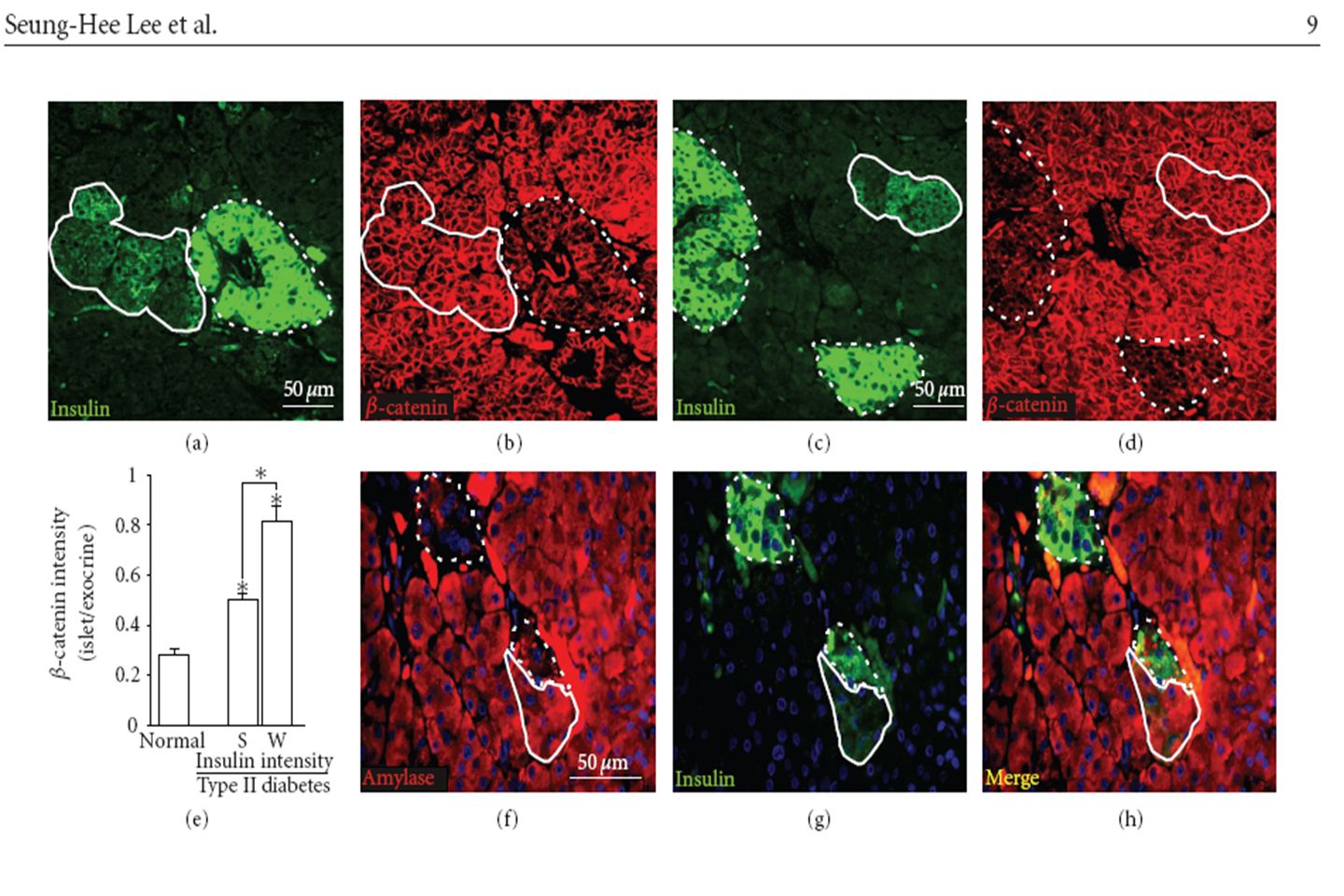
III. Objective: To determine if B-catenin is upregulated in type II diabetes

16
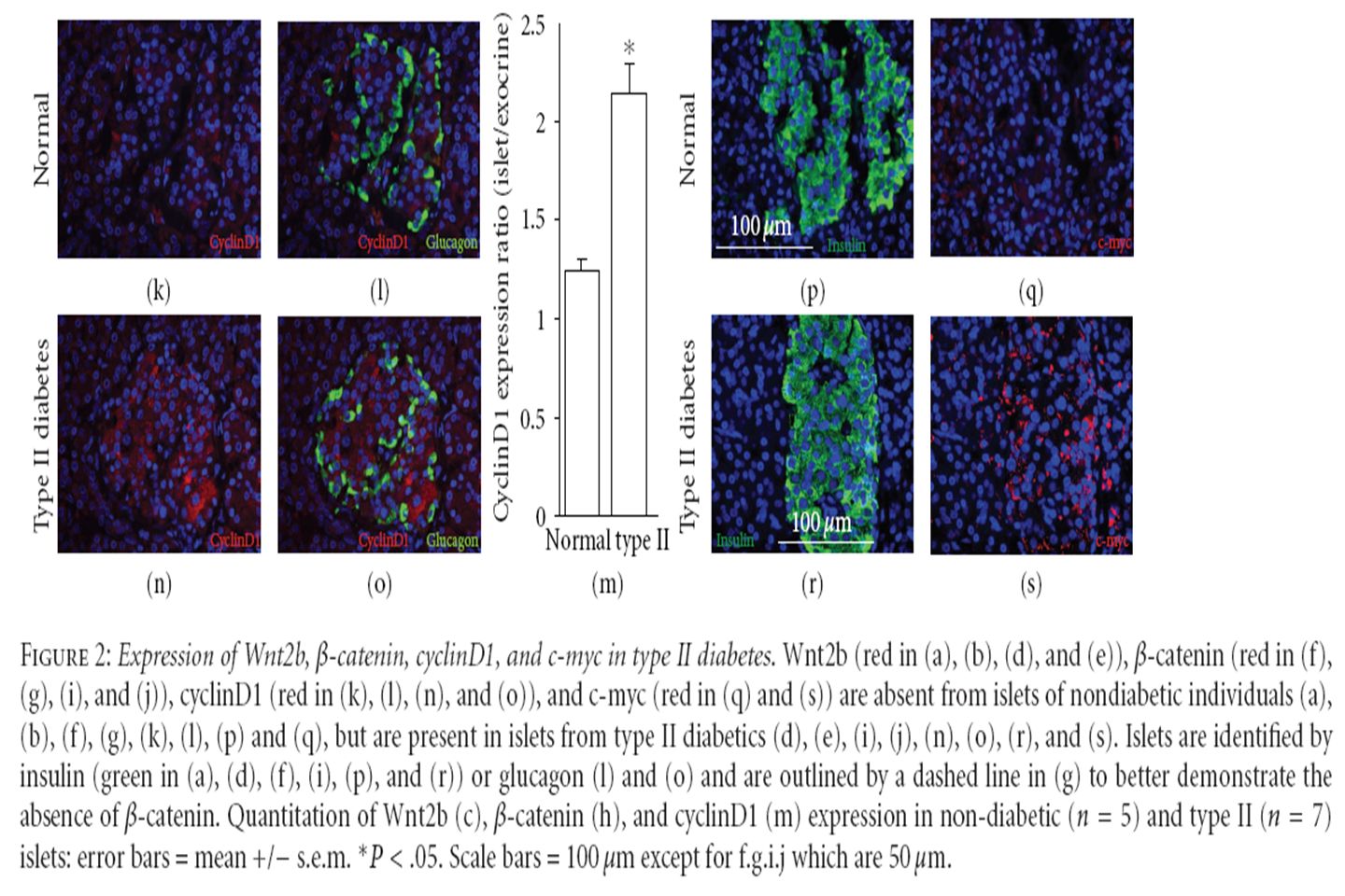
B-Catenin In islets Beta-catenin in human islets of all 5 nondiabetics was markedly lower Than in the surrounding exocrine Tissue where it was strongly Expressed (figs. 2f-2h) Beta-catenin is strongly expres in the islets of all DM T2 to a Level approximately half that of the Surrounding exocrine tissue (figs 2h-2j) insulin islet insulin B-catenin insulin B-catenin DM T2 Normal Human B-cells lack Detectable B-catenin expression but it is highly upregulated in DM T2
 Beta-catenin is strongly expres in the islets of all DM T2 to a Level approximately half that of the Surrounding exocrine tissue (figs 2h-2j) insulin islet insulin B-catenin insulin B-catenin DM T2 Normal Human B-cells lack Detectable B-catenin expression but it is highly upregulated in DM T2.")
17
IV. Objective: To determine if the terminal effectors (c-myc & cyclin D) are upregulated in DM T2 TCF/LEF factors activate a number of terminal effectors Of Wnt signaling (c-myc and cyclinD1)
 are upregulated in DM T2 TCF/LEF factors activate a number of terminal effectors Of Wnt signaling (c-myc and cyclinD1).")
18
c NORMAL DM T2 CyclinD1 In islets C-myc In islets Terminal effectors of Wnt signaling (c-myc & cyclinD1) are upregulated in human type 2 Diabetes
 are upregulated in human type 2 Diabetes")
19
MOUSE MODEL V. Objective: To determine whether Wnt activation was an early or late event in diabetes pathogenesis

20
high-fat diet normal chow Harvested pancreases examined for c-myc expression OBESE BUT WITH NORMAL FBS 12 weeks

21
control High-fat control High-fat control

22
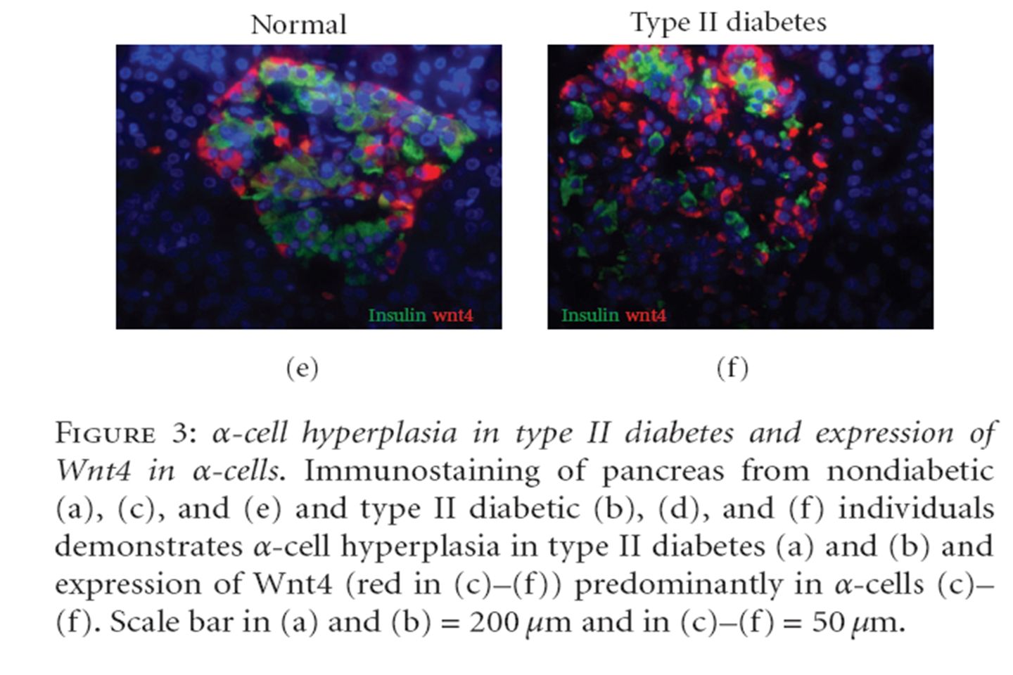
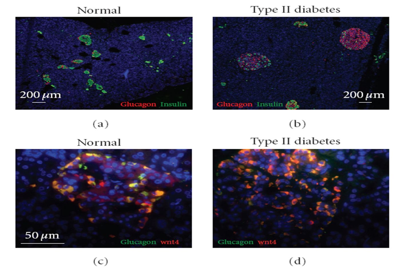
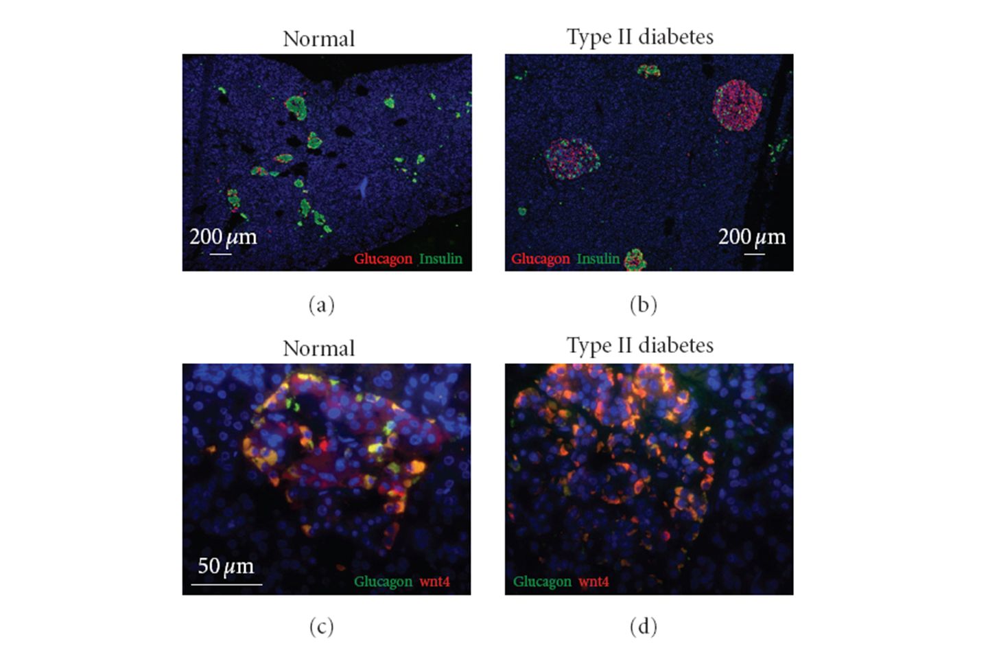
Figure 6: Wnt signaling in high-fat fed mice. In normal mouse pancreas, β -catenin (green in (a) and (b)) is expressed in islets as identified by somatostatin (red in (a)) and colocalizes with insulin (red in (c)). C-myc (red in (d) and (e)) was not expressed in islets of normal mice (marked by dotted lines and glucagon in green in (d)) but was induced in islets and some ducts of high-fat fed mice (islets marked by glucagon in green). C-myc expression is quantitated in (f). B -catenin somatostatin insulin Islets C-myc High-Fat Normal Glucagon Expression of the Wnt target gene c-myc is an early response to high-fat diet
 and (b)) is expressed in islets as identified by somatostatin (red in (a)) and colocalizes with insulin (red in (c)). C-myc (red in (d) and (e)) was not expressed in islets of normal mice (marked by dotted lines and glucagon in green in (d)) but was induced in islets and some ducts of high-fat fed mice (islets marked by glucagon in green). C-myc expression is quantitated in (f). B -catenin somatostatin insulin Islets C-myc High-Fat Normal Glucagon Expression of the Wnt target gene c-myc is an early response to high-fat diet.")
23
Summary 1. B-catenin in human islets of all 5 nondiabetics was markedly lower than in the surrounding exocrine tissue, where it was strongly expressed. 2. Expression of the Wnt2b, B-catenin, TCF7/L2, & terminal effectors (c-myc & cyclinD1) are all upregulated in the islets in type II diabetes 3. The mouse model suggested that obesity alone may be sufficient to induce Wnt activation, which would mark it as an early event in the pathogenesis of type II diabetes.
 are all upregulated in the islets in type II diabetes 3. The mouse model suggested that obesity alone may be sufficient to induce Wnt activation, which would mark it as an early event in the pathogenesis of type II diabetes..")
24
Conclusion Type II Diabetes activates the Wnt signaling pathway specifically in the Beta cells of the islets of Langerhans

25
Thank You!!!

27
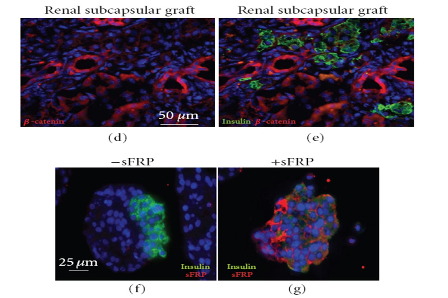
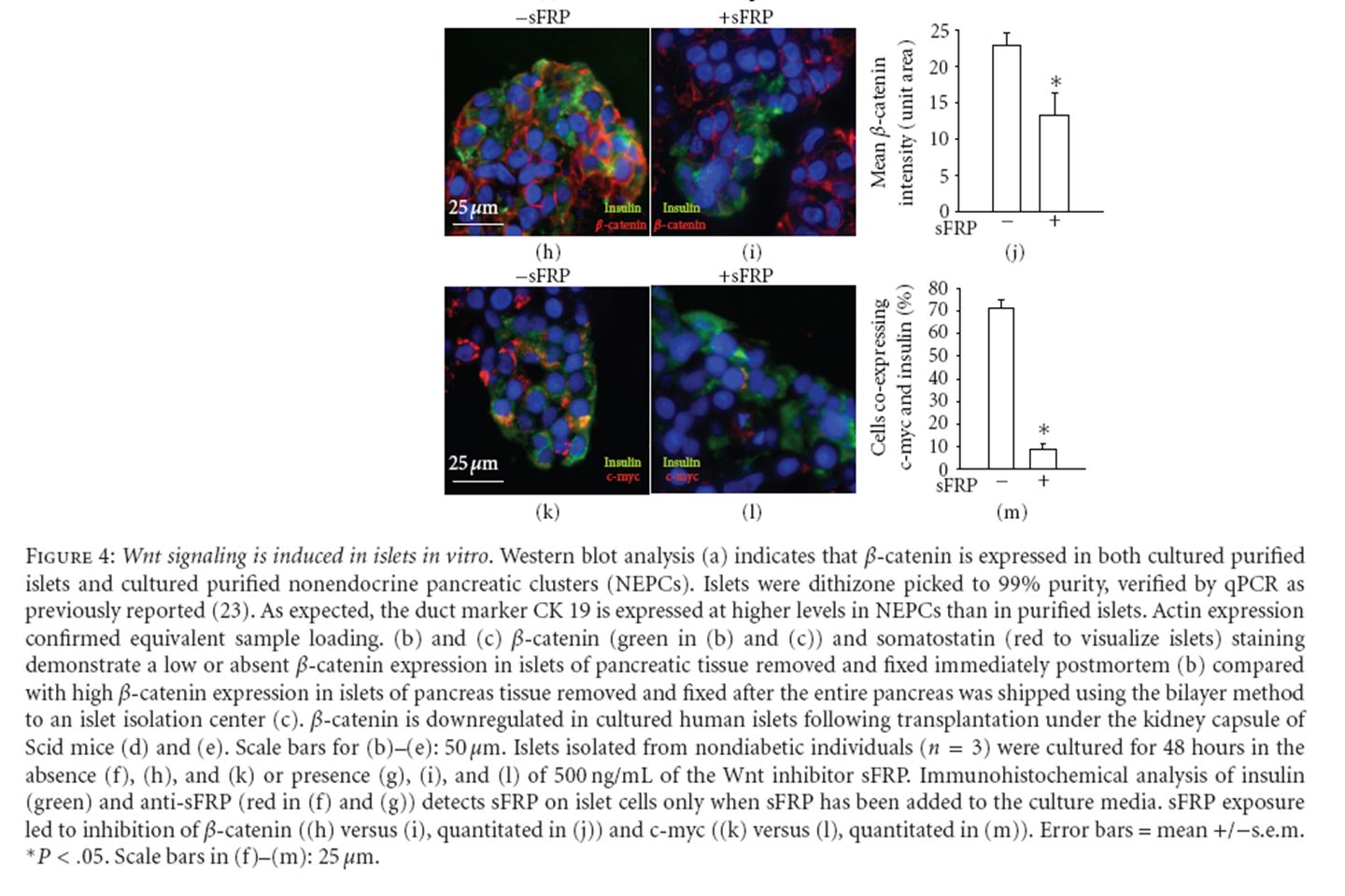
Immunohistochemical analysis of insulin (green) and anti-sFRP (red in (f) and (g)) detects sFRP on islet cells only when sFRP has been added to the culture media. sFRP exposure led to inhibition of β-catenin ((h) versus (i), quantitated in (j)) and c-myc ((k) versus (l), quantitated in (m)). Error bars = mean +/−s.e.m. ∗ P <.05. Scale bars in (f)–(m): 25 μm.
 versus (i), quantitated in (j)) and c-myc ((k) versus (l), quantitated in (m)). Error bars = mean +/−s.e.m. ∗ P <.05. Scale bars in (f)–(m): 25 μm..")
29
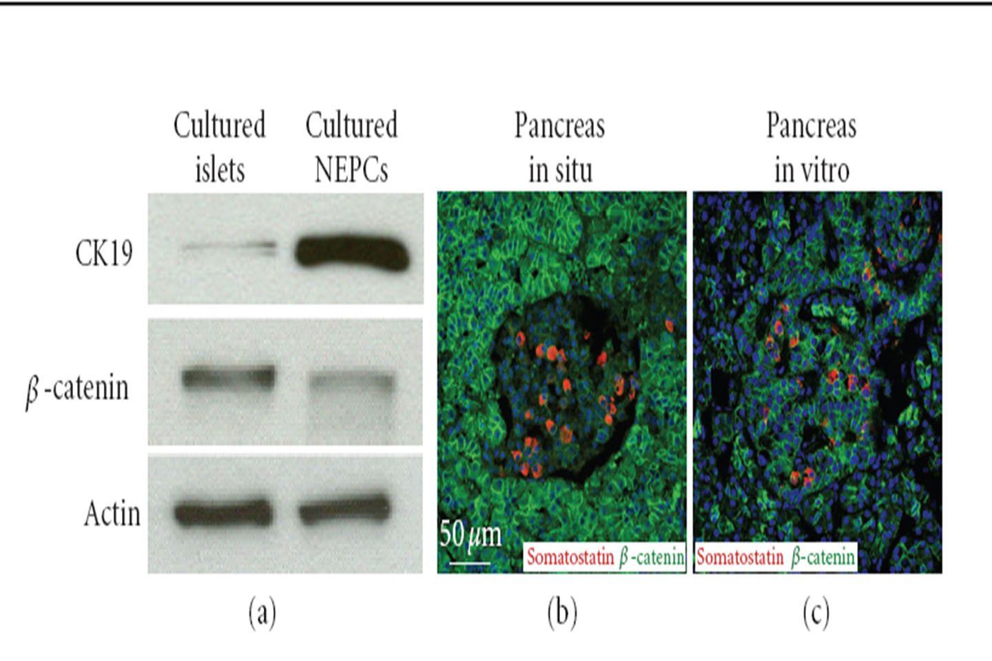
To pursue the role of Wnt activation in the islet, it would be desirable to have an in vitro model. Thus, we examined Wnt activation in isolated islets. Surprisingly, when cultured human islets were examined by Western blotting, β- catenin, which is low or absent in the islet compared with surrounding tissue in situ, was expressed at a higher level than in the nonendocrine pancreatic cells (NEPCs) [23] (Figure 4(a)).
 [23] (Figure 4(a))..")
36
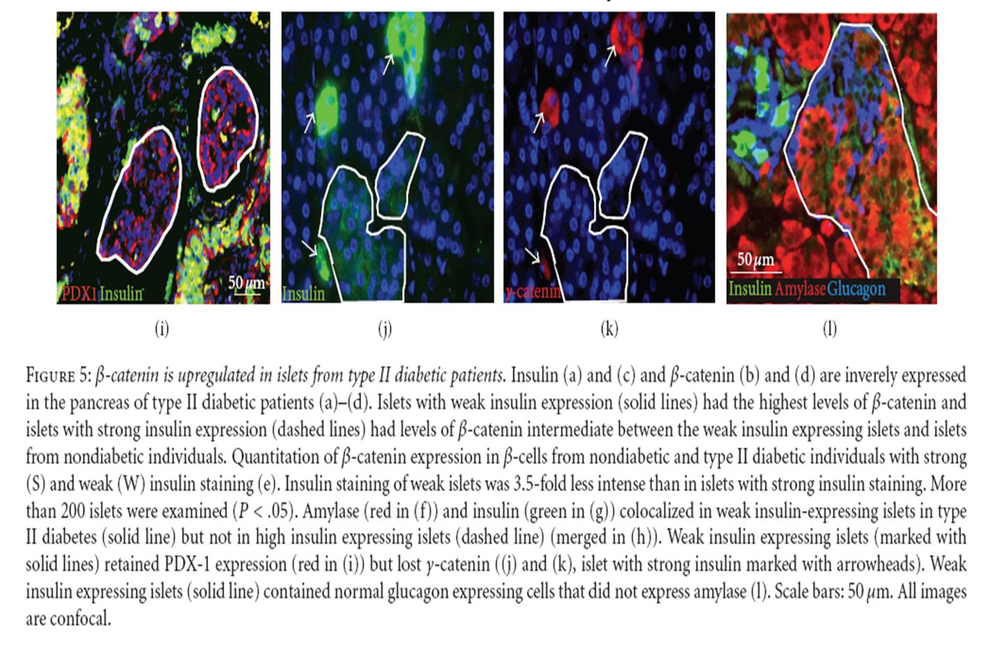
To pursue the finding that β-cells with low- insulin expression had a pattern of catenin expression resembling that in the exocrine pancreas, pancreas sections were immunostained for the acinar marker amylase as well as insulin, revealing that low-insulin β- cells coexpressed amylase (Figures 5(f), 5(g), 5(h)). The insulin/amylase doublepositive cells expressed PDX-1 (Figure 5(i)), which in the adult pancreas is restricted to β-cells and is never expressed in mature acinar cells, indicating that the weak insulin expression was not artifactual.
), which in the adult pancreas is restricted to β-cells and is never expressed in mature acinar cells, indicating that the weak insulin expression was not artifactual..")
38
To further explore whether the areas containing the insulin/amylase double-positive cells arose by alteration of preexisting β-cells or by induction of insulin expression in preexisting exocrine cells, as has been described in some β- cell regeneration models [37–39], we examined those areas for glucagon expression. Consistently, high levels of glucagon and a lack of amylase were observed in all α-cells, whether the islets exhibited high or low insulin expression (Figure 5(l)).
![To further explore whether the areas containing the insulin/amylase double-positive cells arose by alteration of preexisting β-cells or by induction of insulin expression in preexisting exocrine cells, as has been described in some β- cell regeneration models [37–39], we examined those areas for glucagon expression.](http://images.slideplayer.com/24/7451525/slides/slide_38.jpg "Consistently, high levels of glucagon and a lack of amylase were observed in all α-cells, whether the islets exhibited high or low insulin expression (Figure 5(l))..")
40
Figure 1. Schematic representation of WNT/TCF signalling. Secreted WNTs bind to FZD and LRP receptors, which in turn inactivate the degradation complex comprising AXIN, DVL and GSK3B. This results in non-phosphorylated b-catenin entering into the nucleus and binding to TCF7L2, thus activating a wide variety of genes. TCF7L2 could regulate several genes – tissue specifically influencing both insulin secretion and insulin sensitivity. For example, TCF7L2 regulates b-cell growth and differentiation. TCF7L2 also activates the expression of proglucagon gene, which encodes the GLP-1 (glucagons like peptide-1) and thus promotes insulin secretion. Alterations in this pathway (in TCF7L2 risk variants) could lead to reduced secretion of GLP-1 and hence defective insulin secretion. In addition, altered WNT signalling (in TCF7L2 risk variants) could be expected to influence adipose tissue growth and development and thus BMI. Increased pro-inflammatory signals (IL-6, TNF-a) and altered adiponectin from the adipocytes (through their endocrine function) might result in skeletal muscle insulin resistance. With regard to microvascular complications of diabetes, TCF7L2 may also influence mesangial cell expansion and retinal neovascularization.
 and thus promotes insulin secretion. Alterations in this pathway (in TCF7L2 risk variants) could lead to reduced secretion of GLP-1 and hence defective insulin secretion. In addition, altered WNT signalling (in TCF7L2 risk variants) could be expected to influence adipose tissue growth and development and thus BMI. Increased pro-inflammatory signals (IL-6, TNF-a) and altered adiponectin from the adipocytes (through their endocrine function) might result in skeletal muscle insulin resistance. With regard to microvascular complications of diabetes, TCF7L2 may also influence mesangial cell expansion and retinal neovascularization..")
Similar presentations


 Hormones are chemical.>")


