Download presentation
Presentation is loading. Please wait.

1
BACTERIA CLS 212: Medical Microbiology Miss Zeina Alkudmani

2
Prokaryotes Prokaryotic cells possess simpler structures than eukaryotic cells, since they do not have a nucleus or other cytoplasmic organelles. There are two major types of prokaryotes: 1.Bacteria. 2.Archaea (also called archaebacteria) are often found in extreme environments, and while they are clearly prokaryotic, they have evolved separately from bacteria.
 are often found in extreme environments, and while they are clearly prokaryotic, they have evolved separately from bacteria..")
3
History Bacteria were first observed by Anton van Leeuwenhoek in 1676, using a single-lens microscope of his own design. The name bacterium was introduced much later, by Christian Gottfried Ehrenberg in 1838. Robert Koch worked on cholera, anthrax and tuberculosis. In his research into tuberculosis, Koch finally proved the germ theory, for which he was awarded a Nobel Prize in 1905. In 1910, Paul Ehrlich developed the first antibiotic, by changing dyes that selectively stained Treponema pallidum, the spirochaete that causes syphilis, into compounds that selectively killed the pathogen.

4
Introduction Bacteria (plural), Bacterium (singular). Bacteria are unicellular microscopic prokaryotes. The study of bacteria is called: Bacteriology. Bacteria; the good, the bad, and the ugly: Bacteria are vital in recycling nutrients such as the fixation of nitrogen from the atmosphere and decomposition of dead organic materials.

5
Introduction Distribution o Bacteria are the most abundant organisms on earth, found everywhere; air, water, soil, rocks (live bacteria even found in rocks more than a mile below earth's surface) o Billions per gram of fertile soil (will measure this in lab) o Humans contain 1014 bacterial cells, 1013 human cells; 10% of dry weight of humans is bacterial (mostly in large intestine). Feces is 1/3 bacteria.

6
Taxonomy The International Committee on Systematic Bacteriology (ICSB) maintains international rules for the naming of bacteria and taxonomic categories and for the ranking of them in the International Code of Nomenclature of Bacteria. Kingdom Bacteria Phylum Proteobacteria Class Gamma Proteobacteria Order Enterobacteriales Family Enterobacteriaceae Genus Escherichia Species Escherichia coli e.g. Escherichia coli

7
Classification of Bacteria Bacteria can be classified on the basis of cell structure, cellular metabolism or on differences in cell components such as DNA, fatty acids, pigments, and antigens. The most common method to classify pathogenic bacteria is on the basis of Gram Staining and Shape. Bacteria Gram Positive Gram +ve Cocci Gram +ve Bacilli Gram Negative Gram -ve Cocci Gram -ve Bacilli

8
Structure of Bacteria

9
I- Cell Envelope The cell envelope is made up of two to three layers: the interior cytoplasmic membrane the cell wall and in some species of bacteria an outer capsule.

10
Cytoplasmic Membrane A Layer of phospholipids and proteins (lipid bilayer), encloses the interior of the bacterium, regulating the flow of materials in and out of the cell.
, encloses the interior of the bacterium, regulating the flow of materials in and out of the cell.")
11
Cell Wall Each bacterium is enclosed by a rigid cell wall composed of peptidoglycan, a protein-sugar (polysaccharide) molecule. The “glycan” part consist of ulternating units of N- acetylglucosamine (NAG) and N-acetylmuramic acid (NAM), which is located immediately outside of the cytoplasmic membrane. The “peptido” part consist of a short string of amino acids. It cross-links the adjacent polysaccharide strands at the NAM subunit. Peptidoglycan is responsible for the rigidity of the bacterial cell wall and for the determination of cell shape. Several antibiotics (Penicillins and Cephalosporins) stop bacterial infections by interfering with cell wall synthesis, while having no effects on human cells.
 and N-acetylmuramic acid (NAM), which is located immediately outside of the cytoplasmic membrane. The peptido part consist of a short string of amino acids. It cross-links the adjacent polysaccharide strands at the NAM subunit. Peptidoglycan is responsible for the rigidity of the bacterial cell wall and for the determination of cell shape. Several antibiotics (Penicillins and Cephalosporins) stop bacterial infections by interfering with cell wall synthesis, while having no effects on human cells..")
13
Functions of the Cell Wall: 1.The rigid cell wall gives the bacterium its shape and surrounds the cytoplasmic membrane, protecting it from the environment. 2. The strength of the wall is responsible for keeping the cell from bursting when there are large differences in osmotic pressure between the cytoplasm and the environment. 3.It also helps to anchor appendages like the pili and flagella, which originate in the cytoplasmic membrane and protrude through the wall to the outside.

14
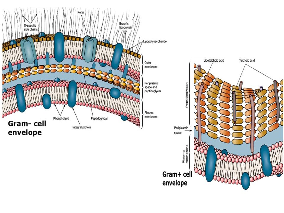
There are two main types of bacterial cell walls, Gram positive and Gram negative, which are differentiated by their Gram staining characteristics. Gram stain Procedure: 1.Crystal violet 2.Iodine 3.Alcohol 4.Saffranine GRAM STAIN

15
Gram stain

16
Gram +ve and Gram –ve Cell Wall The Gram positive cell wall is characterized by the presence of a very thick peptidoglycan layer, which is responsible for the retention of the crystal violet dyes during the Gram staining procedure. Color of Bacteria: Blue-violet The Gram negative cell wall contains a thin peptidoglycan layer, which is responsible for the cell wall's inability to retain the crystal violet stain upon decolourisation with ethanol during Gram staining. Color of Bacteria: RED

18
Capsule Some species of bacteria have a third protective covering, a capsule made up of polysaccharides (complex carbohydrates). Functions of the Capsule: 1.To keep the bacterium from drying out. 2.To protect bacterium from phagocytosis (engulfing) by larger microorganisms. The capsule is a major virulence factor in the major disease- causing bacteria, such as Escherichia coli and Streptococcus pneumoniae. (Noncapsulated mutants of these organisms are avirulent, i.e. they don't cause disease).
 by larger microorganisms. The capsule is a major virulence factor in the major disease- causing bacteria, such as Escherichia coli and Streptococcus pneumoniae. (Noncapsulated mutants of these organisms are avirulent, i.e. they don t cause disease)..")
19
II- Flagella Flagella (singular, flagellum) are long hair-like structures that provide a means of locomotion for those bacteria that have them. They can be found at either or both ends of a bacterium or all over its surface. The flagella beat in a propeller-like motion to help the bacterium move toward nutrients; away from toxic chemicals; or, in the case of the photosynthetic cyanobacteria; toward the light.

21
III- Pili Many species of bacteria have pili (singular, pilus), short hair- like projections emerging from the outside cell surface. These pili assist the bacteria in attaching to other cells and surfaces, such as teeth, intestines, and rocks. Without pili, many disease-causing bacteria lose their ability to infect because they are unable to attach to host tissue.

23
IV- The Cytoplasm The cytoplasm, or protoplasm, of bacterial cells is where the functions for cell growth, metabolism, and replication are carried out. It is a gel-like matrix composed of water, enzymes, nutrients, wastes, and gases and contains cell structures such as ribosomes, a chromosome, and plasmids. The cell envelope encases the cytoplasm and all its components. Unlike the eukaryotic (true) cells, bacteria do not have a membrane enclosed nucleus. The chromosome, a single, continuous strand of DNA, is localized, but not contained, in a region of the cell called the nucleoid. All the other cellular components are scattered throughout the cytoplasm.
 cells, bacteria do not have a membrane enclosed nucleus. The chromosome, a single, continuous strand of DNA, is localized, but not contained, in a region of the cell called the nucleoid. All the other cellular components are scattered throughout the cytoplasm..")
24
The nucleoid The nucleoid is a region of cytoplasm where the chromosomal DNA is located. It is not a membrane bound nucleus, but simply an area of the cytoplasm where the strands of DNA are found. Most bacteria have a single, circular chromosome that is responsible for replication, although a few species do have two or more.

25
Ribosomes They translate the genetic code from the molecular language of nucleic acid to that of amino acids (building blocks of proteins). Differences between Bacteria and Eukaryotes 1.Bacterial ribosomes are similar to those of eukaryotes, but are smaller and have a slightly different composition and molecular structure. 2.Bacterial ribosomes are never bound to other organelles as they sometimes are (bound to the endoplasmic reticulum) in eukaryotes, but are free-standing structures distributed throughout the cytoplasm. 3.Some antibiotics will inhibit the functioning of bacterial ribosomes, but not a eukaryote's, thus killing bacteria but not the eukaryotic organisms they are infecting.
 in eukaryotes, but are free-standing structures distributed throughout the cytoplasm. 3.Some antibiotics will inhibit the functioning of bacterial ribosomes, but not a eukaryote s, thus killing bacteria but not the eukaryotic organisms they are infecting..")
26
Plasmids Plasmids are small extrachromosomal genetic structures carried by many strains of bacteria. Like the chromosome, plasmids are made of a circular piece of DNA. Unlike the chromosome, they are not involved in reproduction. Plasmids replicate independently of the chromosome and, while not essential for survival, appear to give bacteria a selective advantage. Plasmids are passed on to other bacteria through two means. For most plasmid types, copies in the cytoplasm are passed on to daughter cells during binary fission.

27
Plasmids Other types of plasmids form a tubelike structure at the surface called a pilus that passes copies of the plasmid to other bacteria during conjugation, a process by which bacteria exchange genetic information. Plasmids have been shown to be instrumental in the transmission of special properties, such as antibiotic drug resistance, resistance to heavy metals, and virulence factors necessary for infection of animal or plant hosts. The ability to insert specific genes into plasmids have made them extremely useful tools in the fields of molecular biology and genetics, specifically in the area of genetic engineering.

28
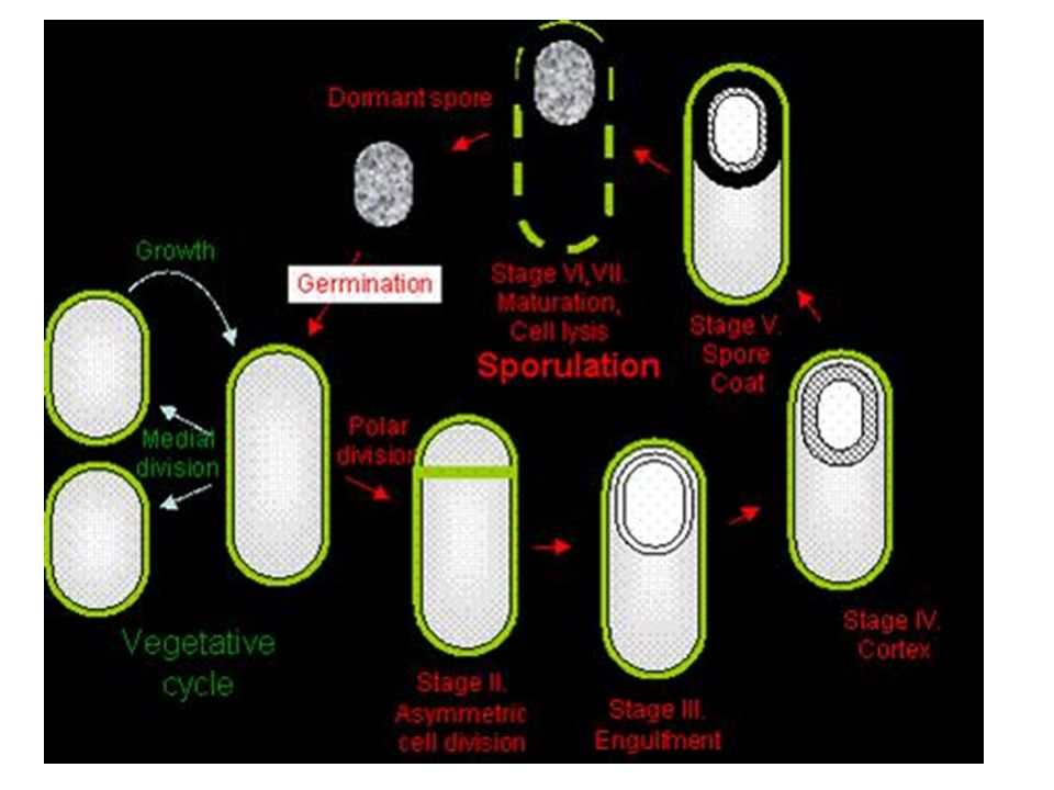
Endospores Endospores are bacterial survival structures that are highly resistant to many different types of chemical and environmental stresses and therefore enable the survival of bacteria in environments that would be lethal for these cells in their normal vegetative form.

30
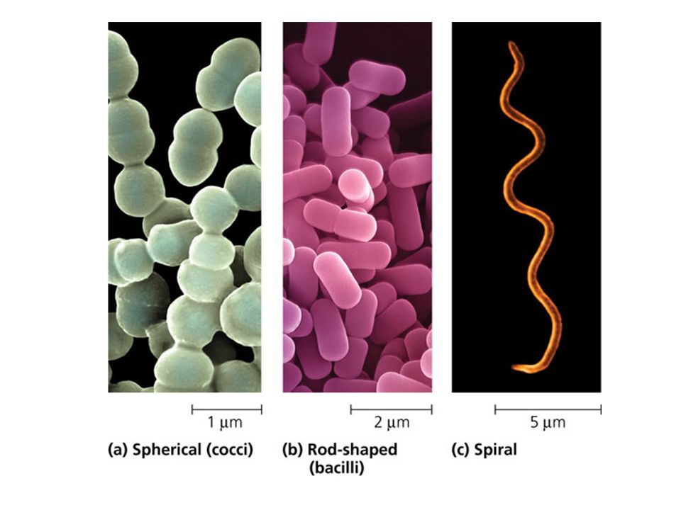
Cell Morphology & Shape of Bacteria 1.Coccus (spherical): Streptococci, Staphylococci 2.Bacillus (rod-like): Enterobacteriacea spp. 3.Spirillum (spiral): Treponema spp.
: Treponema spp..")
33
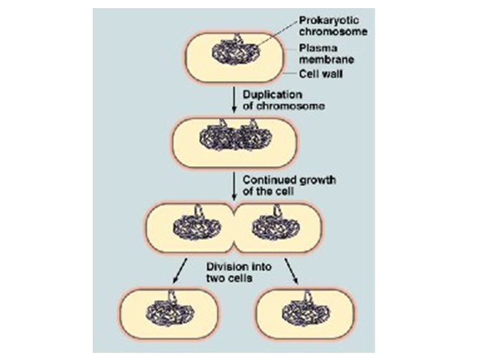
Replication of Bacteria Bacterial cells replicate asexually by a process called: Binary Fission. One cell doubles in size and splits in half to produce two identical daughter cells. These daughter cells can then double in size again to produce four sibling cells and these to produce eight, and so on. Doubling Time: the time it takes for a bacterial cell to grow and divide in two. When nutrients are plentiful, the doubling time of some bacterial species can be as short as 20 minutes. However, most bacterial species show a doubling time between 1-4 hours.

35
Replication of Bacteria The cytoplasm of a bacterial cell contains the DNA molecules that make up the bacterial genome, the transcriptional machinery that copies DNA into ribonucleic acid (RNA), and the ribosomes that translate the messenger RNA information into protein sequence. Since there is no nucleus, all of these processes occur simultaneously. The rapid growth rate of the bacterial cell requires constant DNA replication and ways to segregate the two new chromosomes into the two daughter cells without tangling them.

37
When germ relationship go bad..

Similar presentations


 Cellular Organism EucaryotesProcaryotes: bacteriaVirus Eubacteria: Gram-positive bacteria.>")


2605285.>")


 2.MOLECULE (compounds like carbohydrates &>")


 Compare characteristics of taxonomic groups, including archaea, bacteria, protists, fungi, plants, and animals. 11(C) Summarize.>")


