Download presentation
Presentation is loading. Please wait.

2
Prosthetic group with an iron atom in the middle of a porphyrin ring Ring contains N, alkenes, and carboxylate groups Commonly recognized as parts of hemoglobin (4 per) Iron is responsible for binding oxygen in order to distribute to the rest of the body Uptake of heme is one way the cell can bring in iron
 Iron is responsible for binding oxygen in order to distribute to the rest of the body Uptake of heme is one way the cell can bring in iron")
3
http://omlc.ogi.edu/spectra/hemoglobin/hemestruct/ind ex.html Heme with iron atom bound in the middle of the porphyrin ring

4
Intracellular ◦ Cytochromes (involved in cellular respiration) ◦ Proteins involved in DNA synthesis and cell division ◦ Extracellular Hemoglobin Myoglobin Connective tissue, nervous system, immune system
 ◦ Proteins involved in DNA synthesis and cell division ◦ Extracellular Hemoglobin Myoglobin Connective tissue, nervous system, immune system")
5
http://sandwalk.blogspot.com/2007/08/heme- groups.html

6
Soluble Fe3+ (ferric iron) – retrieved by compounds called siderophores Ferriproteins *Heme Hemoproteins (Hb and Mb) *Hemophores (proteins with high affinity for heme) *discussed in this presentation
 – retrieved by compounds called siderophores Ferriproteins *Heme Hemoproteins (Hb and Mb) *Hemophores (proteins with high affinity for heme) *discussed in this presentation")
7
Entry into cells must be regulated because too much can be toxic Requires a membrane protein because cannot naturally diffuse Active transport mechanism ◦ Energy is derived from proton gradient ◦ Proton gradient formed and energy derived is transduced by proteins Ton B or TonB – related proteins

8
Escherichia Coli

9
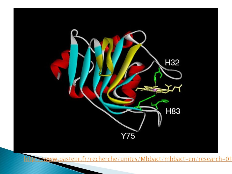
Carrier protein that brings heme to receptor Serratia marcescens hemophore: HasA 188-residue protein Very high affinity for heme Beta sheet layer and 4 alpha helices Heme iron is bound by coordination of His- 32 and Tyr-75 on opposing loops

10
http://www.pasteur.fr/recherche/unites/Mbbact/mbbact-en/research-01.html
11
HasR Can internalize both free heme or that bound to hemophore into periplasm Has a weaker affinity for heme than HasA Binds heme via 2 histidine residues Uses energy derived from proton gradient to move heme to interior of cell

12
HasA= hemophore (carrier protein that brings heme to receptor) HasR = heme transport receptor TonB/HasB = protein complex involved in transduction of energy from proton motive force holoHasA = HasA with heme attached apoHasA = HasA without heme
 HasR = heme transport receptor TonB/HasB = protein complex involved in transduction of energy from proton motive force holoHasA = HasA with heme attached apoHasA = HasA without heme")
13
HasA receives heme Migrates to and docks onto receptor (HasR) Heme transferred from hemophore to receptor Heme passes into periplasmic space and enters cell Hemophore HasA dissociates and can pick up more heme
 Heme transferred from hemophore to receptor Heme passes into periplasmic space and enters cell Hemophore HasA dissociates and can pick up more heme")
14
http://www.pasteur.fr/r echerche/RAR/RAR2003 /Mbbact-en.html

15
HasR can form tight complexes with both hemophores with heme (holoHasA) and those without (apoHasA) Since HasA has high heme affinity, iron uptake can be very high at low [heme] Good because too much reduced iron in the body is harmful
![ HasR can form tight complexes with both hemophores with heme (holoHasA) and those without (apoHasA) Since HasA has high heme affinity, iron uptake can be very high at low [heme] Good because too much reduced iron in the body is harmful](http://images.slideplayer.com/24/7263567/slides/slide_15.jpg " HasR can form tight complexes with both hemophores with heme (holoHasA) and those without (apoHasA) Since HasA has high heme affinity, iron uptake can be very high at low [heme] Good because too much reduced iron in the body is harmful")
16
When holoHasA is bound to HasR, heme is spontaneously transferred to receptor (no energy is required here) Energy from proton motive force required for entry of heme into cell and apoHasA dissociation from HasR HasB (paralog of TonB) transduces energy ◦ Signaling stimulus due to transcriptional autoregulation when HasA and heme bound to receptor
 Energy from proton motive force required for entry of heme into cell and apoHasA dissociation from HasR HasB (paralog of TonB) transduces energy ◦ Signaling stimulus due to transcriptional autoregulation when HasA and heme bound to receptor")
17
http://chemistry.gsu.edu/Dixon.php
18
Determine function of entire heme transport system 2 ternary complexes: HasA-HasR-heme (WT and mutant HasR) Binary complex: HasA-HasR Resolutions: 2.7 angstroms for ternary complexes and 2.8-angstrom for binary complex
 Binary complex: HasA-HasR Resolutions: 2.7 angstroms for ternary complexes and 2.8-angstrom for binary complex")
19
WT ternary solved by MAD (multiwavelength anomalous diffraction) Other two done by difference Fourier methods Final residue counts: ◦ HasR= 752 residues ◦ HasA= 161 residues
 Other two done by difference Fourier methods Final residue counts: ◦ HasR= 752 residues ◦ HasA= 161 residues")
20
HasR contains 22 antiparallel beta-strands like other TonB-dependent receptors Unlike others in the family, HasR has elongated extracellular loops (L2, L6, L9) – bind HasA L7 and C apex used to attain heme
 – bind HasA L7 and C apex used to attain heme")
21
http://strucbio.biologi e.uni- konstanz.de/strucbio/ HasA-HasR-heme complex L6 L9 Heme (green) bound to L7
 bound to L7")
22
Initially, heme-binding site of HasA oriented to face extracellular loops of HasR Heme then binds to the two His residues of HasR (transferred about 9.2 angstroms) ◦ His-603 from L7 and His-189 from apex C of a plug that is common in these receptors ◦ Mutants of these two residues show no heme binding
 ◦ His-603 from L7 and His-189 from apex C of a plug that is common in these receptors ◦ Mutants of these two residues show no heme binding")
23
http://ww w.pnas.org /content/1 06/4/1045.full Superposition of the heme groups attached to holoHasA(blue) and to HasA- HasR-heme (red)
 and to HasA- HasR-heme (red)")
24
Spontaneous transfer from HasA to HasR Transfer is endergonic (non-spont.) Coupling of HasA and HasR is exergonic and exothermic (spontaneous) Latter overrides former
 Coupling of HasA and HasR is exergonic and exothermic (spontaneous) Latter overrides former")
25
During complex formation, heme is not lost to solution HasR-Ile-671 in L8 clashes with heme on holoHasA (Figure A) Without the Ile, heme transport is not possible because the heme rotates to face HasA Mutant with Glycine-671 used (Figure B)
 Without the Ile, heme transport is not possible because the heme rotates to face HasA Mutant with Glycine-671 used (Figure B)")
26
http://www.pnas.org/content/106/4/1 045.full

27
L7 and L8 of HasR displace the loop with HasA-His-32 causing break in coordination between residue and heme Heme and HasA-Tyr-75 (stronger connection) still persists ◦ Stablized by deprotonation of phenol that H-bonds with HasA-His-83
 still persists ◦ Stablized by deprotonation of phenol that H-bonds with HasA-His-83")
28
Later, the His-83 may get protonated and so the coordination is lost Ile-671 displaces heme from HasA Rotation of His-83 side chain prevents sliding back of heme to hemophore

29
Shows that free heme can bind to HasR with apoHasA bound as well There is a channel that goes from between loops 3 and 4 all the way to the heme binding site in which heme can travel

30
http://www.p nas.org/conte nt/106/4/10 45.full

31
Mirrors ABC transport of cargo from bacterial periplasm to inside cell Both have cargo molecule bound to protein that binds to and spontaneously transfers cargo to cis receptor Energy is required (heme-proton motive; ABC-ATP hydrolysis) to get cargo to trans and dissociate protein from receptor
 to get cargo to trans and dissociate protein from receptor")
32
How to get substance to protein with lower affinity? Part of binding energy of donor to ligand is consumed when displacing the first loop (His-32) Ligand transfer occurs when donor-acceptor come together due to steric clash (from Ile- 671)
 Ligand transfer occurs when donor-acceptor come together due to steric clash (from Ile- 671).")
33
Refinement -Resolution, Å 49.2–2.7 (2.73–2.70) 49.4–3.0 (3.03–3.0) 39.2–2.8 (2.83–2.80) -No. of reflections 99,334 (2,329) 77,295 (2,431) 92,482 (2,123) -Completeness, % 95.03 (71) 99.17 (93) 98.1 (71) -Rwork, % 23.7 (34.9) 21.4 (37.4) 22.6 (46.6) -Rfree*, % 27.3 (38.4) 24.3 (39.1) 26.2 (48.3) Model composition -Protein residues 1,850 1,850 1,850 -Heme atoms 86 0 86 -Water molecules 58 19 13 B-factors -Protein 93.5 80.2 110.6 -Heme 84.6 — 120.4 Deviation from ideal values -Bond lengths, Å 0.010 0.010 0.006 -Residues with bad bond lengths †, % 0 0.05 0 -Bond angles, ° 0.61 1.27 1.08 -Residues with bad bond angles †, % 0.22 0.71 0.550 Ramachandran plot † -Favored regions, % 92.4 89.6 89.7 -Allowed regions, % 99.2 99.5 99.1
 77,295 (2,431) 92,482 (2,123) -Completeness, % (71) (93) 98.1 (71) -Rwork, % 23.7 (34.9) 21.4 (37.4) 22.6 (46.6) -Rfree*, % 27.3 (38.4) 24.3 (39.1) 26.2 (48.3) Model composition -Protein residues 1,850 1,850 1,850 -Heme atoms Water molecules B-factors -Protein Heme 84.6 — Deviation from ideal values -Bond lengths, Å Residues with bad bond lengths †, % Bond angles, ° Residues with bad bond angles †, % Ramachandran plot † -Favored regions, % Allowed regions, %")
34
measure of how well refined structure predicts observed data R-factors usually range from 0.2-0.6 Smaller R-factor is better R-factors for the three structures are 0.237,.214,.226 for the WT ternary complex, mutant ternary, and binary complex, resp. Shows well-defined structure

Similar presentations


 Regulates exchange.>")


 Chapter 22 Functions of “Blood” Gas Transport Nutrient Transport Excretory Product Transport Cell Signal Transport Hydraulic.>")


 receptors (eg. pain receptors) transport (ions across membranes, oxygen in blood) molecular motors recognition.>")
 Oxidative phosphorylation. Proton Motive Force ( p ) PMF is the energy of the proton concentration gradient The chemical ( pH=>")
