Download presentation
Presentation is loading. Please wait.

1
John-Henry Corbett Department of clinical imaging science University of the Free State 7/09/2012 CASE PRESENTATION

2
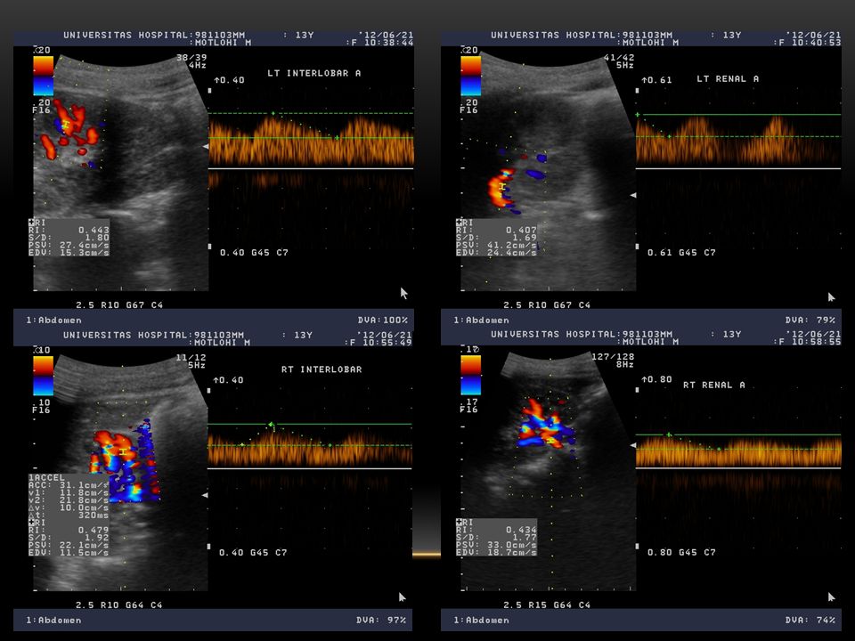
13 year old female patient with hypertension

5
CLINICAL CRITERIA FOR SUSPICION OF RENOVASCULAR HYPERTENSION 1.Epigastric or flank bruit (systolic or diastolic) 2.Accelerated or malignant hypertension 3.Unilateral small kidney discovered with any clinical study 4.Severe hypertension in child or young adult or > 50yrs 5.Sudden development or worsening of hypertension at any age 6.Sudden worsening of renal function in a hypertensive patient 7.Hypertension refractory to an appropriate three drug regimen 8.Impairment of renal function after treatment with an ACE-inhibitor 9.Hypertension and extensive arterial occlusive disease
 2.Accelerated or malignant hypertension 3.Unilateral small kidney discovered with any clinical study 4.Severe hypertension in child or young adult or > 50yrs 5.Sudden development or worsening of hypertension at any age 6.Sudden worsening of renal function in a hypertensive patient 7.Hypertension refractory to an appropriate three drug regimen 8.Impairment of renal function after treatment with an ACE-inhibitor 9.Hypertension and extensive arterial occlusive disease")
6
IMAGING IN RENOVASCULAR HYPERTENSION A) Ultrasound / Doppler Provides only indirect evidence of presence of RAS – screening tool Readily available and cheap Operator and patient dependant, time consuming and cumbersome B) CT angiography Accurate and non-invasive Radiation and iodinated contrast ( in patient with already compromised renal function)
 Ultrasound / Doppler Provides only indirect evidence of presence of RAS – screening tool Readily available and cheap Operator and patient dependant, time consuming and cumbersome B) CT angiography Accurate and non-invasive Radiation and iodinated contrast ( in patient with already compromised renal function)")
7
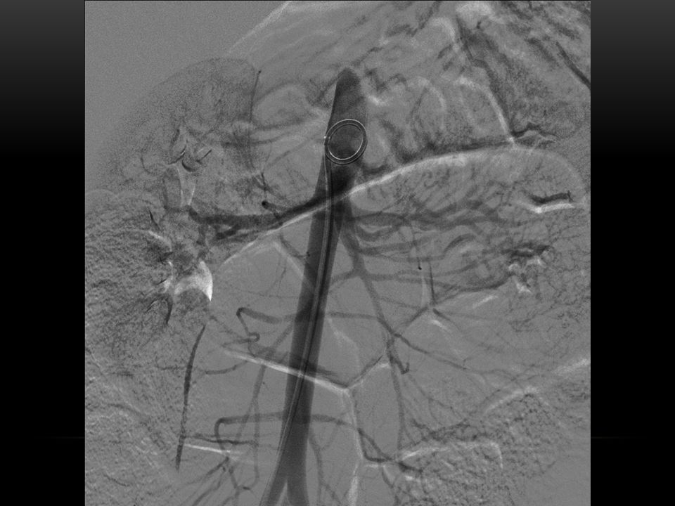
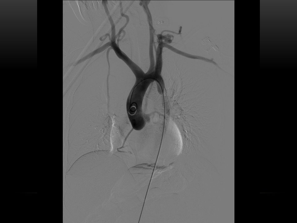
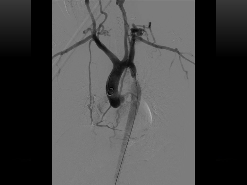
IMAGING IN RENOVASCULAR HYPERTENSION C) MR angiography Accurate information about number of renal arteries, kidney sizes & anatomic variants Accurate detection of main renal artery stenosis or occlusions Overestimation of renal artery stenosis (>10%) Suboptimal for lesions of distal, intrarenal and accessory renal arteries Limited availability and expensive D) Radionuclide renography Acceptance as screening tool for RAS has been hindered by lack of standardized protocols E) Digital subtraction angiography (DSA) Gold standard Benefit of endovascular treatment
 MR angiography Accurate information about number of renal arteries, kidney sizes & anatomic variants Accurate detection of main renal artery stenosis or occlusions Overestimation of renal artery stenosis (>10%) Suboptimal for lesions of distal, intrarenal and accessory renal arteries Limited availability and expensive D) Radionuclide renography Acceptance as screening tool for RAS has been hindered by lack of standardized protocols E) Digital subtraction angiography (DSA) Gold standard Benefit of endovascular treatment")
8
CAUSES OF RENOVASCULAR HYPERTENSION

9
Newborn Thrombosis or embolization related to umbilical artery catheterization Children Fibromuscular dysplasia the most common Aorta coarctation Moyamoya disease, Takayasu arteritis, Kawasaki disease Vasculitis, vascular trauma, renal artery thrombosis

10
ATHEROSCLEROSIS VS FMD IN RENAL ARTERY STENOSIS (RAS)
")
11
ATHEROSCLEROTIC RENAL ARTERY DISEASE Common cause of secondary hypertension in males > 50 yrs Associated with atherosclerosis of aorta, cerebral, coronary and peripheral arteries Eccentric stenosis in proximal 2cm of renal artery Frequently involves orifice Additional renal artery branch stenosis < 10% Bilateral in 1/3 rd of cases

12
ATHEROSCLEROTIC RAS

13
FIBROMUSCULAR DYSPLASIA Focal, irregular thickening of the walls of medium and large muscular arteries by combination of intimal and medial hyperplasia and fibrosis Unknown cause, but probably developmental Most common cause of renovascular hypertension in children and young adults < 30yrs Three times more common in females Associated with fibromuscular dysplasia of other aortic branches Celiac artery, hepatic artery, splenic artery, mesenteric artery Iliac artery, internal carotid artery Affects mid and distal main renal artery and major segmental branches – often bilateral

14
FIBROMUSCULAR DYSPLASIA Types 1.Initmal fibroplasia (1%) – narrow annular radiolucent band in main renal artery 2. Medial fibroplasia (70%) – alternating areas of stenosis and aneurysms in mid & distal renal artery 3.Medial hyperplasia (8-10%) – long smooth tubular narrowing of main renal artery 4.Perimedial fibroplasia (15-20%) – beading without aneurysm formation of distal renal artery 5.Adventitial fibroplasia (1%) – long segmental stenosis of main renal artery and large branches
 – alternating areas of stenosis and aneurysms in mid & distal renal artery 3.Medial hyperplasia (8-10%) – long smooth tubular narrowing of main renal artery 4.Perimedial fibroplasia (15-20%) – beading without aneurysm formation of distal renal artery 5.Adventitial fibroplasia (1%) – long segmental stenosis of main renal artery and large branches.")
15
FIBROMUSCULAR DYSPLASIA

16
TAKAYASU ARTERITIS Granulomatous inflammation of unknown pathogenesis Affects mainly segments of the aorta + major branches and pulmonary arteries (media and adventitia) Stenosis and obstruction are characteristic, but dilatations and aneurysms also occur frequently Usually in persons under 50 years of age Typically young asian women – Females 10 x more common
 Stenosis and obstruction are characteristic, but dilatations and aneurysms also occur frequently Usually in persons under 50 years of age Typically young asian women – Females 10 x more common")
17
TAKAYASU ARTERITIS Early (systemic or pre-pulseless) phase Non-specific constititional symptoms Axial imaging (CT/MRI) may show aortic wall thickening Late (stenotic or pulseless) phase 1-8 years after early phase Four types 1.Classic pulseless disease – involves branches of the aortic arch only 2.Mixed type – classic pulseless + atypical coarctation 3.Atypical coarctation – thoracic and abdominal aorta + branches 4.Dilated type – extensive dilatation of the entire aorta and branches
 phase Non-specific constititional symptoms Axial imaging (CT/MRI) may show aortic wall thickening Late (stenotic or pulseless) phase 1-8 years after early phase Four types 1.Classic pulseless disease – involves branches of the aortic arch only 2.Mixed type – classic pulseless + atypical coarctation 3.Atypical coarctation – thoracic and abdominal aorta + branches 4.Dilated type – extensive dilatation of the entire aorta and branches")
18
TAKAYASU ARTERITIS

19
Four types of late phase Takayasu (alternative classification) 1.Aortic arch and main vessels 2.Descending aorta and abdominal aorta (spares arch) 3.Combination of 1 & 2 4.Pulmonary involvement
 1.Aortic arch and main vessels 2.Descending aorta and abdominal aorta (spares arch) 3.Combination of 1 & 2 4.Pulmonary involvement")
20
TAKAYASU ARTERITIS Termination of left common carotid artery at its origin. Stenosis of both subclavian arteries. Occlusion of the left subclavian artery. Diffuse narrowing of the right common carotid artery. Compensatiory enlargement of both vertebral arteries.

21
COARCTATION OF THE AORTA Discrete narrowing in the proximal descending aorta, adjacent to the arterial duct or ligament Begins late in fetal cardiac development as an endovascular shelf that arises from the posterolateral aspect of the aorta opposite the insertion of the ductus arteriosus 6 th most common form of congenital heart disease One in 3000 live births Associated with bicuspid aortic valve in 75% of cases CoA is also present in 15% of patients with Turner syndrome

22
COARCTATION OF THE AORTA Presentation Most often detected on routine physical examination due to a murmer or hypertension Palpation of brachial and femoral pulses simultaneously will reveal absent or delayed femoral pulses Blood pressure in both arms and one leg must be determined Pressure difference of > 20mmHg in favor of arms = CoA Old classification – in relation to ductus arteriosus Preductile – or infantile type Post ductal – or adult type Important information Age and size of the patient Location and morphology of the coarctation Native or recurrent coarctation

23
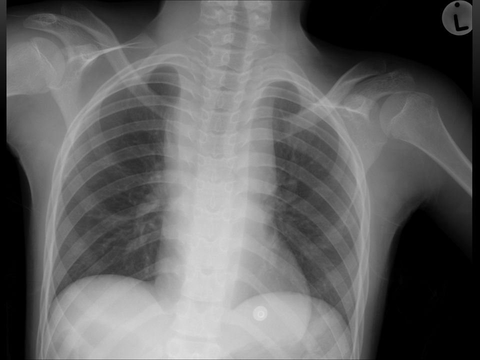
COARCTATION OF THE AORTA CXR Normal or large heart (LVH) Rib notching secondary to collateral vessels “3” sign on frontal radiograph or inverted “3” on barium filled esophagus
 Rib notching secondary to collateral vessels 3 sign on frontal radiograph or inverted 3 on barium filled esophagus")
24
COARCTATION OF THE AORTA LAO Aortagrtam : A) Discrete juxtaductal coarctation B) Long segment coarctation
 Discrete juxtaductal coarctation B) Long segment coarctation")
25
COARCTATION OF THE AORTA Mean age of death without treatment is 34 years Indications for treatment Blood pressure gradient between upper and lower limbs > 20mmHg at rest Signiificant hypertension or cardiac failure Treatment Infants and children <1 year Surgery for native coarctation and balloon angioplasty for recurrent lesions > 1 year up to 30-35kg (9-11years) Surgery or angioplasty for native lesions (no consensus) Balloon angioplasty for recurrent lesions > 35kg Endovascular stent placement for native and recurrent lesions
 Surgery or angioplasty for native lesions (no consensus) Balloon angioplasty for recurrent lesions > 35kg Endovascular stent placement for native and recurrent lesions")
26
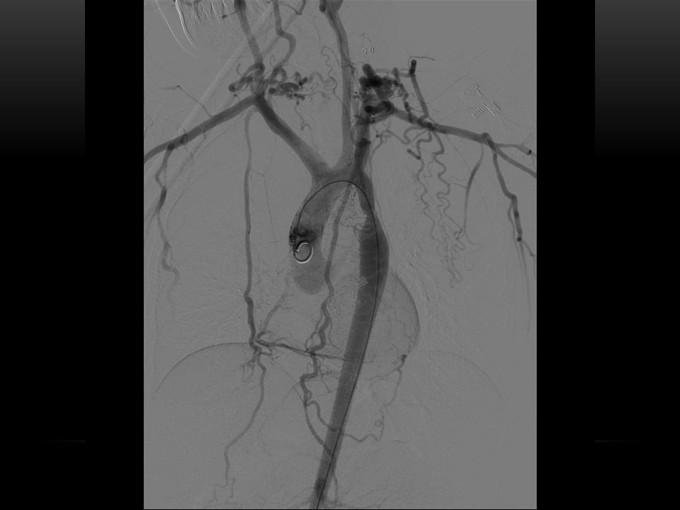
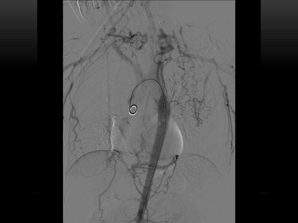
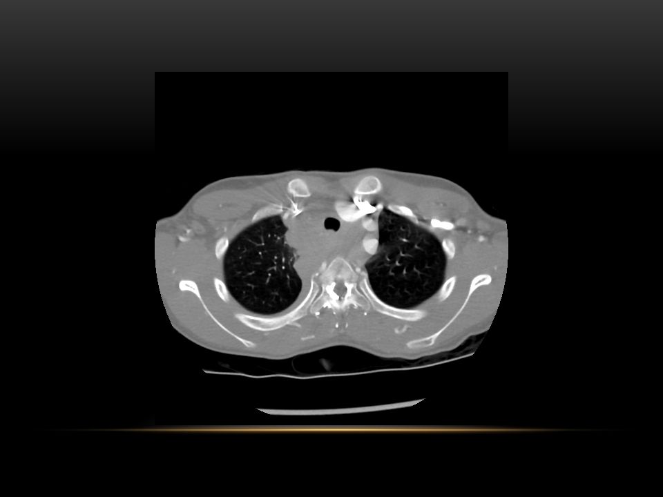
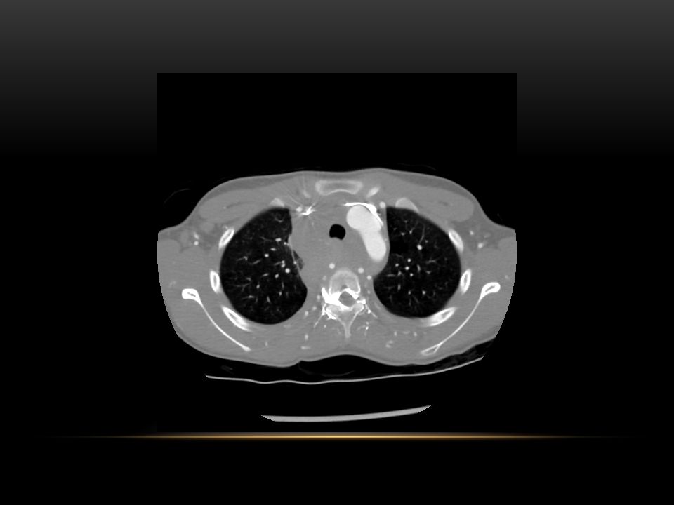
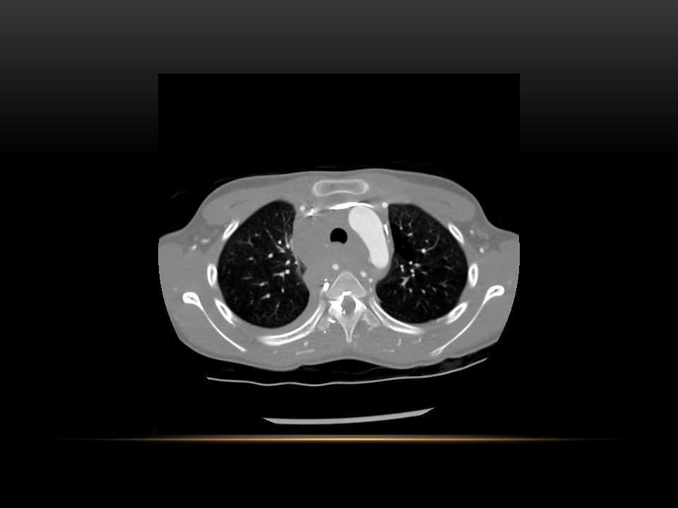
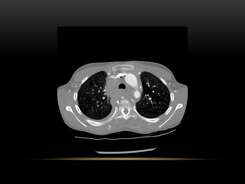
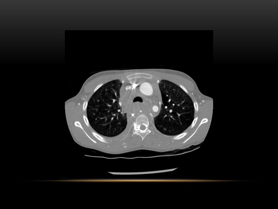
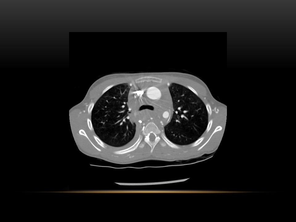
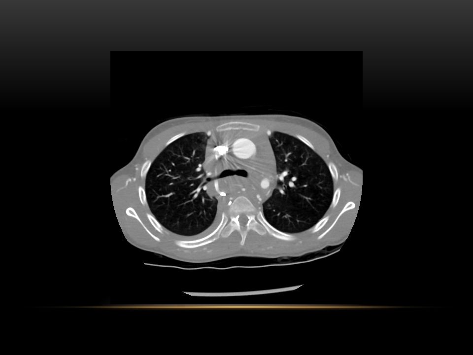
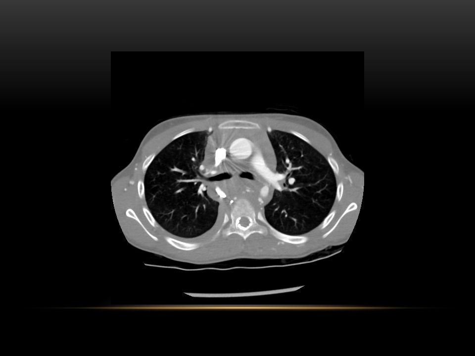
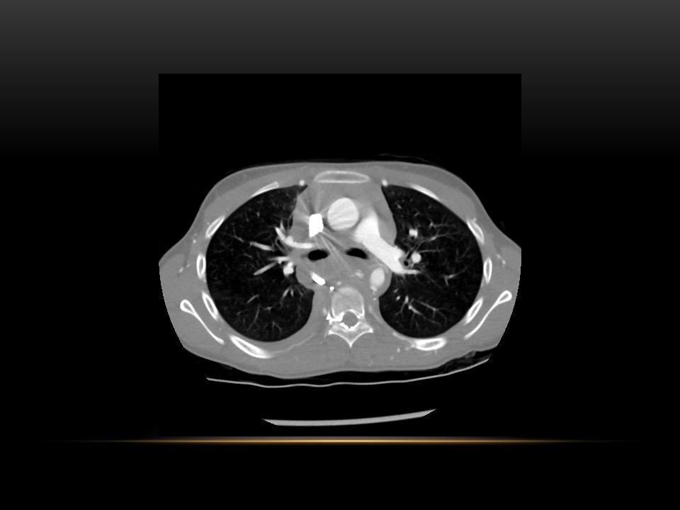
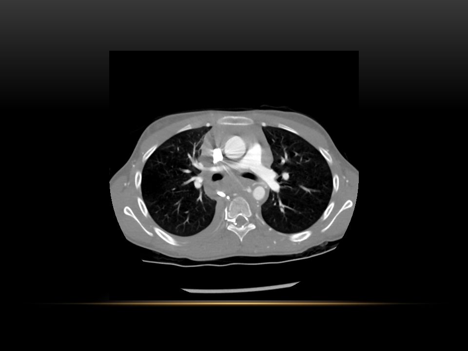
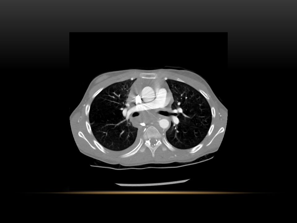
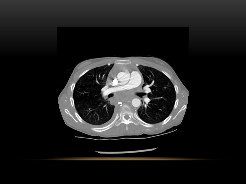
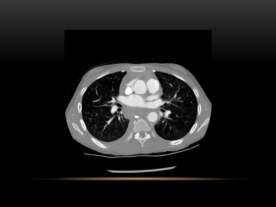
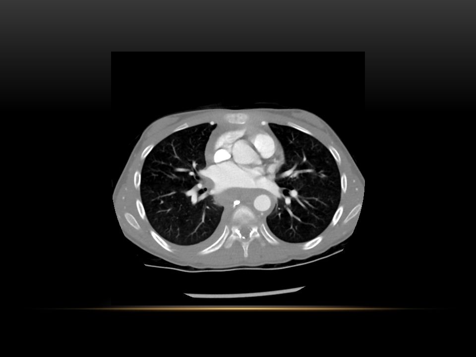
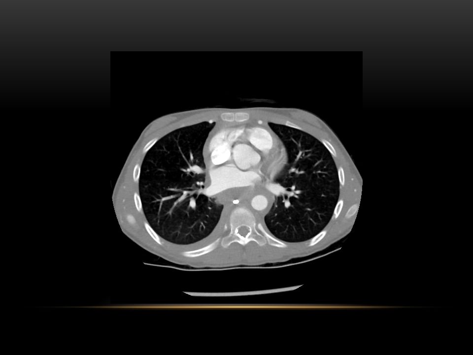
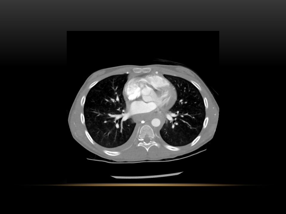
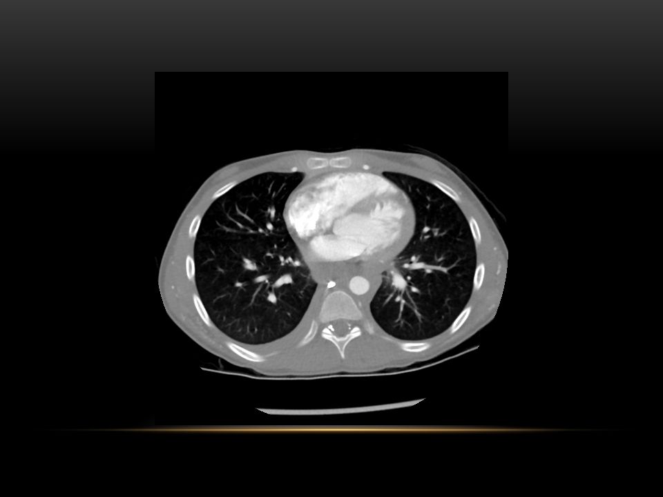
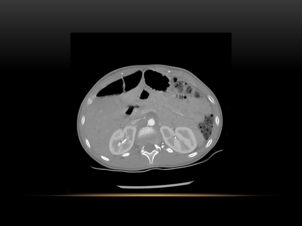
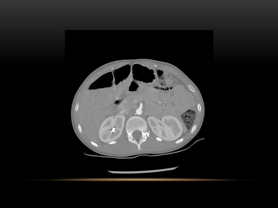
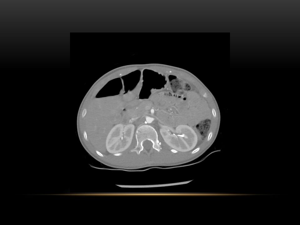
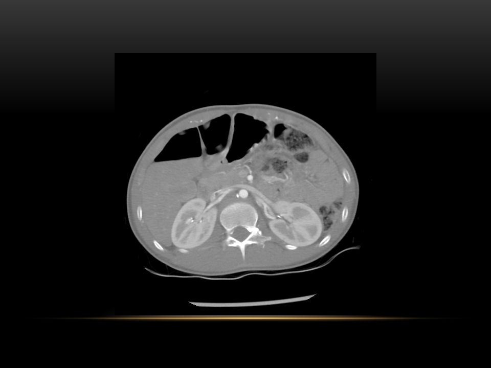
BACK TO OUR 13 YR OLD FEMALE WITH HPT

55
REFERENCES Grainger RG, Allison DJ. Diagnostic Radiology : A textbook of Medical imaging. 5 th Edition. Churchill Livingstone ; 2008. Soulez G, Oliva V, Turpin S et al. Imaging of renovascular hypertension : Respective Values of Renal Scintigraphy, Renal Doppler US, and MR Angiography. Radiographics 2000 ; 20:1355-1368. Schmidt RJ 2012. Renovascular hypertension. Viewed 28/08/2012, http://emedicine.medscape.com/article/245140-overview http://emedicine.medscape.com/article/245140-overview Khan AN 2011. Imaging in renal artery stenosis / Renovascular hypertension. Viewed 28/08/2012, http://emedicine.medscape.com/article/380308-overviewhttp://emedicine.medscape.com/article/380308-overview Tanous D, Benson L, Horlick E. Coarctation of the aorta : Evaluation and management. Current opinion in Cardiology 2009 ; 24: 509-515. Golden AB, Hellenbrand WE. Coarctation of the aorta : Stenting in children and adults. Catheterization an cardiovascular interventions 2007 ; 69:289-299. Syamasundar R. Coarctation of the aorta. Current cardiology reports 2005 ; 7:425-434. Matsunaga N, Hayashi K, Sakamoto I et al. Takayasu Arteritis : Protean Radiologic Manifestations and Diagnosis. Radiographics 1997; 17: 579-594.

Similar presentations


 tricuspid valve 2. Hypoplastic right ventricle 3. Ventricular septal defect 4. Atrial.>")


>")


