Download presentation
Presentation is loading. Please wait.

1
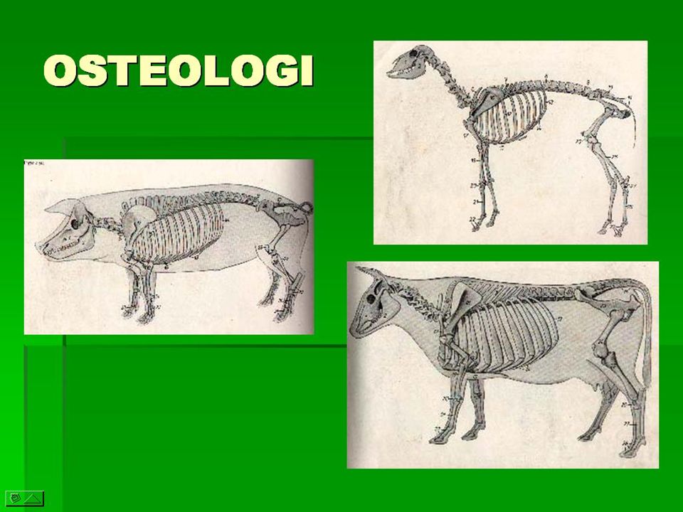
PAV-OSTEOLOGY

2
OSTEOLOGY OSTEOLOGY : is a science which learn about bones and their relation Bones are connected each other term as: skeleton Skeleton: is applied to the frame-work of hard structures which supports and protects the soft tissue of animals.

3
Bones System and their relationship
FERTILISATION ZYGOTE (mitosis: cleavage) MORULA BLASTULA GASTRULA divides into 3: - ectoderm : external layer skin & nerve - endoderm: inner layer viscera - mesoderm: between ecto & endoderm muscles and bones.
 MORULA BLASTULA GASTRULA divides into 3: - ectoderm : external layer skin & nerve. - endoderm: inner layer viscera. - mesoderm: between ecto & endoderm muscles and bones.")
4
FUNCTION OF THE SKELETON
SUPPORT THE BODY MAKE THE SHAPE OF THE BODY PROTECK WEAK ORGANS PASSIVE MOVEMENT ORGAN PLACE FOR FIXING MUSCLE PLACE FOR PRODUCES BLOOD RESERVOIR CHEMICAL AGENT: Ca & P

5
THE SKELETON MAY BE DIVIDED PRIMARY INTO THREE PARTS:
SKELETON/ AXIALIS Collumnar Vertebralis Cranium Sternum Costae, Pelvis SKELETON APPENDICULARIS Ossa Extremitas Cranialis et Caudalis SKELETON VISCERALIS : consists of certain bones developed in the substance of some of the viscera or soft organ Os. Vesali os Hyoideus Os. Penis os Cordis Os. Glandis

6
(1) Collumnar Vertebralis, composes of :
SKELETON AXIALIS (1) Collumnar Vertebralis, composes of : Ossa. Cranii, Cervicalis, Thoracalis, Lumbalis, Sacralis and Coccygealis (2) Ossa Costae, Sternum, Clavicula, Scapula,Coxae (os Coxae, composes of: Ossa pubic, ilium and ischiadicum)
 Collumnar Vertebralis, composes of : Ossa. Cranii, Cervicalis, Thoracalis, Lumbalis, Sacralis and Coccygealis. (2) Ossa Costae, Sternum, Clavicula, Scapula,Coxae. (os Coxae, composes of: Ossa pubic, ilium and ischiadicum)")
7
SKELETON APPENDICULARIS
Extremitas Cranialis : Ossa Humerus, Radius-Ulna, Carpal, Metacarpal, Phalangis/digitalis Extremitas Caudalis : Ossa Femuralis, Tibia-Fibula, Tarsal, Metatarsal, Phalangis/digitalis

10
ACCORDING THEIR BONES SHAPE
Ossa Longa (lonng) Ossa Plana (flat) Ossa Brevia (short) Ossa Irregularis (irregular)
 Ossa Plana (flat) Ossa Brevia (short) Ossa Irregularis (irregular)")
11
os brevia os plana os longa

12
OSSA LONGA (long bone)
")
13
Ossa plana (flat bone)
")
14
OSSA BREVIA (short bone)
")
15
OSSA IRREGULARIA

16
THE AMOUNT OF THE BONES Each breed have different amount
example: horse 205 bones cattle 191 – 193 bones chicken 160 boness human 206 bones (old), 270 (young) This condition is depended by breed (ras) and age.
, 270 (young) This condition is depended by breed (ras) and age.")
17
DEVELOPMENT AND GROWTH OF BONE (osteogenesis)
1.osteogenesis intramembranosa (desmalis = primer): cel-cel mesenchym osteoblast osteocyt matrix become gel & solid (osteoid) calcification punctum ossification. 2. osteogenesis intracartilagenosa (enchondralis = secundair): started by cartilage : cel2 mesenchym chondroblast chondrocyt (fit length) ossification. Osteoblast : destroy bone layer in order to make the shape.
: cel-cel mesenchym osteoblast osteocyt matrix become gel & solid (osteoid) calcification punctum ossification. 2. osteogenesis intracartilagenosa (enchondralis = secundair): started by cartilage : cel2 mesenchym chondroblast chondrocyt (fit length) ossification. Osteoblast : destroy bone layer in order to make the shape.")
18
Punctum ossification DEVELOPMENT : Interstitial development
( from the middle of tissue ) appostitional development (fron the lateral, the tissue change in order to fix bone layer)
 appostitional development. (fron the lateral, the tissue change in order to fix bone layer)")
19
PUNCTUM OSSIFICATION VERTEBRATA post natal
GROUP I Horse nil Cattle nil Sheep nil GROUP II Man Rabbit Dog Cat Pig Guinea pig

20
SEXUAL AND BODY MATURE BREED SEXUAL M BODY M horse 1 year 4-5 years
cattle months years Sheep/goat months years pig months years Dog months ,5-2 year

21
BONE STRUCTURE Bone mainly compose of bone tissue, but as an organ, it have layer term as periosteum, endosteum, medulla ossium, blood vessels and nerves. According to the architecture : 1. compact substance. 2. spongiosa substance In the long bone have medullar cavity.

22
Periosteum: is a membrane which enclose superficial surface of bone, except at the part which is lined by cartilage. Endosteum: is a thin fibrous membrane which enclose medullare cavity and big Haversian canal. Medulla ossium: placed between spongiosum bone and medullar cavity in the long bone. in the adult animals have 2 variation: as red and yellow:

23
In the young animals only have red (medulla ossium rubra), although then a part has change to be yellow (medulla ossium flava) Medulla ossium rubra contains several type characteristic cells and as an substansia to make blood. Medulla ossium flava : contains fatty tissue.

24
BLOOD VESSELS & NERVES composes 2 artery : periosteal and medullaris
nutritional foramen : hole in bone for passing through blood vessels give nutrition. Nerves distribute together with blood vessels. nerve ending in the periosteum (corpuscle Vater-Pacini) may be as sensoris, and probably as connection with muscle sense.
 may be as sensoris, and probably as connection with muscle sense.")
25
BoneStructure macroscopic structure microscopic structure chemical and physical structure

26
MACROSCOPIC STRUCTURE
SUBSTANSIA SPONGIOSA SUBSTANSIA COMPACTA

27
compact and spons bones
spongiosa bone compact bone

28
part os longa DIAPHISA EPIPHYSA

29
Ossa pneumatica SINUS :air spaces within compact substance contain air instead of spongy bone and marrow and, are called pneumatic bones. The cavity are term as sinus, the communicate indirectly with external air SINUS

30
Physic os longa capsula cartilago Osseous epiphysis epiphise plate
metaphisis

31
Physical strength of bone
The bone of just death animal has yellowish white color. If it dip or boil s and give a chemical agent to eliminate stain become white. Berat jenis fresh mass bone: 1,9 The bone is hard & strong enough for pressure Strength pressure pound per suare inch, strength of pulling lb/inch² which more strong than white oak trees.

32
CHEMICAL AND PHYSICAL PROPERTIES OF BONE
Dried bone consists of organic and in organic matter which ratio 1 : 2. Removal of organic matter by heat does not change the general form, but reduces the weight about 1/3 and makes bone very fragile Decalcification, not change the form and size, but renders it soft and pliable. Organic matter (ossein) boils gelatin.
 boils gelatin.")
33
CHEMICAL STRUCTURE & PHYSIC
chemis: organic : inorganic 1:2 composition bone: gelatin ,30% kalsium fosfat ,35% kalsium karbonat ,85% magnesium fosfat ,05% natrium karbonat& klorida 3,45%

34
Parts of Bones Periosteum / endosteum:
has capability to make new bone layer cartilage: its for elascity and pressure absorber joint between 2 bones. Tendo, ligament: fixing tissue between bones or muscles.

35
BONES TERMS Processus : bone elevation usually long
Tuberositas (tuber) :big protruded bone and round form, but it’s not make a joint. Tuberculum: small protruded bone Trochanter :its use for several protruded bone and its not make a joint. Spina (processus spinosus) : high protruded bone, usually flat and sometimes sharp
 :big protruded bone and round form, but it’s not make a joint. Tuberculum: small protruded bone. Trochanter :its use for several protruded bone and its not make a joint. Spina (processus spinosus) : high protruded bone, usually flat and sometimes sharp.")
36
LUMBAL BONE Proc transversus

37
PROC SPINOSUS Proc spinosus

38
Term of form/ structure
cavum sinus Sinus Processus Fissura incissura angulus

39
Crista :elevation of bone which sharp edge.
Linea : very small elevation = line Caput : head = proximal end which enlarger for fitting the joint, probably connected with body/corpus by constricted area term as neck (collum) Collum : neck Condyl (condylus) : bone elevation which cylendrical form for fitting the joint. Epicondylus : cantinuation of condyl which not make joint.
 Collum : neck. Condyl (condylus) : bone elevation which cylendrical form for fitting the joint. Epicondylus : cantinuation of condyl which not make joint.")
40
Trochlea : pulley form (katrol) for mass joint
Cavitas glenoidalis : low concave joint (dangkal). Cavitas cotyloid (acetabulum) : concave joint which is deep. Facet : usually for small joint surface and it’s not so elevated their concave or convex surface. Fossa : concave/ curve Fovea : concave/ curve Alae : wing
. Cavitas cotyloid (acetabulum) : concave joint which is deep. Facet : usually for small joint surface and it’s not so elevated their concave or convex surface. Fossa : concave/ curve. Fovea : concave/ curve. Alae : wing.")
41
Sulcus : long depression like a canal.
Impressio : depression cause of pressure another organ. Foramen : hole for passing blood vessels, nerves, etc. canalis : long hole like a tube Fissura : space/ space within bone. Incissura : cracking bone. Sinus : air spaces within the compact substance, this cavities term as sinuses, are lined with mucous membrane & they communicate indirectly with external air

42
JOINTS / SYNDESMOLOGY Is formed by the union of two or more bones or cartilages by other tissue. The uniting medium is chiefly fibrous fibrous tissue or cartilage, or a mixture of these. Union of parts of the skeleton by muscles term as SYNSARCOSIS

43
CLASSIFICATION 1. Fibrous joint (synsarcosis): the segment are united by fibrous tissue, often term as fixed or immovable joints. 2. cartilaginous joint (amphiarthrosis): the segment are united by fibrocartilages or hyaline cartilage, or combination of the two. 3. synovial joint ( diarthrosis)
: the segment are united by fibrocartilages or hyaline cartilage, or combination of the two. 3. synovial joint ( diarthrosis)")
44
FIBROUS JOINTS 1. SUTURE: the adjacent bones are closely united by fibrous tissue – sutural ligament:- suture serrata: sutura interfrontal - suture squamosa: between temporal and parietal bones sutura plana(harmonia): internasal suture 2. SYNDESMOSIS: the uniting medium is white fibrous or elastic tussue or mixture: exp: metacarpal bones. 3. GOMPHOSIS: implantation of the teeth
: internasal suture. 2. SYNDESMOSIS: the uniting medium is white fibrous or elastic tussue or mixture: exp: metacarpal bones. 3. GOMPHOSIS: implantation of the teeth.")
45
CARTILAGINOUS JOINTS 1. SYNCHONDROSIS (hyaline cartilage joints): it’s a temporary one, then the cartilage is converted into bone: epiphysis. 2. SYMPHYSIS (fibrocartilaginous joints): exp: symphysis pelvis , sternebrae.
: exp: symphysis pelvis , sternebrae.")
46
JOINTS BETWEEN BONES (juncturae ossium)
JOINTS = syndesmology (diarthrosis) requirements: 1. articular surface 2. cartilago 3. capsula synoviale 4. ligamentum 5. discus and meniscus 6. marginal cartilago (labrum gleniodale, acetabulare)
 requirements: 1. articular surface. 2. cartilago. 3. capsula synoviale. 4. ligamentum. 5. discus and meniscus. 6. marginal cartilago (labrum. gleniodale, acetabulare)")
47
CHEMICAL & PHYSICALLY STRUCTURE
chemic: organic : an organic 1:2 composition : gelatin ,30% calcium phosphat ,35% calcium carbonat ,85% magnesium phosphat ,05% natrium carbonat& clorida 3,45%

48
Supporting of the bones
Periosteum / endosteum: it can make a new layer outside and inside of the bone Soft bone (cartilago): it useful for joints shock absorbers between 2 bones. Tendon, ligament: elastic tissue that fix between 2 bones or with muscles.
: it useful for joints shock absorbers between 2 bones. Tendon, ligament: elastic tissue that fix between 2 bones or with muscles.")
Similar presentations


 Joints Cartilages Ligaments Divided into 2 divisions Axial skeleton Appendicular skeleton.>")


 fibers along with water and mineral salts (calcium hydroxide & calcium.>")


 Joints ► Cartilages Ligaments ► Divided.>")