Download presentation
Presentation is loading. Please wait.

1
RED AND WHITE LESIONS OF THE ORAL MUCOSA

2
HEREDITARY WHITE LESIONS
REACTIVE/INFLAMMATORY WHITE LESIONS INFECTIOUS WHITE LESIONS AND WHITE AND RED LESIONS IDIOPATHIC “TRUE” LEUKOPLAKIA BOWEN’S DISEASE ERYTHROPLAKIA ORAL LICHEN PLANUS LICHENOID REACTIONS LUPUS ERYTHEMATOSUS (SYSTEMIC AND DISCOID) DEVELOPMENTAL WHITE LESIONS: ECTOPIC LYMPHOID TISSUE FORDYCE’S GRANULES GINGIVAL AND PALATAL CYSTS OF THE NEWBORN AND ADULT MISCELLANEOUS LESIONS
 DEVELOPMENTAL WHITE LESIONS: ECTOPIC LYMPHOID TISSUE. FORDYCE’S GRANULES GINGIVAL AND PALATAL CYSTS OF THE NEWBORN AND ADULT. MISCELLANEOUS LESIONS.")
3
Leukoedema Etiology Unknown
Benign; common in general population, with racial clustering in Blacks

4
Clinical Presentation
Symmetric, asymptomatic. Buccal mucosa involved by gray-white, diffuse, milky surface with an opalescent quality. Wrinkled surface features at rest. Dissipation of changes with stretching of mucosa. Microscopic examination reveals thickening of the epithelium, with significant intracellular edema of the stratum spinosum.

6
Differential Diagnosis
Clinical recognition is sufficient. Biopsy findings will show marked intracellular edema of spinous layer. Individual cells with clear cytoplasm and compact nuclei. Normal basal cell layer. Differential Diagnosis Cheek chewing Hereditary benign intraepithelial dyskeratosis White sponge nevus Lichen planus Candidiasis

7
Treatment None necessary; no relation to dysplasia /carcinoma Reassurance Prognosis Excellent

8
White Sponge Nevus Etiology
Hereditary (autosomal-dominant) disorder of keratinization affecting nonkeratinizing oral, esophageal, and anogenital mucosal epithelium. Point mutations in the keratin 4 and/or 13 genes
 disorder of keratinization affecting nonkeratinizing oral, esophageal, and anogenital mucosal epithelium. Point mutations in the keratin 4 and/or 13 genes.")
9
Clinical Presentation
Asymptomatic Deeply folded, thickened, white mucosa Buccal mucosa chiefly affected No functional impairment Increased prominence during second decade Microscopic Findings Parakeratosis, acanthosis, intracellular edema Perinuclear condensation of keratin

11
Differential Diagnosis
Clinical appearance Family history Microscopic findings Differential Diagnosis Idiopathic leukoplakia Chemical/thermal burn Chronic low-grade trauma (morsicatio)
")
12
Treatment None required No malignant potential Prognosis Excellent

13
Hereditary Benign Intraepithelial Dyskeratosis
Etiology It is a rare, autosomal dominant hereditary condition. Also known as Witkop’s disease.

14
Clinical Presentation
Early onset of bulbar conjunctivitis and oral white lesions. Preceding the bulbar conjunctivitis are foamy gelatinous plaques that represent the ocular counterpart of the oral mucosal lesions. Oral lesions consist of soft, asymptomatic, white folds and plaques of spongy mucosa. Areas characteristically involved include the buccal and labial mucosa and labial commissures, as well as the floor of the mouth and lateral surfaces of the tongue, gingiva, and palate. The dorsum of the tongue is usually spared. Patients may complain of photophobia, especially in early life. Blindness, secondary to corneal vascularization, has been reported.

16
Differential Diagnosis
Clinical appearance Family history Microscopic findings Differential Diagnosis Idiopathic leukoplakia Chemical/thermal burn Chronic low-grade trauma (morsicatio) Lichen planus Lubus erythematosus
 Lichen planus. Lubus erythematosus.")
17
Treatment Prognosis None required
For evaluation and treatment of the ocular lesions, the patient should be referred to an ophthalmologist. Prognosis Excellent

18
Cheek and Lip Chewing (Morsicatio Buccarum/Labiorum)
Etiology Chronic, low-grade biting habit

19
Clinical Presentation
Shaggy, white, keratotic surface. Surface often appears granular to macerated. More uniform keratotic surface may develop over time if habit continues. Most common sites are lip and buccal mucosa. Microscopic Findings Very irregular, fimbriated surface keratin Surface bacterial colonization No connective tissue changes

21
Differential Diagnosis
Presentation Biopsy Differential Diagnosis Leukoedema Leukoplakia Lichen planus Lichenoid tissue reactions

22
Treatment Elimination of hyperfunction habit Prognosis Excellent

23
Actinic (Solar) Cheilitis
Etiology Chronic, excessive exposure to solar radiation; ultraviolet spectrum (ranging from 290 to 320 nm) most damaging Fair-complexioned people more severely affected than others May progress to cutaneous actinic keratosis and/or squamous cell carcinoma
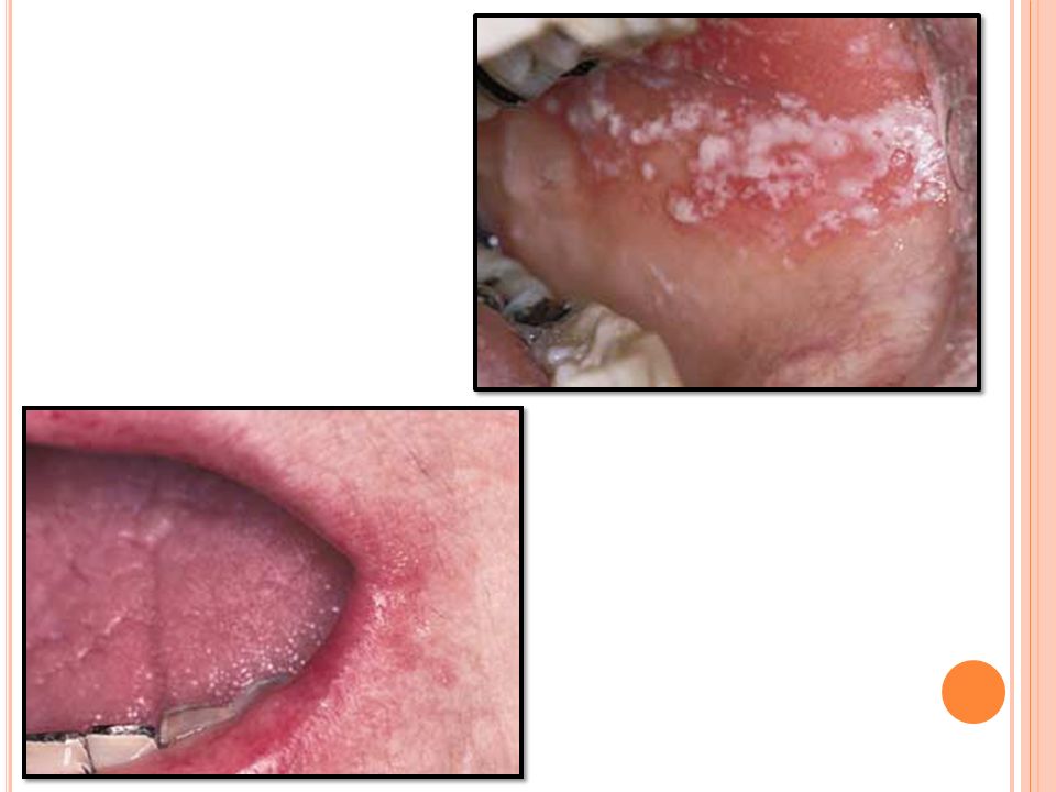
 most damaging. Fair-complexioned people more severely affected than others. May progress to cutaneous actinic keratosis and/or squamous cell carcinoma.")
24
Clinical Presentation
Vermilion portion of lower lip. Pale irregularly opaque (keratotic) surface with intervening red (atrophic) zones. Obfuscated to effaced cutaneous- vermilion border. More advanced lesions are scaly, crusted and/or indurated. Progression to carcinoma often heralded by persistent ulceration or erosion.
 surface with intervening red (atrophic) zones. Obfuscated to effaced cutaneous- vermilion border. More advanced lesions are scaly, crusted and/or indurated. Progression to carcinoma often heralded by persistent ulceration or erosion.")
25
Microscopic Findings Hyperkeratosis. Epithelial atrophy.
Variable degrees of epithelial dysplasia. Amphophilic to basophilic change in submucosa (elastosis). Telangiectasia.
. Telangiectasia.")
27
Differential Diagnosis
Thermal/chemical burn ruled out by history Chronic ultraviolet light exposure Biopsy findings Differential Diagnosis Exfoliative cheilitis Squamous cell carcinoma

28
Treatment Prevention of further damage with sunscreens blocking longwave ultraviolet A (UVA) and short-wave ultraviolet B (UVB) light Biopsy of clinically suspicious areas CO2 laser vermilionectomy Topical 5-fluorouracil or vermilionectomy for severe disease Excision or resection-reconstruction if malignant transformation has occurred

34
Prognosis Lifelong follow-up
Up to 10% develop into squamous cell carcinoma. When carcinoma develops, growth tends to be slow and metastasis occurs late; 85 to 90% long-term survival

35
Smokeless Tobacco Keratosis (Snuff Pouch)
Etiology Persistent habit of holding ground tobacco within the mucobuccal vestibule.

36
Clinical Presentation
Usually in men in Western countries. Powdered snuff use prevalent in Southeast United States often by women. Mucosal pouch with soft, white, fissured appearance. Surface may be pumice-like to verrucous. Leathery surface due to chronic tobacco use over many years

37
Microscopic Findings Hyperkeratosis with parakeratotic “chevron sign” at surface. Increased vascularity. Older lesions with hyalinization in submucosa and minor salivary glands. Epithelial dysplasia and carcinoma may evolve.
![]()
39
Differential Diagnosis
• Clinical appearance • Biopsy Differential Diagnosis Leukoplakia (idiopathic) Mucosal burn (chemical/thermal)
 Mucosal burn (chemical/thermal)")
40
Treatment Prognosis Discontinuation of habit.
If dysplasia is present, stripping of mucosal site. Prognosis Generally good with tobacco cessation. Malignant transformation to squamous cell carcinoma or verrucous carcinoma occurs but less frequently than does smokingrelated carcinoma.

41
Nicotine Stomatitis Etiology
Nicotine Stomatitis is usually found, as the name implies, in pipe-smokers, but also, occasionally, in cigarette- and cigar smokers. The characteristic feature of this condition is the combination of epithelial acanthosis and hyperkeratosis with inflammatory changes in the mucous glands of the palate. As a result of these changes, the palate becomes white and a number of nodules project from the surface, each representing the site of a mucous gland and individually bearing a small red spot at the centre that marks the opening of the duct of the gland.

42
Clinical Presentation
The condition usually appears most marked on the hard palate, although the soft palate may also be involved. This condition is most often found in older males Due to the chronic insult, the palatal mucosa becomes diffusely gray or white. Numerous slightly elevated papules with punctate red centers that represent inflamed and metaplastically altered minor salivary gland ducts are noted.

44
Differential Diagnosis
Clinical appearance Biopsy Differential Diagnosis Leukoplakia (idiopathic)
")
45
Treatment and Prognosis
Discontinuation of tobacco habit Observation and examination of all mucosal sites little risk of malignant transformation in palate, except for reverse smokers.

46
Hairy Leukoplakia Etiology
Probably due to opportunistic Epstein-Barr virus (EBV) infection of epithelial cells. Usually in an immunocompromised or immunosuppressed host.
 infection of epithelial cells. Usually in an immunocompromised or immunosuppressed host.")
47
Clinical Presentation
Usually arises on lateral tongue border. Early lesions are fine, white, vertical streaks with an overall corrugated surface. Later lesions may be thickened to be plaque like. Extensive lesions can involve dorsum of tongue and buccal mucosa. May serve as a pre-AIDS sign.

49
Differential Diagnosis
Incisional biopsy findings show characteristic EBV nuclear inclusions in upper-level keratinocytes. Differential Diagnosis Frictional hyperkeratosis Lichen planus Hyperplastic candidiasis

50
Treatment None necessary; predisposing condition to be investigated Can be suppressed with acyclovir for esthetics Antiviral acyclovir Podophyllin resin topically Prognosis May herald human immunodeficiency virus (HIV) disease in vast majority of cases. Also may be present after AIDS is established.
 disease in vast majority of cases. Also may be present after AIDS is established.")
51
Candidiasis Etiology Infection with a fungal organism of the Candida species, usually Candida albicans. Associated with predisposing factors: most commonly, immunosuppression, diabetes mellitus, antibiotic use, or xerostomia (due to lack of protective effects of saliva).
.")
52
Clinical Presentation
Acute (thrush) Pseudomembranous. Painful white plaques representing fungal colonies on inflamed mucosa. Erythematous (acute atrophic): painful red patches caused by acute Candida overgrowth and subsequent stripping of those colonies from mucosa.
 Pseudomembranous. Painful white plaques representing fungal colonies on inflamed mucosa. Erythematous (acute atrophic): painful red patches caused by acute Candida overgrowth and subsequent stripping of those colonies from mucosa.")
53
Clinical Presentation
Chronic Atrophic (erythematous): painful red patches; organism difficult to identify by culture, smear, and biopsy. “Denture-sore mouth” : a form of atrophic candidiasis associated with poorly fitting dentures; mucosa is red and painful on denture-bearing surface. Median rhomboid glossitis: a form of hyperplastic candidiasis seen on midline dorsum of tongue anterior to circumvallate papillae. Perleche: chronic Candida infection of labial commissures; often co-infected with Staphylococcus aureus. Hyperplastic/chronic hyperplastic: a form of hyperkeratosis in which Candida has been identified; usually buccal mucosa near commissures; cause and effect not yet proven. Syndrome associated: chronic candidiasis may be seen in association with endocrinopathies.
: painful red patches; organism difficult to identify by culture, smear, and biopsy. Denture-sore mouth : a form of atrophic candidiasis associated with poorly fitting dentures; mucosa is red and painful on denture-bearing surface. Median rhomboid glossitis: a form of hyperplastic candidiasis seen on midline dorsum of tongue anterior to circumvallate papillae. Perleche: chronic Candida infection of labial commissures; often co-infected with Staphylococcus aureus. Hyperplastic/chronic hyperplastic: a form of hyperkeratosis in which Candida has been identified; usually buccal mucosa near commissures; cause and effect not yet proven. Syndrome associated: chronic candidiasis may be seen in association with endocrinopathies.")
56
Differential Diagnosis
Microscopic evaluation of lesion smears Potassium hydroxide preparation to demonstrate hyphae Periodic acid–Schiff (PAS) stain Culture on proper medium (Sabouraud’s, corn meal, or potato agar) Biopsy with PAS, Gomori’s methenamine silver (GMS), or other fungal stain of microscopic sections Differential Diagnosis Allergic or irritant contact stomatitis Atrophic lichen planus
 stain. Culture on proper medium (Sabouraud’s, corn meal, or potato agar) Biopsy with PAS, Gomori’s methenamine silver (GMS), or other fungal stain of microscopic sections. Differential Diagnosis. Allergic or irritant contact stomatitis. Atrophic lichen planus.")
57
Treatment Prognosis Topical or systemic antifungal agents
For immunocompromised patients: routine topical agents after control of infection is achieved, usually with systemic azole agents Correction of predisposing factor, if possible Some cases of chronic candidiasis may require prolonged therapy (weeks to months). Prognosis Excellent in the immunocompetent host
. Prognosis. Excellent in the immunocompetent host.")
58
Identify and correct provocative factors.
Topical therapy Nystatin oral suspension (100,000 units/mL); rinse 5 mL and swallow 4 times/d Clotrimazole (Lotrimin) solution 1%; rinse 5 mL and swallow 4 times/d Clotrimazole troches (Mycelex) 10 mg; dissolve 1 troche in mouth 5 times/d Clotrimazole vaginal tablets 1/2 of 500 mg tablet dissolved in mouth bid Systemic therapy Fluconazole (Diflucan) 100 mg #15; 2 tablets on the first day, 1 tablet days 2–7, 1 tablet every other day for days 8–21 Ketoconazole (Nizoral) 200 mg #21; 1 tablet every day with breakfast × 21 d Itraconazole (Sporanox) 200 mg #21; 1 tablet every day with breakfast × 21 d May use shorter duration for less severe infections
; rinse 5 mL and swallow 4 times/d. Clotrimazole (Lotrimin) solution 1%; rinse 5 mL and swallow 4 times/d. Clotrimazole troches (Mycelex) 10 mg; dissolve 1 troche in mouth 5 times/d. Clotrimazole vaginal tablets 1/2 of 500 mg tablet dissolved in mouth bid. Systemic therapy. Fluconazole (Diflucan) 100 mg #15; 2 tablets on the first day, 1 tablet days 2–7, 1 tablet every other day for days 8–21. Ketoconazole (Nizoral) 200 mg #21; 1 tablet every day with breakfast × 21 d. Itraconazole (Sporanox) 200 mg #21; 1 tablet every day with breakfast × 21 d. May use shorter duration for less severe infections.")
59
Leukoplakia Etiology Essentially unknown, although many cases related to use of tobacco or areca nut in its various formulations Other possible factors include nutritional deficiency (iron, vitamin A) and infection (Candida albicans, human papillomavirus).
 and infection (Candida albicans, human papillomavirus).")
61
Clinical Presentation
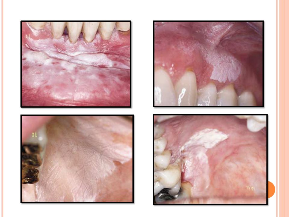
An idiopathic white (sometimes white-and-red) patch Most common on lip, gingiva, buccal mucosa Increased risk of dysplasia or carcinoma when occurring on tongue, floor of mouth, vermilion portion of lip Clinical subsets include homogeneous, verrucous, speckled, and proliferative verrucous leukoplakia (proliferative form may be multiple and persistent) Cases may advance or regress unpredictably— reflective of a dynamic process Most occur in the fifth decade and beyond Progress to dysplasia or malignancy may occur with little or no change in clinical appearance.
 patch. Most common on lip, gingiva, buccal mucosa. Increased risk of dysplasia or carcinoma when occurring on tongue, floor of mouth, vermilion portion of lip. Clinical subsets include homogeneous, verrucous, speckled, and proliferative verrucous leukoplakia (proliferative form may be multiple and persistent) Cases may advance or regress unpredictably— reflective of a dynamic process. Most occur in the fifth decade and beyond. Progress to dysplasia or malignancy may occur with little or no change in clinical appearance.")
64
Diagnosis Performance of a biopsy is mandatory after elimination of any suspected causative factors Multiple biopsies of large lesions are needed to be performed due to microscopic heterogeneity within a single lesion.

65
Differential Diagnosis
Other white lesions √ Frictional keratosis √ Burn (thermal/chemical) √ Hyperplastic candidiasis √ Lichen planus Genetic alterations √ White sponge nevus √ Hereditary benign intra- √ Dyskeratosis epithelial dyskeratosis
 √ Hyperplastic candidiasis √ Lichen planus. Genetic alterations. √ White sponge nevus √ Hereditary benign intra- √ Dyskeratosis epithelial dyskeratosis.")
66
Treatment Excision modalities (surgery, laser ablation, cryosurgery) Option to observe lesions diagnosed as benign hyperkeratosis or mild dysplasia Possibly photodynamic therapy Topical cytotoxic drugs (bleomycin) remain experimental. Recurrences common following apparent complete excision Prognosis Guarded Observation with repeat biopsies to be performed
 remain experimental. Recurrences common following apparent complete excision. Prognosis. Guarded. Observation with repeat biopsies to be performed.")
67
Prevention Recurrences may be reduced by systemic retinoid therapy.
Elimination of tobacco use and heavy alcohol consumption Recurrences may be reduced by systemic retinoid therapy. Possible dietary measures

68
Erythroplakia Etiology
Unknown: a red patch that cannot be clinically attributed to another condition Contributing factors include tobacco use, alcohol consumption

69
Clinical Presentation
Red, often velvety, well-defined patch(es). Most common on floor of mouth, retromolar trigone area, lateral tongue. Usually asymptomatic. May be smooth to nodular. Chiefly in males.
. Most common on floor of mouth, retromolar trigone area, lateral tongue. Usually asymptomatic. May be smooth to nodular. Chiefly in males.")
71
Differential Diagnosis
Appearance; history of tobacco/alcohol use Biopsy results differentiate from inflammatory and atrophic lesions Differential Diagnosis Erythematous (atrophic) candidiasis Kaposi’s sarcoma Ecchymosis Contact stomatitis Vascular malformation Squamous cell carcinoma Geographic tongue/ erythema migrans
 candidiasis. Kaposi’s sarcoma. Ecchymosis. Contact stomatitis. Vascular malformation. Squamous cell carcinoma. Geographic tongue/ erythema migrans.")
72
Treatment Prognosis Surgical excision if proven dysplastic/ malignant
Fair to good depending upon microscopic diagnosis Almost all cases are premalignant to malignant upon initial discovery.

73
Lichen Planus Etiology Unknown.
Autoimmune T cell–mediated disease targeting basal keratinocytes (antigen unknown). Lichenoid changes associated with galvanism, graft-versus-host disease (GVHD), certain drugs, contact allergens.
. Lichenoid changes associated with galvanism, graft-versus-host disease (GVHD), certain drugs, contact allergens.")
75
Clinical Presentation
Up to 3 to 4% of population have oral lichen planus 0.5 to 1% of population have cutaneous lichen planus; 50% also have oral lesions (25% with oral lesions have concomitant skin lesions) White females (60%) Occurs in fourth to eighth decades Variants: reticular (most common oral form); erosive (painful); atrophic, papular, plaque types; bullous (rare) Bilateral and often symmetric distribution Oral site frequency: buccal mucosa (most frequent), then tongue, then gingiva, then lips (least frequent) Skin sites: forearm, shin, scalp, genitalia
 White females (60%) Occurs in fourth to eighth decades. Variants: reticular (most common oral form); erosive (painful); atrophic, papular, plaque types; bullous (rare) Bilateral and often symmetric distribution. Oral site frequency: buccal mucosa (most frequent), then tongue, then gingiva, then lips (least frequent) Skin sites: forearm, shin, scalp, genitalia.")
78
Differential Diagnosis
Examination of oral mucosa, skin, genitalia Negative ocular mucosa history; no history of blistering Use of drugs, galvanism, GVHD to be ruled out Biopsy Direct immunofluorescence–fibrinogen and cytoid bodies at interface help confirm Differential Diagnosis • Lichenoid drug eruptions • Erythema multiforme • Lupus erythematosus • Contact stomatitis • Mucous membrane pemphigoid

79
Treatment of Oral Lichen Planus
Mild to moderate: topical corticosteroids Severe: systemic immunosuppression, chiefly with prednisone Corticosteroid-sparing drugs with prednisone Topical tacrolimus ointment Prognosis Control, not cure, can be expected. Good prognosis; rare malignant transformation (0.5–3%) May be cyclic; may last for years/decades Tends to be chronic
 May be cyclic; may last for years/decades. Tends to be chronic.")
Similar presentations


 Pathogenesis` (Mechanisms:inflammation) Clinical Features (Signs and Symptoms) Fever,>")


