Download presentation
Presentation is loading. Please wait.

1
Technological advances in Brachytherapy
Ekkasit Tharavichitkul, MD The Division of Therapeutic Radiology and Oncology, Faculty of Medicine, Chiang Mai University

2
History Greek word = short –Interstitial brachytherapy
–Contact brachytherapy → surface mould BT → intracavitary BT → endolumina BT

3
Brachytherapy history
1896: Becquerel 1898: MarieSklodowska-Curie 1901: Danlosand Block: Paris 1905Abbe: US Radium implantations

4
Different empirical methods and rules
•Stockholm method for Gyne (1914) •Paris method for Gyne (1919) •Manchester system (1934) Paterson-Parker, Meredith •Paris System for IS : Pierquin, Chassagne, Dutreix
 •Paris method for Gyne (1919) •Manchester system (1934) Paterson-Parker, Meredith. •Paris System for IS : Pierquin, Chassagne, Dutreix.")
5
Discovery of artificial radioactive isotopes
• 1934 Irene Curie -FrédericJoliot • 1958 Iridium-I92: U. Henschke Development of afterloading concept • U. Henschke-D. Chassagne Developmentof 3D dosimetry and fundamental rules of dosimetry • B. Pierquin-D. Chassagne-A. Dutreix

6
Brachytherapy

7
Developments in BT Source and loading methods Imaging developments
Applicator developments Planning developments Clinical research developments

8
Source and loading method

9
Radioisotope sources and loading methods
From Radium --- Iridium From LDR --- HDR --- PDR From manual loading to remote after- loading

10
Radium--Iridium

12
Manual and remote loading

13
LDR vs. HDR พารามิเตอร์ LDR HDR Dose rate < 2 Gy/ชั่วโมง
Problem of radiation hazard + - Discomfort +++ Unexpect shift of applicators Ward Number of patient per day 1 มากกว่า 1 ราย Time for loading ชั่วโมง นาที Cost of machine

14
Manual or remote control afterloading
Gynecological applicators Guide needles: straight and curved Plastic tubes Moulds Hypodermic needles Silk wires Endo-luminal catheters • Remote control afterloaders

15
Imaging

16
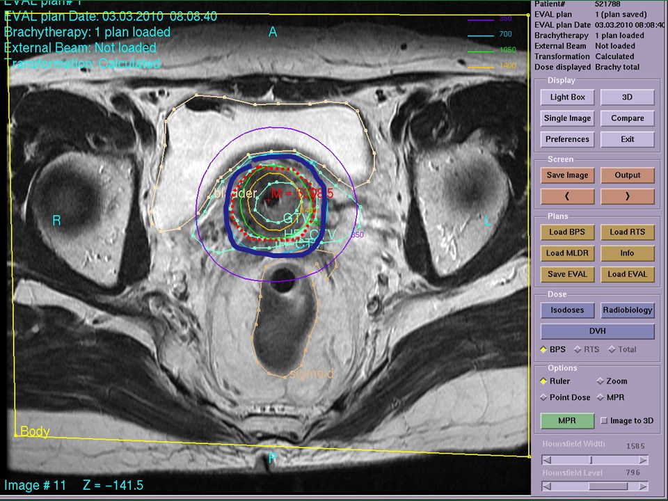
Imaging developments Modern imagingtechniques: -US, CT, MRI
• 3D dosimetry -More accurate dose distribution -DVH relation to outcome for target + OAR

17
X-ray base

18
Ultrasound guidance

19
U/S guidance

21
Applicators

22
Applicator developments
More compatible with imaging CT/MR applicator Gynecological cancers Plastic catheter Breast cancer Prostate cancer

23
Standard applicators Nucletron.com

24
Scatter; metallic applicator

25
CT/Applicator

26
Breast BT mammosite Multicatheter Clearpath Polgar, 2009

27
Volume concepts

28
Volume concepts 1985 ICRU 38 :Gynecological brachytherapy
1997 ICRU 58 :Interstitial and intraluminal brachytherapy 2000 GEC-ESTRORec: Prostate Permanent Implants 2001 GEC-ESTRO Rec: Endovascular brachytherapy 2005 GEC-ESTRORec: Prostate Temporary Implants 2005 GEC-ESTRORec: 3D-GYNE

29
Ultrasound

30
CTV prostate GEC-ESTRO handbook,2002

31
Target volume: HDR

33
Volume concepts of cervix
CHM, 2005

34
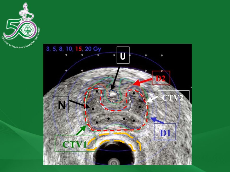
Diagram of CTVs

35
35

37
HR-CTV Bladder Rectum Sigmoid

38
D90 HR-CTV D2cc B D2cc R D2cc S

39
Chiang Mai

40
Breast cancer Polgar, 2009

41
Planning developments

42
Planning developments
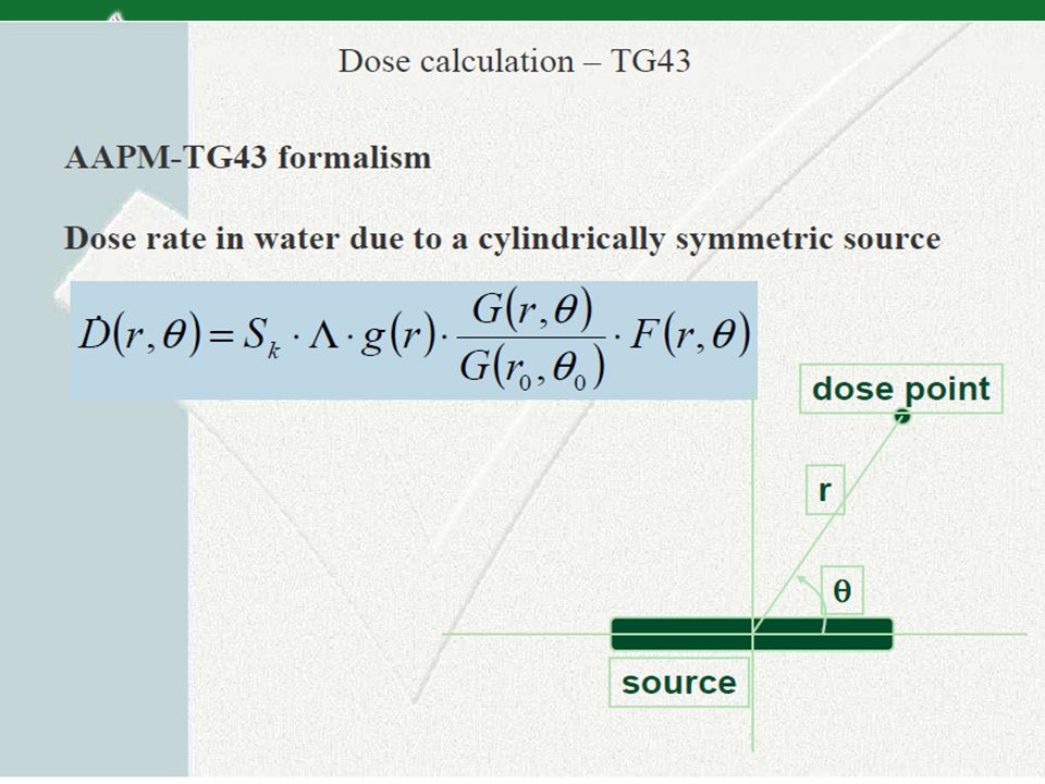
Shifting from 2D to 3D Target volume definition Algorithm: AAPM TG43 to Monte Carlo Inverse planning IPSA (Inverse Planning Simulated Annealing) HIPO (the Hybrid Inverse Planning and Optimization)
 HIPO (the Hybrid Inverse Planning and Optimization)")
44
Clinical studies

45
Cervix

47
Breast BT Polgar, 2009

48
Clinical studies

49
EVL, Estro course 2008

50
Toxicity profile Niehoff

51
Clinical developments
All RCTs NSABP B-39/RTOG 0413 (4800 pts enrolled) WBI 50 Gy plus boost to Gy versus Multicatheter (34Gy)/Mammosite(34 Gy) /3D- CRT(38.5Gy) GEC-ESTRO working group trial (1170 pts enrolled) WBI 50 Gy plus boost 10 Gy versus HDR and PDR Pending for results 51
 WBI 50 Gy plus boost to Gy versus Multicatheter (34Gy)/Mammosite(34 Gy) /3D- CRT(38.5Gy) GEC-ESTRO working group trial (1170 pts enrolled) WBI 50 Gy plus boost 10 Gy versus HDR and PDR. Pending for results. 51.")
52
Conclusions Modern brachytherapy which is high Ballistic selectivity and adaptivity is a competitive tool in the multidisciplinary treatment of cancer patients A strong collaboration between -Radiation oncologists -Organ specialists -Medical physicists -Radiation technologists is necessary to obtain optimal results for the patient(s)
")
53
Our researches: CT From July 2008 - Dec 2009
16 pts in CT-based planning in EBRT and BT BT 6.5 Gy x Fx GEC-ESTRO recommendations concepts Image-guided planning (optimized plan) can reduced the dose to the bladder and sigmoid colon with compromised dose to the target
 can reduced the dose to the bladder and sigmoid colon with compromised dose to the target.")
54
Our research:MRI From Feb 2009- nowadays
Planned 14 pts will be enrolled MRI guided treatment: Dx, 1st BT, after treatment With GEC-ESTRO recommendations Now 6 patients finished Pending results

Similar presentations


: Brachytherapy Techniques IAEA Training Material on Radiation Protection in.>")


 Extremity / Superficial Trunk STS (n=141) LSS Alone.>")

: The optimal indication for operable tumors in inoperable patients D.Katsochi 1, S.Kosmidis 1, A.Fotopoulou.>")


