Download presentation
Presentation is loading. Please wait.

1
Introduction to Microbial Genetics
Microbiology 221

3
The scientists who provided the clues to the nature of DNA
A Historical Overview The scientists who provided the clues to the nature of DNA Friederich Meischer – DNA isolated Luria and Delbruck – Bacteriophages Stanely Giffiths( 1928) The idea of the transforming “ substance” – Avery, MacLoed, and McCarty( 1944) – the nature of transformation Hershey and Chase – Bacteriophage – DNA as the hereditary material Chargaff – A= T and C=G Maurice Wilkins and Rosalind Franklin – x-ray crystallography of DNA Watson and Crick – Double helix
 The idea of the transforming substance – Avery, MacLoed, and McCarty( 1944) – the nature of transformation. Hershey and Chase – Bacteriophage – DNA as the hereditary material. Chargaff – A= T and C=G. Maurice Wilkins and Rosalind Franklin – x-ray crystallography of DNA. Watson and Crick – Double helix.")
4
Griffiths

5
Luria and Delbruck at Cold Spring Harbor in 1953
Luria and Delbruck studied bacterial mutations and resistance to infection with bacteriophages The characterized the virus and its life cycle

7
Alfred Hershey and Martha Chase and the Blender Experiment
Hershey and Chase wanted to verify that DNA was the hereditary material They used a bacteriophage for their study They labeled the DNA with Radioactive P( P32) and the protein with radioactive sulfur( S35)
 and the protein with radioactive sulfur( S35)")
8
Results of the Experiment
Proved that the radioactivity from the labeled DNA was present in the progeny phage produced from infection of the bacteria.

9
The Race for the Double Helix
Rosalind Franklin and Maurice Wilkins at Kings College Studied the A and B forms of DNA Rosalind’s famous x-ray crystallography picture of the B form held the secret, but she didn’t realize its significance

10
The Race for the Double Helix
Watson and Crick formed an unlikely partnership A 22 year old PhD and a 34 year old “want to be” PhD embarked on a model making venture at Cambridge Used the research of other scientists to determine the nature of the double helix

11
Nucleic Acid Composition DNA and RNA
DNA – Basic Molecules Purines – adenine and guanine Pyrmidines – cytosine and thymine Sugar – Deoxyribose Phosphate phosphate group - DNA background

12
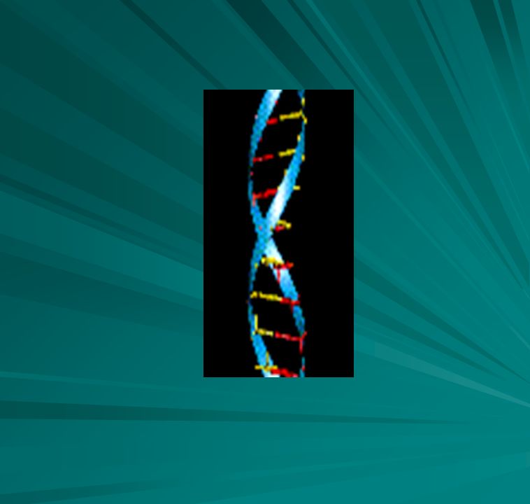
Double Helix Two polynucleotide strands joined by phosphodiester bonds( backbone) Complementary base pairing in the center of the molecule A= T and C G – base pairing. Two hydrogen bonds between A and T and three hydrogen bonds between C and G. A purine is bonded to a complementary pyrimidine Bases are attached to the 1’ C in the sugar At opposite ends of the strand – one strand has the 3’hydroxyl, the other the 5’ hydroxyl of the sugar molecule

13
DNA Structure - DNA structure

14
Double helix ( continued)
The double helix is right handed – the chains turn counter-clockwise. As the strand turn around each other they form a major and minor groove. The is a distance of .34nm between each base The distance between two major grooves is 2.4nm or 10 bases The diameter of the strand is 2nm

15
Complementary Base Pairing
Adenine pairs with Thymine Cytosine pairs with Guanine

16
The end view of DNA This view shows the double helix and the outer backbone with the bases in the center. An AT base pair is highlighted in white

17
Double helix and anti-parallel
DNA is a directional molecule The complementary strands run in opposite directions One strand runs 3’-5’ The other strand runs 5’ to 3’ ( the end of the 5’ has the phosphates attached, while the 3’ end has a hydroxyl exposed)
")
18
RNA structure Polynucleotide – nucleic acid - Single stranded molecule that can coil back on itself and produce complementary base-pairing ( t- RNA) Four bases in RNA are Adenine and Guanine ( purines) and Cytosine and Uracil( pyrimidines) Sugar – ribose Phosphates
 and Cytosine and Uracil( pyrimidines) Sugar – ribose. Phosphates.")
19
RNA Three types of RNA Messenger Transfer Ribosomal
nc- non coding RNA’s

20
Prokaryote DNA Tightly coiled
Coiling maintained by molecules similar to the coiling in eukaryotes Circular ds molecule Borrelia burgdoferi ( Lyme Disease )has a linear chromosome Other bacteria have multiple chromosomes Agrobacterium tumefaciens ( Produces Crown Gall disease in plants) has both circular and linear
has a linear chromosome. Other bacteria have multiple chromosomes. Agrobacterium tumefaciens ( Produces Crown Gall disease in plants) has both circular and linear.")
21
Prokaryote chromosomes
Circular DNA

23
E. coli – most often studied in molecular biology of prokaryotes
The genes of E. coli are located on a circular chromosome of 4.6 million basepairs. This 1.6 mm long molecule is compressed into a highly organized structure which fits inside the 1-2 micrometer cell in a format which can still be read by the gene expression machinery. Bacterial DNA is supercoiled by DNA gyrase. Chemical inhibition of gyrase without allowing the cells to reprogram gene expression relaxes supercoiling and expands the nucleoid, suggesting that supercoiling is one of the tools used to compress the genome

24
Coiling Coiling maintained by Gyrase
Relaxation of the coils by Topoisomerase

25
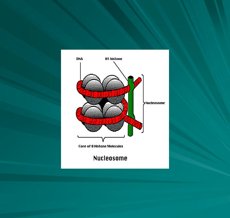
Nucleosome formation DNA is more highly organized in eukaryote cells
The DNA is associated with proteins called histones.( eukaryotes) These are small basic proteins rich in the amino acids lysine and/or arginine There are five histones in eukaryote cells, H1, H2A, H2B,H3 and H4. .
 These are small basic proteins rich in the amino acids lysine and/or arginine. There are five histones in eukaryote cells, H1, H2A, H2B,H3 and H4. .")
27
Beads on a String The DNA coils around the ellipsoid approximately 1 ¾ turns or 166 base pairs before proceeding to the next. The DNA + the histone proteins arranged in this formation are referred to as a nucleosome. The stretch of DMA between the beads varies in length from 14 to 100 base pairs. H1 appears to associate with the linker regions to enable the nucleosome to supercoil When folding of the structure reaches a maximum, the chromosomes can be visualized

28
Chromosome structure

29
Eukaryote replication
The nature of DNA replication was elucidated by Meselson and Stahl

30
Meselson and Stahl experiment
Grew bacteria in heavy Nitrogen – N-15 Transferred bacteria to N-14 Before bacteria reproduce in new media, all bacteria contain heavy DNA Samples were taken after one round of replication and two round of replication

31
Semiconservative replication
Each original strand serves a template or pattern for the replication of the new strand. The new strand contains one original and a newly synthesized strand

32
Eukaryote replication
Multiple linear chromosomes Each chromosome has more than one origin of replication Approximately 1400 x as long as bacterial DNA Multiple replicons on a chromosome Oris along the length – every 10 to 100 um Replication forks and bubbles are formed. Replication proceeds bidirectionally until the bubbles meet This shortens the length of time necessary to replicate eukaryote chromosomes The process of elongation occurs at a speed of base pairs/minute as compared to 750 to 1000 base pairs/ minute

33
The origin of replication and replication forks

34
Eukaryote replication
During the S phase, there are 100 replication complexes and each one contains as many as 300 replication forks. These replication complexes are stationary. The DNA threads through these complexes as single strands and emerges as double strands.

35
DNA Polymerases Fourteen DNA polymerases have been observed in human beings as compared to three in E. coli.

36
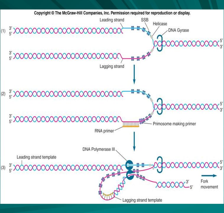
Prokaryote Replication

37
Bacterial topoisomerase I (v protein) I E. coli
Enzyme Type Source Properties Bacterial topoisomerase I (v protein) I E. coli Relaxation of negative but not positive supercoils Vaccinia virus topoisomerase I Vaccinia virus Relaxation of positive and negative supercoils Eukaryotic topoisomerase I Calf thymus Reverse gyrase Thermophilic bacteria Introduces positive supercoils into DNA Topoisomerase V Hyperthermophilic bacteria Relaxation of positive supercoils DNA gyrase II Introduces negative supercoils into DNA Topoisomerase IV DNA relaxation and potent decatenation T4 topoisomerase II Bacteriophage T4 Relaxation of positive and negative supercoils and decatenation Eukaryotic topoisomerase II S. cerevisiae
 I. E. coli. Relaxation of negative but not positive supercoils. Vaccinia virus topoisomerase I. Vaccinia virus. Relaxation of positive and negative supercoils. Eukaryotic topoisomerase I. Calf thymus. Reverse gyrase. Thermophilic bacteria. Introduces positive supercoils into DNA. Topoisomerase V. Hyperthermophilic bacteria. Relaxation of positive supercoils. DNA gyrase. II. Introduces negative supercoils into DNA. Topoisomerase IV. DNA relaxation and potent decatenation. T4 topoisomerase II. Bacteriophage T4. Relaxation of positive and negative supercoils and decatenation. Eukaryotic topoisomerase II. S. cerevisiae.")
38
Bidirectional replication
There is an origin of replication Two replication forks are formed Replication occurs around the circle until they have opened and copied the entire chromosome Replicon- contains an origin and is replicated as a unit

39
Ori – Origin of replication
Characteristics used to define Origins: The position on the DNA at which replication start points (see right) are found. A DNA sequence that when added to a non-replicating DNA causes it to replicate. A DNA sequence whose mutation abolishes replication. A DNA sequence that in vitro is the binding target for enzyme
 are found. A DNA sequence that when added to a non-replicating DNA causes it to replicate. A DNA sequence whose mutation abolishes replication. A DNA sequence that in vitro is the binding target for enzyme.")
40
Topoisomerases Topoisomerase
When the double helix of DNA, which is composed of two strands, separates, helicase makes these two strands rotate around each other. The DnaB protein is the helicase most involved in replication, but the n’ protin may also participate in unwinding. The single stranded binding proteins SSBP help to keep the strand open But there is a problem due to the topological reason that the unreplicated part ahead of the replication fork will rotate around its helical axis when the two strands separate at the replication fork

41
Topoisomerase action It causes strong strain in the helix (1). Thus, it is impossible to unlink the double helical structure of DNA without disrupting the continuity of the strands. In order to perform unraveling of a "compensating winding up" DNA, enzymes are required (1). Topoisomerase changes the linking number as well as catalyzes the interconversionn of other kinds of topological isomers of DNA (2).
. Thus, it is impossible to unlink the double helical structure of DNA without disrupting the continuity of the strands. In order to perform unraveling of a compensating winding up DNA, enzymes are required (1). Topoisomerase changes the linking number as well as catalyzes the interconversionn of other kinds of topological isomers of DNA (2).")
42
Initiation Initiation a. oriC - origin of chromosomal replication Recognized by DnaA protein - only recognizes if GATC sites are fully methylated Binding of DnaA allows DnaB to open complex b. DnaB is the replication helicase c. Strand separation by helicase d. SSB (single-stranded binding) protein keeps strands apart e. DNA gyrase - a topoisomerase - puts swivel in DNA which allows strands to rotate and relieve strain of unwinding
 protein keeps strands apart e. DNA gyrase - a topoisomerase - puts swivel in DNA which allows strands to rotate and relieve strain of unwinding.")
43
Explanation Recall that DNA double helix is tightly wound structure and that bases lie between the two backbones. If these bases are the template for new strand, how do the appropriate enzymes reach these bases? By the unwinding of the helix. An enzyme called helicase catalyzes the unwinding of short DNA segments just ahead of the replication fork. The reaction is driven by the hydrolysis of ATP.

44
Explanation continued
As soon as duplex is unwound, SSB (single-stranded binding protein) binds to each of the separated strands to prevent them from base-pairing again. Therefore, the bases are exposed to the replication system. The unwinding of the duplex would cause the entire DNA molecule to swivel except for the action of a topoisomerase (DNA gyrase) which introduce breaks in the DNA just ahead of the unwinding duplex. These breaks are then rejoined after a few revolutions of the duplex.
 binds to each of the separated strands to prevent them from base-pairing again. Therefore, the bases are exposed to the replication system. The unwinding of the duplex would cause the entire DNA molecule to swivel except for the action of a topoisomerase (DNA gyrase) which introduce breaks in the DNA just ahead of the unwinding duplex. These breaks are then rejoined after a few revolutions of the duplex.")
45
The need for a primer When DNA template is exposed, DNA synthesis must begin. But DNA polymerases not only need a template but also a primer for replication to proceed. Where does the primer come from? After observations that RNA synthesis is required for DNA synthesis, it was discovered that the synthesis of DNA fragments requires a short length of RNA as a primer. Primosome (complex of 20 polypeptides) makes RNA primers in E. coli
 makes RNA primers in E. coli.")
46
Formation of the Primer
Primosome contains primase Primosome moves along DNA duplex in 3'>5' direction (with respect to lagging strand; follows replication fork) even though primer is made in 5'>3' direction (Note: The symbol ">" indicates the direction; that is, the primer is made from 5' to 3'.) n' protein removes SSB in front of primosome DnaB protein organizes some components of primosome and prepares DNA for primase Primase forms the primer
 even though primer is made in 5 >3 direction (Note: The symbol > indicates the direction; that is, the primer is made from 5 to 3 .) n protein removes SSB in front of primosome. DnaB protein organizes some components of primosome and prepares DNA for primase Primase forms the primer.")
47
DNA POLYMERASE III Holoenzyme
Complex that synthesizes most of the DNA copy contains the DNA polymerase enzyme and other proteins The gamma delta complex and the B subunits of the holoenzyme bind it to the template and the primer The alpha subunit carries out the actual polymerization reaction All of the proteins form a huge complex called the replisome

48
DNA polymerase III This is a stationary complex that probably attached to the plasma membrane. The DNA moves through the replisome and is copied

49
Elongation of the chain
dCTP dCMP + PPi Energy is supplied for biosynthesis by the cleaving of the phosphate bond

50
Elongation( continued)
Elongation proceeds in 5' > 3' direction and requires 1) all 4 deoxyribonucleoside 5'-triphosphates (dATP, dGTP, dCTP, dTTP), 2) Mg+ ions, 3) a primer made of nucleic acid, and 4) a DNA template. Rate of elongation = nucleotides per second Rate of formation of initiation complex = 1-2 minutes
 all 4 deoxyribonucleoside 5 -triphosphates (dATP, dGTP, dCTP, dTTP), 2) Mg+ ions, 3) a primer made of nucleic acid, and 4) a DNA template. Rate of elongation = nucleotides per second Rate of formation of initiation complex = 1-2 minutes.")
51
Elongation Elongation DNA polymerase I, II and III in E .coli DNA polymerase III holoenzyme - complex of 7 polypeptides Replisome - primosome and 2 DNA polymerase III - synthesizes DNA on both strands simultaneously without dissociating from DNA DNA polymerase III catalyzes the addition of deoxyribonucleotide units to end of the DNA strand with release of inorganic pyrophosphate (PPi) (DNA)n residues + dNTP <> (DNA)n + 1 residues + PPi Attachment of new units is by their a-phosphate groups to a free 3'-hydroxyl end of preexisting DNA chain.
 (DNA)n residues + dNTP <> (DNA)n + 1 residues + PPi Attachment of new units is by their a-phosphate groups to a free 3 -hydroxyl end of preexisting DNA chain.")
54
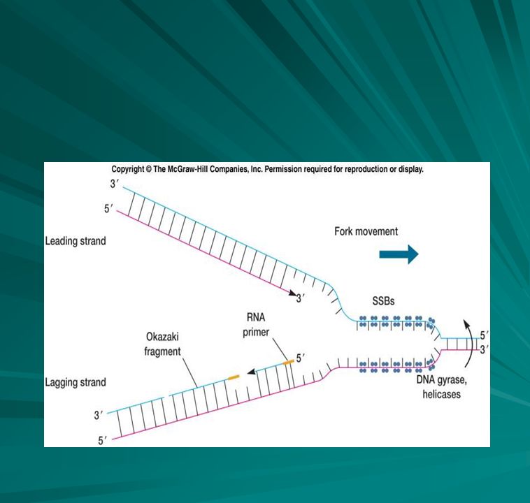
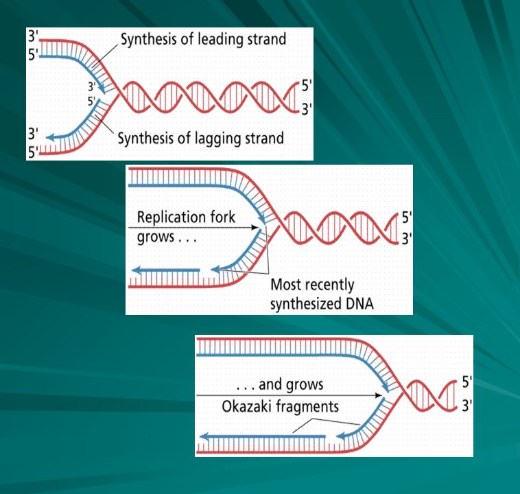
The lagging strand and discontinuous replication
The replication on the 5’ to 3’ strand differs The template strand still must be read from 3’ to 5’ The reading begins at the replication fork Occurs at the same time as the synthesis of the lagging strand Same steps in synthesis of DNA But DNA is synthesized in pieces about 1000 to 2000 bases in length. These are known as Okazaki fragments

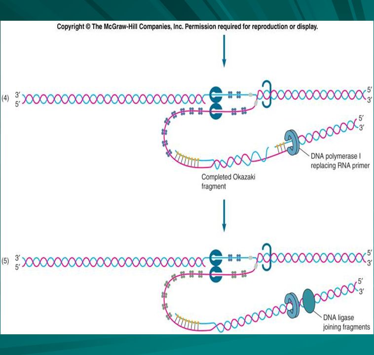
55
Okazaki fragments After the lagging strand has been duplicated by the formation of Okazaki fragments, DNA Polymerase I or RNase H removes the RNA primer. Polymerase I synthesizes the complementary DNA to fill the gap resulting from the RNA delection. The polymerase removes one nucleotide at a time and then replaces it AMP( RNA nucleotide) replaced by dAMP( DNA nucleotide)
 replaced by dAMP( DNA nucleotide)")
56
DNA ligase Ligase can catalyze the formation of a phosphodiester bond given an unattached but adjacent 3'OH and 5'phosphate. This can fill in the unattached gap left when the RNA primer is removed and filled in. The DNA polymerase can organize the bond on the 5' end of the primer, but ligase is needed to make the bond on the 3' end.

59
The End of Replication DNA replication stops when the polymerase complex reaches a termination site on the DNA in E. coli The Tus protein binds to the ter site and halts replication. In many prokaryotes the replication process stops when the replication forks meet

60
Plasmid replication ColE1 is a naturally occurring plasmid of E. coli. Its replication is controlled independently of the replication of the host chromosome. Two plasmids with the same origin of replication can not coexist in the same cell. The ColE1 origin, defined by molecular genetic methods, is in a region from which two RNAs are transcribed. An active RNase H gene is required for ColE1 replication. RNase H cleaves the RNA II transcript. The remaining RNA serves as primer for initiation of replication. RNA I binds to 5' sequences of RNA II via pseudoknots and regular complementary pairing. This binding is stabilized by the ROP or ROM protein. The binding prevents changes in the conformation of RNA II that would otherwise result in RNAse H cleavage.

61
Rolling Circle Replication – Occurs in Conjugation in E. coli.

62
How can one account for the high fidelity of replication?
The answer is based on the fact that DNA Polymerase absolutely requires 3'-OH end of base-paired primer strand on which to add new nucleotides. DNA polymerase III has 3' > 5' exonuclease activity. It was discovered that DNA polymerase III actually proofreads the newly synthesized strand before continuing with replication. When incorrect nucleotide is incorporated, DNA polymerase III, by means of the 3' > 5' exonuclease activity, "backs up" and hydrolyzes off the incorrect nucleotide. The correct nucleotide is then added to the chain and elongation is resumed. All 3 DNA polymerases have 3'>5' exonuclease activity Proofreading ability - 1 error in 10 million

63
Exonucleases and repair
DNA polymerase I also has 5'>3' exonuclease activity which removes RNA primer and 5'>3' polymerase activity which fills in the gap This causes a single-stranded break in the DNA - called a nick DNA ligase repairs nick by creating a phosphodiester bond

64
Genes and Gene Expression
Genes are written in a code consisting of groups of three letters called triplets. There are four letters in the DNA alphabet. There are 64 possible arrangements of the four letters in groups of three The triplets specify amino acids for the synthesis of proteins from the information contained in the gene Genes can also specify t- RNA or r- RNAs The gene begins with a start triplet and ends with a stop. The bases between the start and the stop are called an open reading frame, ORF. The information in the gene is transcribed by RNA polymerase. It reads the gene from 3’ to 5’ The template strand is now referred to as the CRICK strand and the nontemplate strand is now known as the WATSON strand DNA sequences are stored in data bases as the WATSON strand Reference - COLD SPRING HARBOR

65
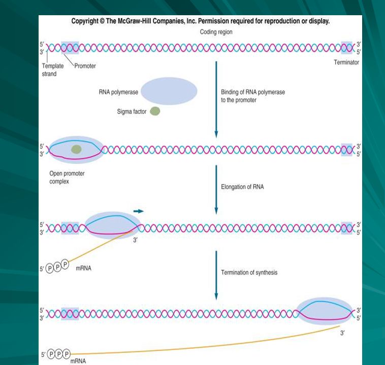
Promoters are at the beginning of the Gene
RNA polymerase recognizes a binding site in front of the gene. This is referred to as upstream of the gene. The direction of transcription is referred to as downstream Different genes have different promoters. IN E. coli the promoters have two functions The RNA recognition site for transcription which is the consensus sequence for prokaryotes is 5’ TTGACA3’ ( Watson strand) which means on the reading strand 3’ AACTGT5’ ( Crick strand)
 which means on the reading strand 3’ AACTGT5’ ( Crick strand)")
66
The Pribnow Box and Shane -Dalgarno
The RNA binding site has a consensus sequence of 5’ TATAAT 3’ ( -) and 3’ ATATTA 5’ (+) This is where the DNA begins to become unwound for transcription The initially transcribed sequence of the gene may not reflect doing but may be a leader sequence. The prokaryotes usually contain a consensus sequence known as the Shane Delgarno which is complememtary to the 16s rRNA on the ribosome ( small subunit ) The leader sequence also may regulate transcription
 and 3’ ATATTA 5’ (+) This is where the DNA begins to become unwound for transcription. The initially transcribed sequence of the gene may not reflect doing but may be a leader sequence. The prokaryotes usually contain a consensus sequence known as the Shane Delgarno which is complememtary to the 16s rRNA on the ribosome. ( small subunit ) The leader sequence also may regulate transcription.")
67
The structure of a prokaryote gene

68
Prokaryote Genes are Continuous
They do not contain introns like eukaryote genes The gene consists of codons that will determine the sequence of amino acids in the protein At the end of the gene there is a terminator sequence rather than an actual stop The terminator may be at the end of a trailer sequence located downstream from the actual coding region of the gene

69
The Gene begins with DNA is read 3’ to 5’ and m RNA is synthesized 5’ to 3’ 3’ TAC is the start triplet This produces a complementary mRNA message 5’ AUG 3’ – Groups of three bases in the messenger RNA formed are referred to as CODONS

70
RNA POLYMERASE

72
Wobble There is wobble in the DNA code – This is a protection from mutations More than one codon can specify the same amino acid Note arginine - CGU, CGC,CGA, CGG all code for arginine – only the third base in the codon changes There are two additional codons for arginine as well AGA and AGG these reflect the degenerate nature of the code

73
Codon chart

74
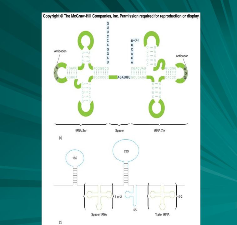
Genes for t RNAs and r RNAs
The genes for t RNAs have a promoter and transcribed leader and trailer sequence that are removed prior to their utilization in translation. Genes coding for tRNA may code for more than a single tRNA molecule The segments coding for r RNAs are separated by spacer sequencs that are removed after transcription.

76
t-RNA The acceptor stem includes the 5' and 3' ends of the tRNA.
The 5' end is generated by RNase P The 3' end is the site which is charged with amino acids for translation. Aminoacyl tRNA synthetases interact with both the acceptor 3' end and the anticodon when charging tRNAs. The anticodon matches the codon on mRNA and is read 3’ to 5’

77
t- RNA Found in the cytoplasm
Amino acyl t- RNA synthetase is an enzyme that enables the amino acid to attach to t-RNA Also activates the t- RNA Clover leaf has a stem for attachment to the amino acid and an anticodon on the bottom of the clover leaf

78
t- RNA Common Features a CCA trinucleotide at the 3' end, unpaired
four base-paired stems, and One loop containing a T-pseudoU-C sequence and another containing dihydroU.

79
tRNA tRNAs attach to a specific amino acid and carry it to the ribosome There are 20 amino acids 61 different codons for these amino acids and 61 tRNAs The anticodon is complementary to the codon Binds to the codon with hydrogen bonds

80
Ribosomal genes Very similar to the structure of protein genes

81
tRNA and rRNA genes The genes for rRNA are also similar to the organization of genes coding for proteins All rRNA genes are transcribed as a large precursor molecule that is edited by ribonucleases after transcription to yield the final r RNA products

82
Ribosomal RNA Combines with specific proteins to form ribosomes
Serves as a site for protein synthesis Associated enzymes and factors control the process of translation

83
Prokaryote ribosomes Ribosomes are small, but complex structures, roughly 20 to 30 nm in diameter, consisting of two unequally sized subunits, referred to as large and small which fit closely together as seen below. A subunit is composed of a complex between RNA molecules and proteins; each subunit contains at least one ribosomal RNA (rRNA) subunit and a large quantity of ribosomal proteins. The subunits together contain up to 82 specific proteins assembled in a precise sequence.
 subunit and a large quantity of ribosomal proteins. The subunits together contain up to 82 specific proteins assembled in a precise sequence.")
84
Approximate number of nucleotides
Prokaryote ribosomal RNA Type of rRNA Approximate number of nucleotides Subunit Location 16s 1,542 30s 5s 120 50s 23s 2,904

85
Prokaryote ribosomes – polysomes- the process of translation

86
Prokaryote transcription and translation
Prokaryote transcription and translation take place in the cytoplasm All necessary enzymes and molecules are present for the transcription and translation to take place

87
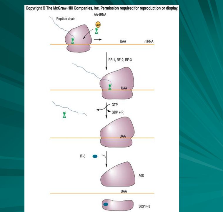
Translation A molecule of messenger RNA binds to the 30S ribosome ( small ribosomal unit) at the Shine Dalgarno sequence This insures the correct orientation for the molecule The large ribosomal sub unit locks on top

88
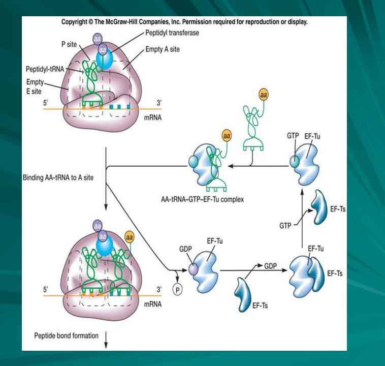
The Ribosome There are four significant positions on the ribosome EPAT When the 5’ AUG 3’ of the mRNA is on the P site the t-RNA with the anticodon, 5’UAG3’ forms a temporary bond to begin translation

92
From Gene to polypeptide

93
E. Coli Gene Map

94
Mutations in DNA May be characterized by their genotypic or phenotypic change Mutations can alter the phenotype of a microorganisms in different ways Mutations can involve a change in the cellular or colonial morphology

95
Types of Mutations Conditional mutations are those mutations that are expressed only under specific environmental conditions ( temperature) Biochemical mutations are those that can cause a change in the biochemistry of the cell ( these may inactivate a biochemical pathway) These mutants are referred to as auxotrophs because they cannot grow on minimal media Prototrophs are usually wild type strains capable of growing on minimal media
 These mutants are referred to as auxotrophs because they cannot grow on minimal media. Prototrophs are usually wild type strains capable of growing on minimal media.")
96
Two types of mutations Spontaneous mutations – These occur without a causative agent during replication Induced mutations are the result of a substance referred to as a mutagen Cairns reports that a mutant E. coli strain unable to use lactose is able to regain its ability to use the sugar again – should this be referred to as adaptive mutation?

97
Hypermutation One possible explanation is hypermutation
A starving bacterium has the ability to generate multiple mutations with special mutator genes that enable them to form bacteria with the ability to metabolize lactose This is an interesting theory still under investigation

98
Spontaneous mutations
Types A purine substitutes for a purine or a pyrimidine substitutes of a pyrimidine. This type of mutation is referred ta as a transition. Most of these can be repaired by proofreading mechanisms A pyrimidine substituted for by a purine is referred to as a transversion. These are rarer due to steric problems in the DNA molecule such as pairing purines with purines. Insertions or deletions cause frame shifts – the code shifts over the number of bases inserted or deleted

99
Mutation Types Erors in replication due to base tautomerization
AT and CG pairs are formed when keto groups participate in hydrogen bonds In contrast enol tautomers produce AC and GT base pairing

100
Spontaneous mutations – another cause
Depurination A purine nucleotide can lose its base It will not base pair normally It will probably lead to a transition type mutation after the next round of replication. Cytosine can be deaminated to uracil which can then create a problem

101
Frame Shifts Additions and deletions change the reading frame.
The hypothetical origin of deletions and insertions may occur during replication If the new strand slips an insertion or addition may occur If the parental slips a deletion may occur

102
Mutagenesis Any agent that directly damages DNA, alters its chemistry, or interferes with repair mechanisms will induce mutations Base analogs Specific mispairing Intercalating agents Ionizing radiation Base analogs are structurally similar to normal nitrogenous bases and can be incorporated into the growing polynucleotide chain during replication.

103
The expression of mutations
Forward mutations – a mutation from the wild type to a mutant form is called a forward mutation Reversion-If the organism regains its wild type characteristics through a second mutation Back mutation – The actual nucleotide sequence is converted back to the original Suppressor mutation – overcomes the effects of the first mutation

104
More on mutations Point mutations – caused by the change in one DNA base Silent mutations – mutations can occur which cause no effect – this is due to the degeneracy of the code ( more than one base coding for the same amino acid) Missense mutation – changes a codon for one amino acid into a codon for another amino acid Nonsense – In eukaryotes the substitution of a stop into the sequence of a normal gene
 Missense mutation – changes a codon for one amino acid into a codon for another amino acid. Nonsense – In eukaryotes the substitution of a stop into the sequence of a normal gene.")
105
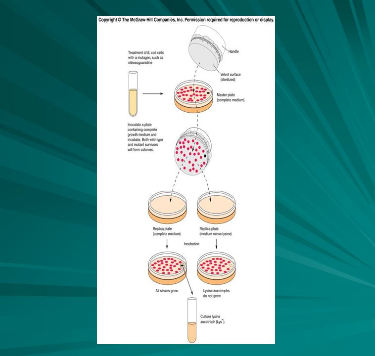
Detection and isolation of mutants
Requires a sensitive system Mutations are rare One in about every 107 – 1011 Replica plating is a technique that is used to detect auxotrophs It distinguishes between wild type and mutants because of their ability to grow in the absence of a particular biosynthetic end product Replica plating allows plating on minimal media and enriched media from the same master plate

107
The selection of auxotorph revertants
The lysine auxotrophs ( Lys-) are treated with a mutagen such as nitroquanidine or uv light to produce revertants
 are treated with a mutagen such as nitroquanidine or uv light to produce revertants.")
108
Ames Test Developed by Bruce Ames Used to test for carcinogens
A mutational reversion assay based upon mutants of Salmonella typhimurium

109
DNA repair mechanisms Type I -Excision repair
Corrects damage which causes distortions in the double helix A repair endonuclease or uvr ABC endonuclease removes the damaged bases along with some bases on either side of thee lesion The usual gap is about 12 nucleotides long. It is filled by DNA polymerase and ligase joins the fragments. This can remove Thymine-Thymine dimers A special type of repair utilizes glycosylases to remove damaged or unnatural bases yielding the results discussed above

110
Mutations and repair Type II – Removal of lesion
Thymine dimers and alkylated bases are often repaired directly Photoreactivation is the repair of thymine dimers by splitting them apart into separate thymines with the aid of visible light in a photochemical reaction catalyzed by the enzyme photolyase Light repair -phr gene - codes for deoxyribodipyrimidine photolyase that, with cofactor folic acid, binds in dark to T dimer. When light shines on cell, folic acid absorbs the light and uses the energy to break bond of T dimer; photolyase then falls off DNA

112
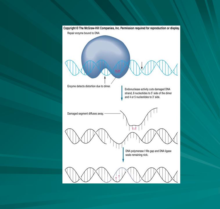
Dark repair of mutations
Dark repair Three types 1) UV Damage Repair (also called NER - nucleotide excision repair) Excinuclease (an endonuclease; also called correndonuclease [correction endo.]) that can detect T dimer, nicks DNA strand on 5' end of dimer (composed of subunits coded by uvrA, uvrB and uvrC genes). UvrA protein and ATP bind to DNA at the distortion. UvrB binds to the UvrA-DNA complex and increases specificity of UvrA-ATP complex for irradiated DNA. UvrC nicks DNA 8 bases upstream and 4 or 5 bases downstream of dimer. UvrD (DNA helicase II; same as DnaB used during replication initiation) separates strands to release 12-bp segment. DNA polymerase I now fills in gap in 5'>3' direction and ligase seals.
 UV Damage Repair (also called NER - nucleotide excision repair) Excinuclease (an endonuclease; also called correndonuclease [correction endo.]) that can detect T dimer, nicks DNA strand on 5 end of dimer (composed of subunits coded by uvrA, uvrB and uvrC genes). UvrA protein and ATP bind to DNA at the distortion. UvrB binds to the UvrA-DNA complex and increases specificity of UvrA-ATP complex for irradiated DNA. UvrC nicks DNA 8 bases upstream and 4 or 5 bases downstream of dimer. UvrD (DNA helicase II; same as DnaB used during replication initiation) separates strands to release 12-bp segment. DNA polymerase I now fills in gap in 5 >3 direction and ligase seals.")
113
The Effects of uv light

114
Post replication repair
If T dimer not repaired, DNA Pol III can't make complementary strand during replication. Postdimer initiation - skips over lesion and leaves large gap (800 bases). Gap may be repaired by enzymes in recombination system - lesion remains but get intact double helix. Successful post replication depends upon the ability to recognize the old and newly replicated DNA strands This is possible because the newly replicated DNA strand lack methyl groups on their bases, whereas the older DNA has methyl groups on the bases of both strands. The DNA repair system cuts out the mismatch from the non- methylated strand
. Gap may be repaired by enzymes in recombination system - lesion remains but get intact double helix. Successful post replication depends upon the ability to recognize the old and newly replicated DNA strands. This is possible because the newly replicated DNA strand lack methyl groups on their bases, whereas the older DNA has methyl groups on the bases of both strands. The DNA repair system cuts out the mismatch from the non- methylated strand.")
115
Recombination repair The DNA repair for which there is no remaining template is restored RecA protein cuts a piece of template DNA from a sister molecule and puts it into the gap or uses it to replace a damaged strand Rec A also participates in a type of inducible repair known as SOS repair. If the DNA damage is so great that synthesis stops completely leaving many gaps, the Rec A will bind to the gaps and initiate strand exchange. It takes on a proteolytic funtion that destroys the lexA repressor protein which regulates genes involved in DNA repair and synthesis

Similar presentations


 Harmless bacteria (rough colonies) Heat-killed, disease- causing bacteria (smooth colonies) Control (no growth)>")


 Two strands of DNA run antiparallel.>")
 and Replication of DNA (chapter 12, pages 318 – 334)>")


