Download presentation
Presentation is loading. Please wait.

1
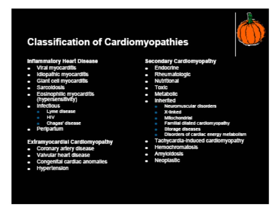
Cardiomyopathies Myocardial diseases “of unknown cause” – excludes hypertensive, valvular, ischaemic cardiomyopathies.

3
Cardiomyopathy Dilated (congestive)
Progressive congestive failure with a dilated heart ( need to consider ischaemia, toxins, viruses) Nutritional deficiency (protein, thiamine (B1), other vitamins) Some cases apparently familial
 Nutritional deficiency (protein, thiamine (B1), other vitamins) Some cases apparently familial.")
4
Dilated cardiomyopathy or DCM, also known as congestive cardiomyopathy, is a condition in which the heart becomes weakened and enlarged, and cannot pump blood efficiently. The decreased heart function can affect the lungs, liver, and other body systems. In DCM a portion of the myocardium is dilated, often without any obvious cause. Left and/or right ventricular systolic pump function of the heart is impaired, leading to progressive cardiac enlargement andhypertrophy, a process called remodeling.

5
Dilated cardiomyopathy is the most common form of cardiomyopathy.
It occurs more frequently in men than in women, and is most common between the ages of 20 and 60 years. About one in three cases of CHF is due to dilated cardiomyopathy. Dilated cardiomyopathy also occurs in children.

6
Causes: Although no cause (etiology) is apparent in many cases, dilated cardiomyopathy is probably the end result of damage to the myocardium produced by a variety of toxic,metabolic, or infectious agents. It may be the late sequel of acute viral myocarditis, possibly mediated through an immunologic mechanism. Autoimmune mechanisms are also suggested as a cause for dilated cardiomyopathy. A reversible form of dilated cardiomyopathy may be found with alcohol abuse,pregnancy,thyroid disease, stimulant use, and chronic uncontrolled tachcardia. Many cases of dilated cardiomyopathy are described as idiopathic - meaning that the cause is unknown.
 is apparent in many cases, dilated cardiomyopathy is probably the end result of damage to the myocardium produced by a variety of toxic,metabolic, or infectious agents. It may be the late sequel of acute viral myocarditis, possibly mediated through an immunologic mechanism. Autoimmune mechanisms are also suggested as a cause for dilated cardiomyopathy. A reversible form of dilated cardiomyopathy may be found with alcohol abuse,pregnancy,thyroid disease, stimulant use, and chronic uncontrolled tachcardia. Many cases of dilated cardiomyopathy are described as idiopathic - meaning that the cause is unknown.")
7
Genetics About 20-40% of patients have familial forms of the disease, with mutations of genes encoding cytoskeletal,contractile, or other proteins present in myocardial cells. The disease is genetically heterogeneous, but the most common form of its transmission is an autosomal dominant pattern. Autosomal recessive, as found, for example, in Alstrom syndrome, X-linked, and mitochondrial inheritance of the disease is also found. Relatives of dilated cardiomyopathy patients have been found to show preclinical, asymptomatic heart-muscle changes. Although the disease is more common in African-Americans than in whites, it may occur in any patient population.

8
DCM Morphology:

9
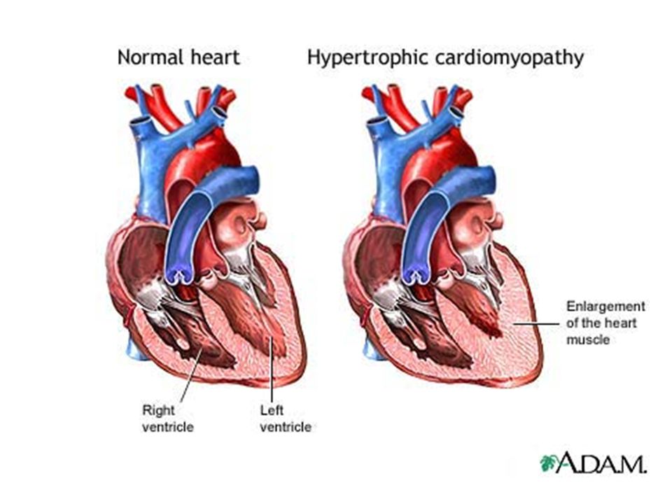
Cardiomyopathy Hypertrophic cardiomyopathy
Asymmetric left ventricular hypertrophy Affects septum Associated with sudden death Often familial with structural protein abnormalities (tropomyosin)
")
11
Hypertrophic cardiomyopathy (HCM) is a genetic disorder that is typically inherited in an autosomal dominant fashion with variable penetrance and variable expressivity. The disease has complex symptomatology and potentially devastating consequences for patients and their families. The disorder has a variable presentation and carries a high incidence of sudden death. In fact, HCM is the leading cause of sudden cardiac death in both preadolescent and adolescent children. The hallmark of the disorder is myocardial hypertrophy that is inappropriate, often asymmetric, and occurs in the absence of an obvious inciting hypertrophy stimulus. This hypertrophy can occur in any region of the left ventricle but frequently involves the interventricular septum, which results in an obstruction of flow through the left ventricular (LV) outflow tract.
 outflow tract.")
12
Microscopy: Microscopically, there is a characteristic whorled pattern of disorganized muscle bundles. There are abnormalities in the cell-to-cell arrangement and in the myofibrillar architecture within individual cells. There is fibrosis and scar formation. Most patients have abnormal intramural coronary arteries in affected areas, with thickened vessel walls and luminal narrowing, and these abnormalities may contribute to myocardial ischemia in these patients

13
Myocardium in HCM:

14
Pathophysiology: Most patients have abnormalities of diastolic function due to high filling pressures. A minority of patients exhibit a sub-aortic pressure gradient, which is thought to be due to abnormal anterior motion of the mitral valve towards the hypertrophied septum during systole, "systolic anterior motion" (SAM). Many exhibit myocardial ischemia, probably related to abnormally narrowed intramural vessels, increased oxygen demand, and increased intraventricular pressures resulting in subendocardial ischemia.
. Many exhibit myocardial ischemia, probably related to abnormally narrowed intramural vessels, increased oxygen demand, and increased intraventricular pressures resulting in subendocardial ischemia.")
15
Symptoms: Most patients with hypertrophic cardiomyopathy are asymptomatic. The most common symptom is dyspnea. angina, fatigue, and syncope are also common. Palpitations, paroxysmal nocturnal dyspnea, congestive heart failure, and dizziness are less frequent. Syncope may result from inadequate cardiac output with exertion or be a result of arrhythmias.

16
Hypertrophic cardiomyopathy

17
Restrictive cardiomyopathy
A “stiff” heart with reduced filling in diastole Dilated atria Endomyocardial fibrosis (EMF,tropical) Subendocardial fibrosis with thrombosis Loeffler endomyocarditis Similar to EMF but with eosinophil infiltrate (possibly related to parasite infection) Amyloid heart disease has similar features
 Subendocardial fibrosis with thrombosis. Loeffler endomyocarditis. Similar to EMF but with eosinophil infiltrate. (possibly related to parasite infection) Amyloid heart disease has similar features.")
18
Definition of RCM: Restrictive cardiomyopathy refers to a group of disorders in which the heart chambers are unable to fill with blood properly because of stiffness of the heart. In restrictive cardiomyopathy, the heart is normal in size or only slightly enlarged, but it cannot relax normally during diastole (that is, the time between heartbeats in which the blood returns from the body to the heart). Later in the disease, the heart may not pump blood efficiently. The abnormal heart function can affect the lungs, liver, and other body systems. Restrictive cardiomyopathy may affect either or both ventricles and may or may not be associated with a disease of the heart muscle.
. Later in the disease, the heart may not pump blood efficiently. The abnormal heart function can affect the lungs, liver, and other body systems. Restrictive cardiomyopathy may affect either or both ventricles and may or may not be associated with a disease of the heart muscle.")
19
Causes The most common causes of restrictive cardiomyopathy are amyloidosis and idiopathic myocardial fibrosis (a scarring of the heart of unknown cause). It frequently occurs after a heart transplant. Other causes of restrictive cardiomyopathy include sarcoidosis, hemochromatosis, radiation fibrosis, and various tumor infiltrations of the heart. More rarely, restrictive cardiomyopathy is caused by diseases of the endocardium such as endomyocardial fibrosis and Loeffler's syndrome.
. It frequently occurs after a heart transplant. Other causes of restrictive cardiomyopathy include sarcoidosis, hemochromatosis, radiation fibrosis, and various tumor infiltrations of the heart. More rarely, restrictive cardiomyopathy is caused by diseases of the endocardium such as endomyocardial fibrosis and Loeffler s syndrome.")
20
Symptoms Excessive tiredness (fatigue) .
Swelling of the feet and ankles. Cough. Difficulty breathing: especially with exertion at night when lying flat Easily fatigued (poor tolerance of exercise). Swelling of the abdomen.
. Swelling of the abdomen.")
21
Diagnosis : Restrictive cardiomyopathy may be hard to differentiate from constrictive pericarditis. A biopsy of the heart muscle may be used to confirm the diagnosis.

22
Prognosis: People with restrictive cardiomyopathy may be candidates for heart transplant. Prognosis is dependent on the underlying cause but it is usually poor. Average (mean) survival after diagnosis is 9 years.
 survival after diagnosis is 9 years.")
23
Possible Complications:
Progressive heart failure. Mitral regurgitation. Tricuspid regurgitation.

24
Endomyocardial fibrosis:Histologic Findings:
The heart size is not usually enlarged in EMF. The ventricular cavities are frequently laden with thrombi and, in severe cases, may be nearly totally obliterated by endocardial thickening and thrombosis. The histologic findings of EMF are characterized by reactive fibrosis associated with a selective increase in type I collagen deposition, subendocardial infarction and fibrosis, and thrombus formation. Additionally, specific features of other diseases, such as those associated with hemochromatosis or glycogen storage disease, are notably absent.

25
RCM due to hemochromatosis:
Prussian Blue reaction shows iron deposits in myocardium.

26
Restrictive cardiomyopathy with amyloidosis

Similar presentations


 Deterioration of heart muscle Becomes enlarged, thick or rigid Scar tissue Pumping blood.>")


>")


 F.R.C.P.(E) F.R.C.P.(LONDON) F.A.C.C. DESIGNED AT A.V. DEPTT F.J.M.C. BY RABIA KAZMI.>")