Download presentation
Presentation is loading. Please wait.

1
Biology 323 Human Anatomy for Biology Majors Lecture 10 Dr. Stuart S. Sumida Heart: Structure, Function, Development

2
The first blood vessels of the embryo form inside the embryonic disc even before somites appear. They form near the edge of the yolksac (a primitive condition inherited from macrolecithal organisms that stored yolk for food).
..")
3
Angiogenetic cell clusters extend in an arc around the head end of the ventral opening of the yolk sac. Initially, this means that the angiogenetic cell clusters (and the blood vessel that forms from them) have the pattern of a "horseshoe" if viewed from a dorsal or ventral perspective.
 have the pattern of a horseshoe if viewed from a dorsal or ventral perspective..")
4
An important point to understand is that the coelom runs up and down either side of the body. At the head end, right underneath the developing pharynx, the coelom on the left communicates with the coelom on the right. Thus, the coelom cuts across the midline here.

5
The brain grows at an incredible rate. It grows so fast that it makes the head bend around under the embryo's body. This is why the heart winds up on the VENTRAL SIDE of the body.

6
The part of the heart ventral to the gut tube is a single tube itself. The tube exiting the heart at its cranial end is the ventral aorta. However, the heart cannot remain a simple tube (like a fish), so it must be subdivided into a right and left side. A septum subdivides the heart into a left and right side.
, so it must be subdivided into a right and left side. A septum subdivides the heart into a left and right side..")
7
The tube exiting the heart at its cranial end is the ventral aorta. It also subdivides: The right side connects with the lungs. The left side supplies the body. (More later…)
.")
8
Anatomy of the Postnatal Heart

9
Heart in VENTRAL view. (You see mostly right ventricle!)
")
10
Heart in DORSAL view. (You see mostly left ventricle.)
")
11
HEART The real thing in ventral view. Lungs have been removed.

12
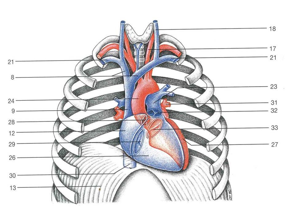
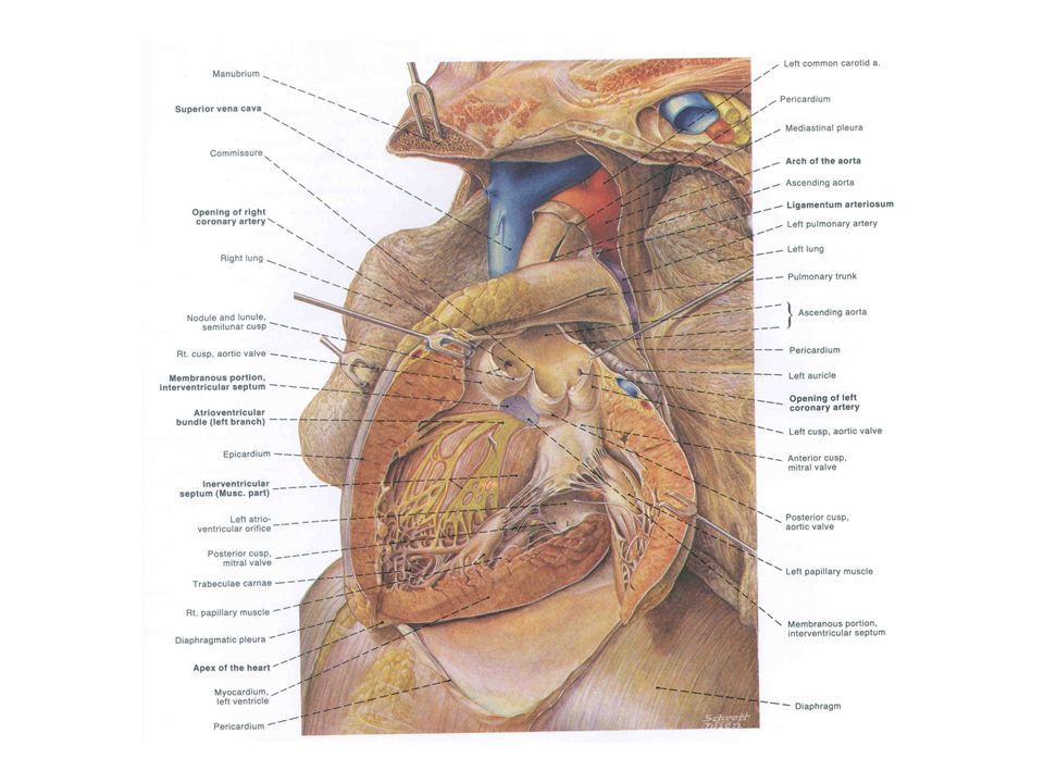
Gross Anatomy of Heart Right Atrium: Receives deoxygenated blood from body. Left Atrium: Receives oxygenated blood from lungs. Right Ventricle: Receives deoxygenated blood from right atrium and sends it to lungs. Left Ventricle: Receives oxygenated blood from left atrium and sends it to body.

14
Walls of the ventricles: Left wall is thicker!

15
Trabeculae carnae Papillary muscles Bicuspid valve Chordae Tendonae

16
Find: 1. Walls of the ventricles 2. Auricles 3. Inner walls of the atria 4. Fossa ovalis 5. Trabeculae carnae 6. Atrioventricular valve (a) "Bicuspid valve" (b) "Tricuspid valve" 7. Chordae tendonae 8. Papillary muscles 9. Aortic & pulmonary valves
 Bicuspid valve (b) Tricuspid valve 7. Chordae tendonae 8. Papillary muscles 9. Aortic & pulmonary valves.")
17
Blood Supply of the Heart Wall 1. Coronary arteries (a) Left coronary artery (b) Right coronary artery (c) Interventricular branches (d) Right marginal branch 2. Cardiac veins
 Left coronary artery (b) Right coronary artery (c) Interventricular branches (d) Right marginal branch 2. Cardiac veins.")
18
Coronary arteries are the FIRST branches of the aorta! 1. Coronary arteries (a) Left coronary artery (b) Right coronary artery (c) Interventricular branches (d) Right marginal branch 2. Cardiac veins
 Left coronary artery (b) Right coronary artery (c) Interventricular branches (d) Right marginal branch 2. Cardiac veins.")
19
Function of the Heart & Control of Heartbeat 1. Contracts spontaneously; does not need nervous stimulation to contract. 2. Motor nerves that supply the human heart = modulate heart rate. 3. Sympathetic motor impulses speed up heart rate & parasympathetic motor impulses slow it down. SYMPATHETIC: UPPER THORACIC SEGMENTS (T3-T4) GO UP TO THE NECK, AND COME BACK DOWN TO THE HEART. Why would it do this?!? PARASYMPATHETIC: VAGUS NERVE (X)
 GO UP TO THE NECK, AND COME BACK DOWN TO THE HEART. Why would it do this !. PARASYMPATHETIC: VAGUS NERVE (X).")
20
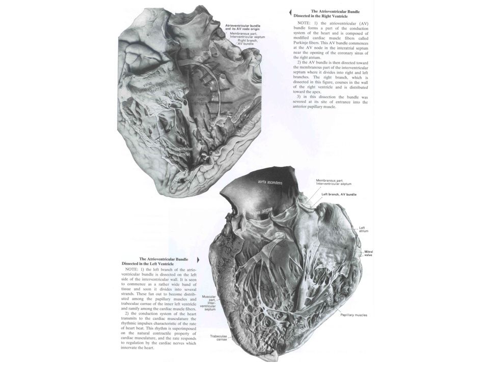
Intrinsic regulation of heart beat System made up of cells called Purkinje fibers (insulated from surrounding cells of heart. Sinoatrial node is PACEMAKER OF HEART, and beginning of process. Geenrates periodic impulses that initiate contraction of right atrium. Signal then runs to Atrioventricular node. Message is passed along a track of Purkinje fibers called the... Atrioventricular bundle. Atrioventricular bundle then splits into right and left limbs/branches that pass to individual inner ventricular walls on right and left.

21
Intrinsic regulation of heart beat 1. Sinoatrial node is PACEMAKER OF HEART, and beginning of process. Geenrates periodic impulses that initiate contraction of right atrium. 2. Signal then runs to Atrioventricular node. Message is passed along a track of Purkinje fibers called the... 3. Atrioventricular bundle. Atrioventricular bundle then splits into right and left limbs/branches that pass to individual inner ventricular walls on right and left. 1 2 3 A-V bundle path shown with blue arrows

22
Intrinsic regulation of heart beat 1. Sinoatrial node is PACEMAKER OF HEART, and beginning of process. Geenrates periodic impulses that initiate contraction of right atrium. 2. Signal then runs to Atrioventricular node. Message is passed along a track of Purkinje fibers called the... 3. Atrioventricular bundle. Atrioventricular bundle then splits into right and left limbs/branches that pass to individual inner ventricular walls on right and left. 1 2 3 A-V bundle path shown with blue arrows

23
Intrinsic regulation of heart beat 1. Sinoatrial node is PACEMAKER OF HEART, and beginning of process. Geenrates periodic impulses that initiate contraction of right atrium. 2. Signal then runs to Atrioventricular node. Message is passed along a track of Purkinje fibers called the... 3. Atrioventricular bundle. Atrioventricular bundle then splits into right and left limbs/branches that pass to individual inner ventricular walls on right and left. 1 2 3 A-V bundle path shown with blue arrows

26
The Great Vessels of the thorax are a logical extension of the heart Embryonic Origin of Great Vessels: They are derivatives of the aortic arches.

29
This is in your lab manual!

30
Aortic Arch Summary: Arch I: Mostly disappears ( a small part becomes a bit of the maxillary artery).
.")
31
Aortic Arch Summary: Arch II: DISAPPEARS

32
Aortic Arch Summary: Arch III: CAROTID ARCH – becomes part of carotid arteries.

33
Aortic Arch Summary: Arch IV: AORTIC ARCH -- Right side disappears. Left side becomes ARCH OF AORTA.

34
Aortic Arch Summary: Arch v: DISAPPEARS

35
Aortic Arch Summary: Arch VI: PULMONARY ARCH – Becomes pulmonary artery to lungs.

36
Great Veins of the Thorax 1. Venous blood dumps in the right atrium of the heart. (a) Blood from the cranial region enters via superior vena cava (b) Body blood enters via inferior vena cava 2. Inferior vena cava - passes through the diaphragm after receiving blood from the abdominal gut. 3. Superior vena cave & its 3 tributaries: (a) Azygous vein (b) Right brachiocephalic vein (c) Left brachiocephalic vein
 Blood from the cranial region enters via superior vena cava (b) Body blood enters via inferior vena cava 2. Inferior vena cava - passes through the diaphragm after receiving blood from the abdominal gut. 3. Superior vena cave & its 3 tributaries: (a) Azygous vein (b) Right brachiocephalic vein (c) Left brachiocephalic vein.")
37
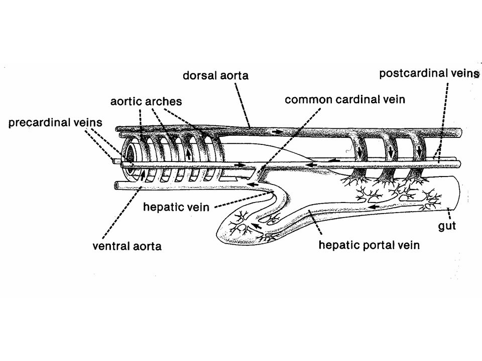
The embryological Cardinal Veins look like a big letter “H”. H Head Right anterior cardinal Left anterior cardinal Right common cardinal Sinus left common cardinal venosus Right posterior cardinal Left posterior cardinal (ventral view)
.")
38
The embryological Cardinal Veins look like a big letter “H”. H Head Right anterior cardinal Left anterior cardinal Right common cardinal Sinus left common cardinal venosus Right posterior cardinal Left anterior cardinal (ventral view) new Becomes left brachiocephalic vein. Becomes hemiazygous vein. Becomes superior vena cava. Becomes azygous vein (last tributary of SVC). Becomes part of wall of right atrium. Left posterior cardinal
 new Becomes left brachiocephalic vein. Becomes hemiazygous vein. Becomes superior vena cava. Becomes azygous vein (last tributary of SVC). Becomes part of wall of right atrium. Left posterior cardinal.")
39
Superior Vena Cava Azygous Vein Hemiazygous Vein

41
Great Veins of the Thorax 1. Venous blood dumps in the right atrium of the heart. (a) Blood from the cranial region enters via superior vena cava (b) Body blood enters via inferior vena cava 2. Inferior vena cava - passes through the diaphragm after receiving blood from the abdominal gut. 3. Superior vena cave & its 3 tributaries: (a) Azygous vein (b) Right brachiocephalic vein (c) Left brachiocephalic vein
 Blood from the cranial region enters via superior vena cava (b) Body blood enters via inferior vena cava 2. Inferior vena cava - passes through the diaphragm after receiving blood from the abdominal gut. 3. Superior vena cave & its 3 tributaries: (a) Azygous vein (b) Right brachiocephalic vein (c) Left brachiocephalic vein.")
Similar presentations


 in thorax, in inferior mediastinum>")


