Download presentation
Presentation is loading. Please wait.

1
Thrombosis, Embolism and Infarction

2
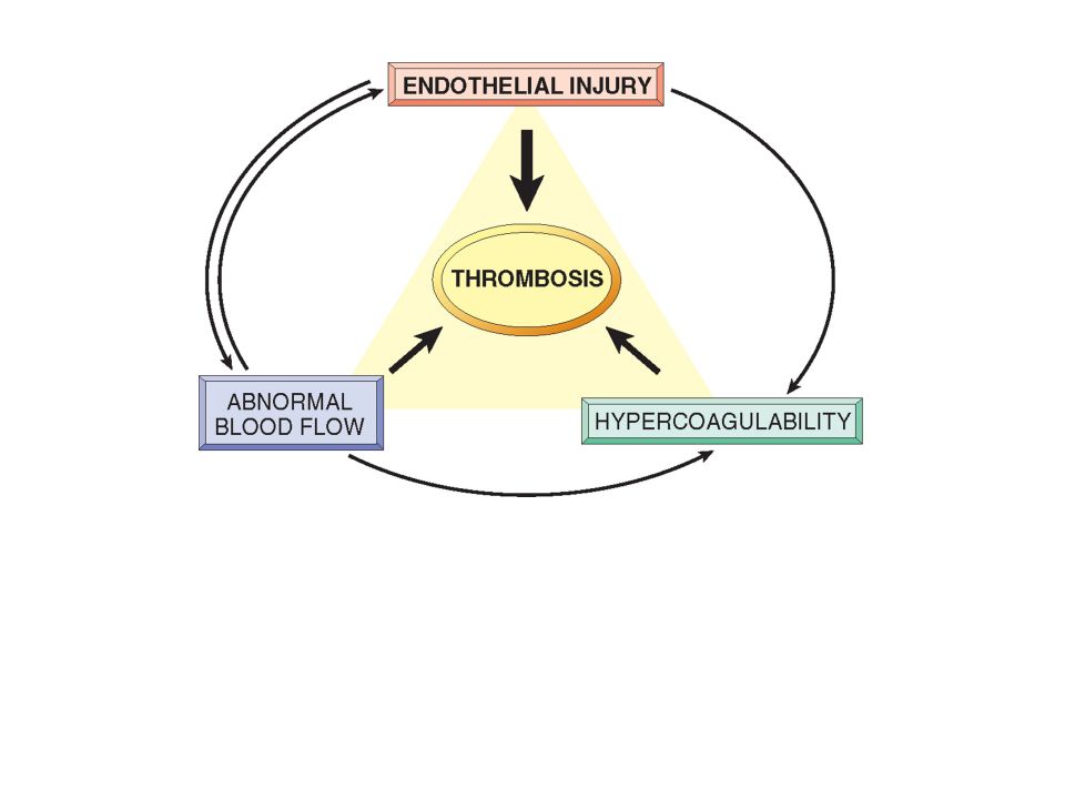
Thrombus formation (called Virchow's triad): (1)endothelial injury, (2) stasis or turbulent blood flow (3) hypercoagulability of the blood THROMBOSIS
: (1)endothelial injury, (2) stasis or turbulent blood flow (3) hypercoagulability of the blood THROMBOSIS")
3
Endothelial injury is particularly important for thrombus formation in the heart or the arterial circulation, where the normally high flow rates might otherwise impede clotting by preventing platelet adhesion and washing out activated coagulation factors. Thus, thrombus formation within cardiac chambers (e.g., after endocardial injury due to myocardial infarction), over ulcerated plaques in atherosclerotic arteries, or at sites of traumatic or inflammatory vascular injury (vasculitis) is largely a consequence of endothelial cell injury. Endothelial Injury
, over ulcerated plaques in atherosclerotic arteries, or at sites of traumatic or inflammatory vascular injury (vasculitis) is largely a consequence of endothelial cell injury. Endothelial Injury.")
4
Date of download: 3/11/2013 Copyright © 2012 American Medical Association. All rights reserved. From: Prospects for Cardiovascular Research JAMA. 2001;285(5):581-587. doi:10.1001/jama.285.5.581 Chronic endothelial injury, inflammation, and oxidative stress arecentral to the development of atherosclerosis. Endothelial injury resultsfrom a variety of factors including tobacco use, hypercholesterolemia, interventionaltherapies with angioplasty or coronary stents, and from ulceration or fissuringof atherosclerotic plaques. At sites of endothelial injury, production ofendothelial-derived substances (nitric oxide [NO], tissue plasminogen activator[tPA], and prostacyclin [PGI2]) is decreased, creating a prothromboticenvironment characterized by increased platelet and leukocyte adhesion, increasedpermeability to plasma lipoproteins, myointimal hyperplasia, and vasoconstriction.Ulceration or fissuring of the atherosclerotic plaque results from degradationof collagen matrix in the fibrous cap by metalloproteases released from macrophages.Exposure of the subendothelium after plaque ulceration or fissuring leadsto platelet adhesion and aggregation and local accumulation of largely platelet- derivedmediators (thromboxane A2, serotonin, adenosine diphosphate [ADP],thrombin, platelet activating factor [PAF], oxygen- derived free radicals,tissue factor, and endothelin) that promote thrombus growth, fibroproliferation,and vasoconstriction. LDL indicates low-density lipoprotein. Figure Legend :
: doi: /jama Chronic endothelial injury, inflammation, and oxidative stress arecentral to the development of atherosclerosis. Endothelial injury resultsfrom a variety of factors including tobacco use, hypercholesterolemia, interventionaltherapies with angioplasty or coronary stents, and from ulceration or fissuringof atherosclerotic plaques. At sites of endothelial injury, production ofendothelial-derived substances (nitric oxide [NO], tissue plasminogen activator[tPA], and prostacyclin [PGI2]) is decreased, creating a prothromboticenvironment characterized by increased platelet and leukocyte adhesion, increasedpermeability to plasma lipoproteins, myointimal hyperplasia, and vasoconstriction.Ulceration or fissuring of the atherosclerotic plaque results from degradationof collagen matrix in the fibrous cap by metalloproteases released from macrophages.Exposure of the subendothelium after plaque ulceration or fissuring leadsto platelet adhesion and aggregation and local accumulation of largely platelet- derivedmediators (thromboxane A2, serotonin, adenosine diphosphate [ADP],thrombin, platelet activating factor [PAF], oxygen- derived free radicals,tissue factor, and endothelin) that promote thrombus growth, fibroproliferation,and vasoconstriction. LDL indicates low-density lipoprotein. Figure Legend :.")
5
Physical loss of endothelium can lead to exposure of the subendothelial ECM, adhesion of platelets, release of tissue factor, and local depletion of PGI2 and plasminogen activators. However, it should be emphasized that endothelium need not be denuded or physically disrupted to contribute to the development of thrombosis; any perturbation in the dynamic balance of the prothombotic and antithrombotic activities of endothelium can influence local clotting events. Thus, dysfunctional endothelial cells can produce more procoagulant factors (e.g., platelet adhesion molecules, tissue factor, PAIs) or may synthesize less anticoagulant effectors (e.g., thrombomodulin, PGI2, t-PA). Endothelial dysfunction can be induced by a wide variety of insults, including hypertension, turbulent blood flow, bacterial endotoxins, radiation injury, metabolic abnormalities such as hypercholesterolemia. Endothelial Injury
 or may synthesize less anticoagulant effectors (e.g., thrombomodulin, PGI2, t-PA). Endothelial dysfunction can be induced by a wide variety of insults, including hypertension, turbulent blood flow, bacterial endotoxins, radiation injury, metabolic abnormalities such as hypercholesterolemia. Endothelial Injury.")
6
Turbulence contributes to arterial and cardiac thrombosis by causing endothelial injury or dysfunction, as well as by forming countercurrents and local pockets of stasis; stasis is a major contributor in the development of venous thrombi. Normal blood flow is laminar such that the platelets (and other blood cellular elements) flow centrally in the vessel lumen, separated from endothelium by a slower moving layer of plasma. Stasis and turbulence therefore: Alterations in Normal Blood Flow http://www.oocities.org/venkatej/mech/fluid_mechanics/LaminarFlowProfile.gif
 flow centrally in the vessel lumen, separated from endothelium by a slower moving layer of plasma. Stasis and turbulence therefore: Alterations in Normal Blood Flow")
7
http://www.phlebolymphology.org/2009/07/recent-findings-in-the-pathogenesis-of-venous-wall-degradation/ http://content.onlinejacc.org/data/Journals/JAC/23086/02059_gr4.jpeg

8
Promote endothelial activation, enhancing procoagulant activity and leukocyte adhesion. In part through flow-induced changes in endothelial cell gene expression. Disrupt laminar flow and bring platelets into contact with the endothelium. Prevent washout and dilution of activated clotting factors by fresh flowing blood and the inflow of clotting factor inhibitors. Alterations in Normal Blood Flow

9
Ulcerated atherosclerotic plaques cause turbulence. Aortic and arterial dilations called aneurysms result in local stasis and are therefore fertile sites for thrombosis. Acute myocardial infarctions result in areas of noncontractile myocardium and sometimes cardiac aneurysms; both are associated with stasis and flow abnormalities. Hyperviscosity (such as is seen with polycythemia vera) increases resistance to flow and causes small vessel stasis; the deformed red cells in sickle cell anemia ( Chapter 14) cause vascular occlusions, with the resulting stasis also predisposing to thrombosis. Turbulence and stasis contribute to thrombosis
 increases resistance to flow and causes small vessel stasis; the deformed red cells in sickle cell anemia ( Chapter 14) cause vascular occlusions, with the resulting stasis also predisposing to thrombosis. Turbulence and stasis contribute to thrombosis.")
10
http://www.doctortipster.com/wp-content/uploads/2011/08/Deep- venous-thrombosis-causes.jpg How John C. Lincoln's advanced deep vein thrombosis treatment works: Once guided to the blood clot, an AngioJet catheter creates a powerful fluid flow, drawing the clot toward the inflow windows. Inside the catheter, saline jets break the clot into microscopic particles, which are removed from the body.

11
http://www.aafp.org/afp/2012/1115/afp20121115p913-f1.gif http://www.stoptheclot.org/images/natt_other_ar t/acute%20left%20leg%20DVT;%20postthormb otic%20syndrome%20right%20leg.jpg

12
Hypercoagulability http://ars.els-cdn.com/content/image/1-s2.0-S0268960X08000362-gr1.jpg

13
PRIMARY (GENETIC) Common Factor V mutation (G1691A mutation; factor V Leiden) Prothrombin mutation (G20210A variant) 5,10- Methylenetetrahydrofolate reductase (homozygous C677T mutation) Increased levels of factors VIII, IX, XI, or fibrinogen.
 Common Factor V mutation (G1691A mutation; factor V Leiden) Prothrombin mutation (G20210A variant) 5,10- Methylenetetrahydrofolate reductase (homozygous C677T mutation) Increased levels of factors VIII, IX, XI, or fibrinogen.")
14
http://www.med.illinois.edu/hematology/Pics,%20etc/Factor%20V%20Leiden%20Main.JPG Factor V mutation (G1691A mutation; factor V Leiden) In the normal person, factor V functions as a cofactor to allow factor Xa to activate thrombin. Thrombin in turn cleaves fibrinogen to form fibrin. Activated protein C (aPC) is a natural anticoagulant that acts to limit the extent of clotting by cleaving and degrading factor V. (http://en.wikipedia.org/wiki/Factor_V_Leiden)
 is a natural anticoagulant that acts to limit the extent of clotting by cleaving and degrading factor V. (")
15
http://www.ismaap.org/uploads/pics/faktor1_eng.jpg

16
Rare Antithrombin III deficiency Protein C deficiency Protein S deficiency Very Rare Fibrinolysis defects Homozygous homocystinuria (deficiency of cystathione β-synthetase) Hypercoagulability http://www.practical-haemostasis.com/images/Images-2/Thrombophilia%20Tests/pc_ps_pathway.jpg
 Hypercoagulability")
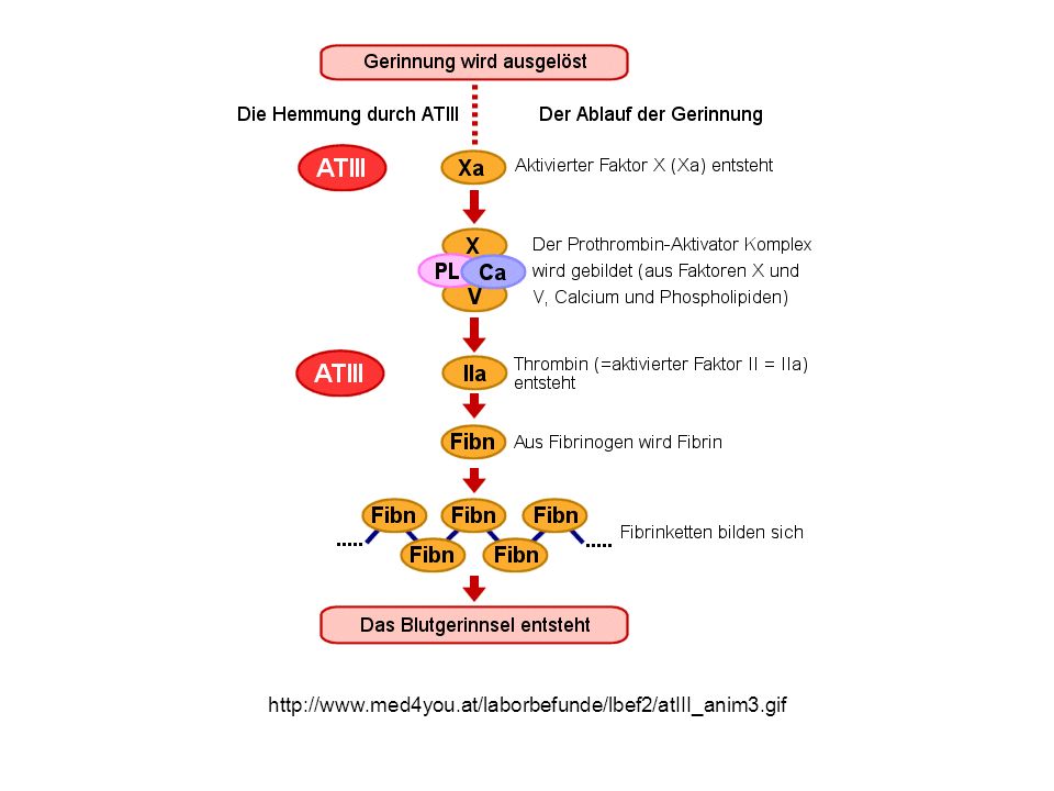
17
http://www.med4you.at/laborbefunde/lbef2/atIII_anim3.gif
18
Thrombomodulin (TM) CD141 or BDCA-3 is an integral membrane protein expressed on the surface of endothelial cells and serves as a cofactor for thrombin. It reduces blood coagulation by converting thrombin to an anticoagulant enzyme from a procoagulant enzyme. http://en.wikipedia.org/wiki/Thrombomodulin

19
http://www.neurology.org/content/78/3/157/F1.large.jpg APC= activated Protein C

20
Fig. 1: Activated protein C anticoagulant pathway: 1. Bloomenthal D et al. CMAJ 2002;167:48-54 ©2002 by Canadian Medical Association

21
High Risk for Thrombosis Prolonged bedrest or immobilization Myocardial infarction Atrial fibrillation Tissue injury (surgery, fracture, burn) Cancer Prosthetic cardiac valves Disseminated intravascular coagulation Heparin-induced thrombocytopenia Antiphospholipid antibody syndrome Hypercoagulability (secondary)
 Cancer Prosthetic cardiac valves Disseminated intravascular coagulation Heparin-induced thrombocytopenia Antiphospholipid antibody syndrome Hypercoagulability (secondary)")
22
Lower Risk for Thrombosis Hyperestrogenic states (pregnancy and postpartum) Oral contraceptive use Cardiomyopathy Nephrotic syndrome Sickle cell anemia Smoking Hypercoagulability (secondary) http://hcp.obgyn.net/image/image_gallery?img_id=2082561&t=1339545353297
 Oral contraceptive use Cardiomyopathy Nephrotic syndrome Sickle cell anemia Smoking Hypercoagulability (secondary) img_id= &t=")
23
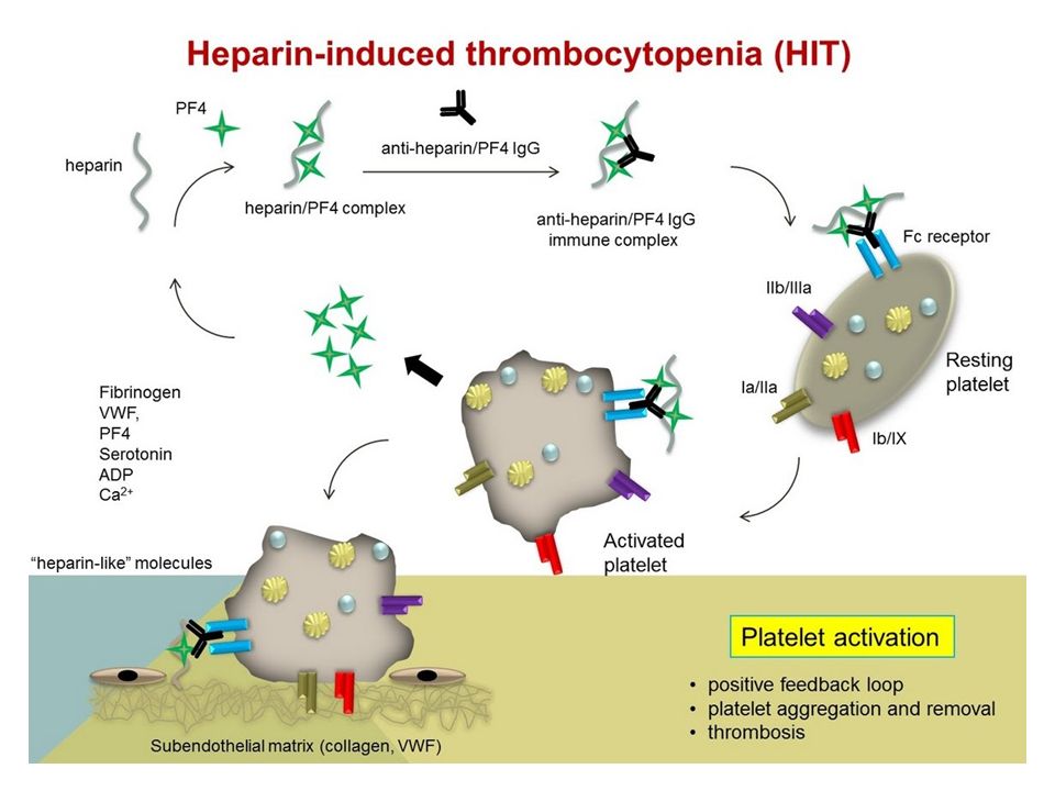
Among the acquired thrombophilic states, two that are particularly important. Heparin-induced thrombocytopenia (HIT) syndrome. Antiphospholipid antibody syndrome (previously called the lupus anticoagulant syndrome).
 syndrome. Antiphospholipid antibody syndrome (previously called the lupus anticoagulant syndrome)..")
24
Heparin-induced thrombocytopenia (HIT) syndrome HIT occurs following the administration of unfractionated heparin, which may induce the appearance of antibodies that recognize complexes of heparin and platelet factor 4 on the surface of platelets, as well as complexes of heparin- like molecules and platelet factor 4-like proteins on endothelial cells. Binding of these antibodies to platelets results in their activation, aggregation, and consumption (hence the thrombocytopenia in the syndrome name). Effect on platelets and endothelial damage combine to produce a prothrombotic state, even in the face of heparin administration and low platelet counts. Newer low-molecular weight heparin preparations induce antibody formation less frequently, but still cause thrombosis if antibodies have already formed. Other anticoagulants such as fondaparinux (a pentasaccharide inhibitor of factor X) also cause a HIT-like syndrome on rare occasions.
. Effect on platelets and endothelial damage combine to produce a prothrombotic state, even in the face of heparin administration and low platelet counts. Newer low-molecular weight heparin preparations induce antibody formation less frequently, but still cause thrombosis if antibodies have already formed. Other anticoagulants such as fondaparinux (a pentasaccharide inhibitor of factor X) also cause a HIT-like syndrome on rare occasions..")
26
Antiphospholipid antibody syndrome Antiphospholipid antibodies are a heterogeneous group of auto-antibodies (IgG, IgM, and IgA) This syndrome has protean clinical manifestations, including recurrent thromboses, repeated miscarriages, cardiac valve vegetations, and thrombocytopenia. Depending on the vascular bed involved, the clinical presentations can include pulmonary embolism (following lower extremity venous thrombosis), pulmonary hypertension (from recurrent subclinical pulmonary emboli), stroke, bowel infarction, or renovascular hypertension. Fetal loss is attributable to antibody-mediated inhibition of t- PA activity necessary for trophoblastic invasion of the uterus. Antiphospholipid antibody syndrome is also a cause of renal microangiopathy, resulting in renal failure associated with multiple capillary and arterial thromboses.
, pulmonary hypertension (from recurrent subclinical pulmonary emboli), stroke, bowel infarction, or renovascular hypertension. Fetal loss is attributable to antibody-mediated inhibition of t- PA activity necessary for trophoblastic invasion of the uterus. Antiphospholipid antibody syndrome is also a cause of renal microangiopathy, resulting in renal failure associated with multiple capillary and arterial thromboses..")
27
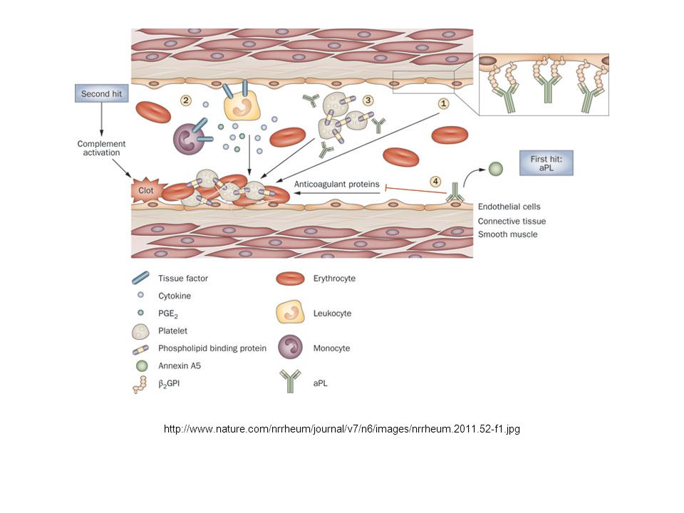
Figure 3 Endothelial cell activation by anti ‑ β 2 GPI autoantibodies Meroni, P. L. et al. (2011) Pathogenesis of antiphospholipid syndrome: understanding the antibodies Nat. Rev. Rheumatol. doi:10/1038/nrrheum.2011.52 http://www.acponline.org/graphics/bioterro/canthrax/anti_lipid.jpg beta2 Glycoprotein 1
 Pathogenesis of antiphospholipid syndrome: understanding the antibodies Nat. Rev. Rheumatol. doi:10/1038/nrrheum beta2 Glycoprotein 1.")
28
http://www.nature.com/nrrheum/journal/v7/n6/images/nrrheum.2011.52-f1.jpg
30
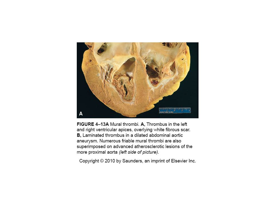
Thrombi can develop anywhere in the cardiovascular system (e.g., in cardiac chambers, on valves, or in arteries, veins, or capillaries). The size and shape of thrombi depend on the site of origin and the cause. Arterial or cardiac thrombi usually begin at sites of turbulence or endothelial injury. Venous thrombi characteristically occur at sites of stasis. Thrombi are focally attached to the underlying vascular surface; arterial thrombi tend to grow retrograde from the point of attachment, while venous thrombi extend in the direction of blood flow (thus both propagate toward the heart). The propagating portion of a thrombus is often poorly attached and therefore prone to fragmentation and embolization.
. The propagating portion of a thrombus is often poorly attached and therefore prone to fragmentation and embolization..")
33
Arterial vs venous thrombi Grow retrograde to flow Begin at site of injury or turbulence Frequently occlusive Occur in coronary, cerebral, femoral arteries Grow with direction of flow Begin at site of stasis Occlusive Occur in lower extremities 90%, also upper extremities, periprostatic plexus, ovarian or periuterine veins

34
Thrombi often have grossly and microscopically apparent laminations called lines of Zahn; these represent pale platelet and fibrin deposits alternating with darker red cell–rich layers. Such laminations signify that a thrombus has formed in flowing blood; their presence can therefore distinguish antemortem thrombosis from the bland nonlaminated clots that occur postmortem http://811699.net/wp-content/uploads/2011/03/LUNG065.jpg http://i48.tinypic.com/2vmvqcj.jpg

35
Fate of the Thrombus Propagation. Thrombi accumulate additional platelets and fibrin. Embolization. Thrombi dislodge and travel to other sites in the vasculature. Dissolution. Result of fibrinolysis, which can lead to the rapid shrinkage and total disappearance of recent thrombi. (Extensive fibrin deposition and crosslinking in older thrombi renders them more resistant to lysis.) Natural and Therapeutic. Organization and recanalization. Older thrombi become organized by the ingrowth of endothelial cells, smooth muscle cells, and fibroblasts. Capillary channels eventually form that re-establish the continuity of the original lumen, albeit to a variable degree.
 Natural and Therapeutic. Organization and recanalization. Older thrombi become organized by the ingrowth of endothelial cells, smooth muscle cells, and fibroblasts. Capillary channels eventually form that re-establish the continuity of the original lumen, albeit to a variable degree..")
36
Organized arterial thrombus

37
http://www.geocities.ws/m4pathology/Osce/Slides/histsch03.jpg

38
http://farm3.static.flickr.com/2791/4337748744_3a92ba6583.jpg Recanilizatuion

39
Aortic thrombi from electrical injury

40
Lung hilum thromboembolus with lines of Zahn

41
Right atrial mural thrombus with lines of Zahn

42
Clinical Consequences. Thrombi are significant because they cause obstruction of arteries and veins, and are sources of emboli. Which effect predominates depends on the site of the thrombosis. Venous thrombi can cause congestion and edema in vascular beds distal to an obstruction, but they are far more worrisome for their capacity to embolize to the lungs and cause death (see below). Conversely, although arterial thrombi can embolize and cause downstream infarctions, a thrombotic occlusion at a critical site (e.g., a coronary artery) can have more serious clinical consequences.
. Conversely, although arterial thrombi can embolize and cause downstream infarctions, a thrombotic occlusion at a critical site (e.g., a coronary artery) can have more serious clinical consequences..")
43
Embolisms Detached intravasular solid, liquid, gasous mass carried by the blood Pulmonary embolisms Often arise from deep vein thromboses Associated with immobilization, hypercoagulability Frequently small, silent, becoming organized Right heart failure, cor pulmonale, when >60% pulmonary circulation obstructed Rupture of obstructed arteries causes bleeding without infarction due to blood supply Multiple emboli lead to hypertension and right ventricular failure

44
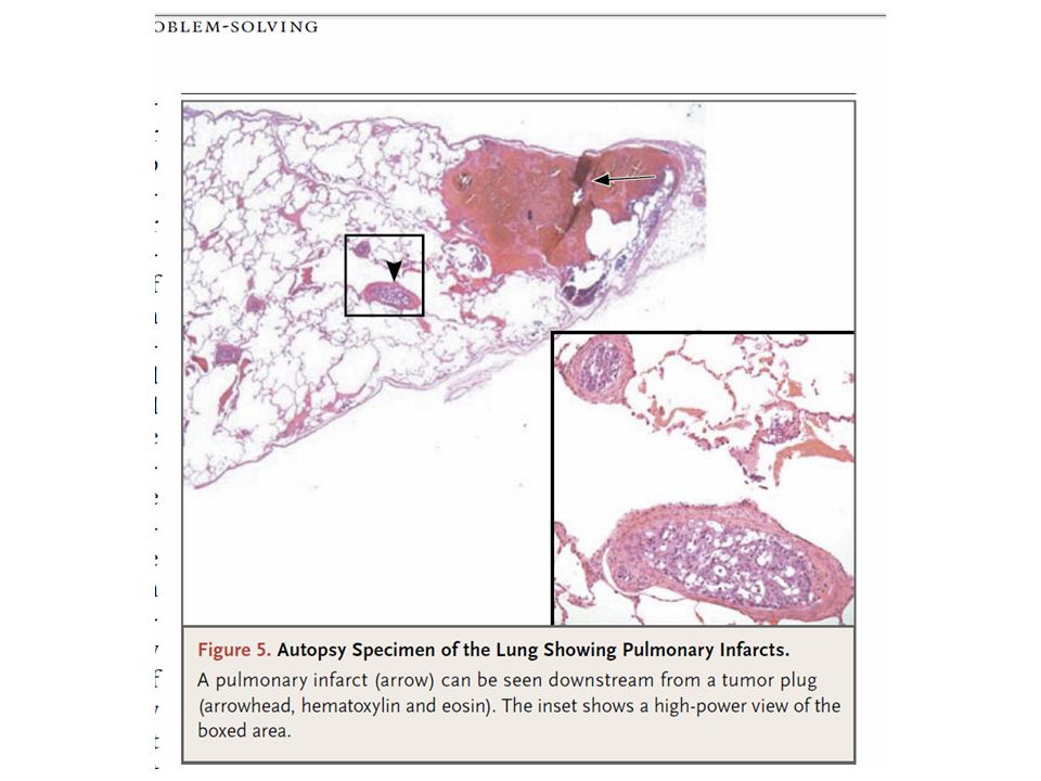
An embolus is a detached intravascular solid, liquid, or gaseous mass that is carried by the blood to a site distant from its point of origin. The term embolus was coined by Rudolf Virchow in 1848 to describe objects that lodge in blood vessels and obstruct the flow of blood. Almost all emboli represent some part of a dislodged thrombus, hence the term thromboembolism. Rare forms of emboli include fat droplets, nitrogen bubbles, atherosclerotic debris (cholesterol emboli), tumor fragments, bone marrow, or even foreign bodies. However, unless otherwise specified, emboli should be considered thrombotic in origin. Inevitably, emboli lodge in vessels too small to permit further passage, causing partial or complete vascular occlusion; a major consequence is ischemic necrosis (infarction) of the downstream tissue. Depending on where they originate, emboli can lodge anywhere in the vascular tree; the clinical outcomes are best understood based on whether emboli lodge in the pulmonary or systemic circulations.
, tumor fragments, bone marrow, or even foreign bodies. However, unless otherwise specified, emboli should be considered thrombotic in origin. Inevitably, emboli lodge in vessels too small to permit further passage, causing partial or complete vascular occlusion; a major consequence is ischemic necrosis (infarction) of the downstream tissue. Depending on where they originate, emboli can lodge anywhere in the vascular tree; the clinical outcomes are best understood based on whether emboli lodge in the pulmonary or systemic circulations..")
45
http://www.news.vcu.edu/images/image.aspx?id=2904&w=400

46
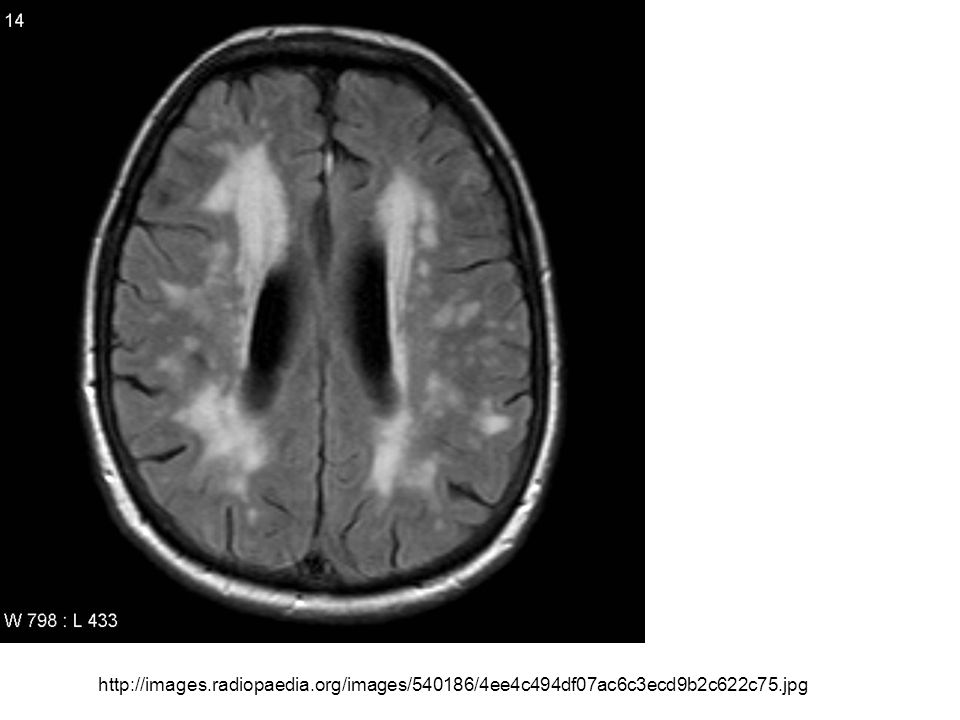
Systemic thromboembolism Emboli in arterial circulation Arise from intracardiac mural thrombi 60% associated with left ventricular wall infarcts 25% associated with atrial dilation or fibrillation Remainder originate from aneurysms, valvular vegetation Deposit in lower extremities or brain Consequences depend on caliber of occluded vessel, redundant blood supply

47
http://images.radiopaedia.org/images/540186/4ee4c494df07ac6c3ecd9b2c622c75.jpg
48
An infarct is an area of ischemic necrosis caused by occlusion of either the arterial supply or the venous drainage. Tissue infarction is a common and extremely important cause of clinical illness. Roughly 40% of all deaths in the United States are caused by cardiovascular disease, and most of these are attributable to myocardial or cerebral infarction. Pulmonary infarction is also a common complication in many clinical settings, bowel infarction is frequently fatal, and ischemic necrosis of the extremities (gangrene) is a serious problem in the diabetic population.
 is a serious problem in the diabetic population..")
49
Nearly all infarcts result from thrombotic or embolic arterial occlusions. Occasionally infarctions are caused by other mechanisms, including local vasospasm, hemorrhage into an atheromatous plaque, or extrinsic vessel compression (e.g., by tumor). Rarer causes include torsion of a vessel (e.g., in testicular torsion or bowel volvulus), traumatic rupture, or vascular compromise by edema (e.g., anterior compartment syndrome) or by entrapment in a hernia sac. Although venous thrombosis can cause infarction, the more common outcome is just congestion; in this setting, bypass channels rapidly open and permit vascular outflow, which then improves arterial inflow. Infarcts caused by venous thrombosis are thus more likely in organs with a single efferent vein (e.g., testis and ovary).
. Rarer causes include torsion of a vessel (e.g., in testicular torsion or bowel volvulus), traumatic rupture, or vascular compromise by edema (e.g., anterior compartment syndrome) or by entrapment in a hernia sac. Although venous thrombosis can cause infarction, the more common outcome is just congestion; in this setting, bypass channels rapidly open and permit vascular outflow, which then improves arterial inflow. Infarcts caused by venous thrombosis are thus more likely in organs with a single efferent vein (e.g., testis and ovary)..")
50
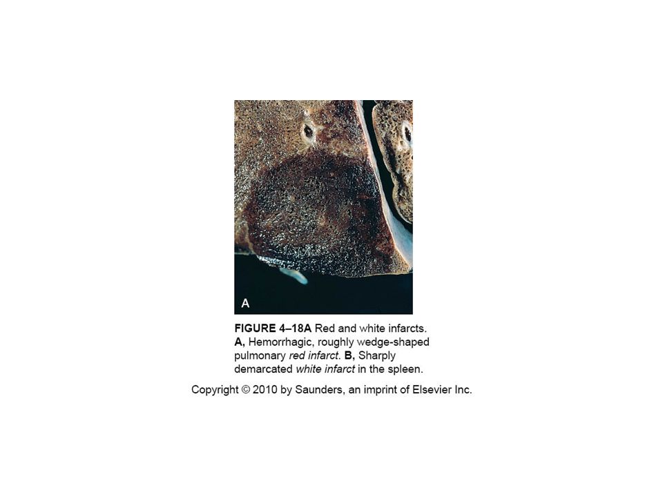
White infarcts occur with arterial occlusions in solid organs with end-arterial circulation (e.g., heart, spleen, and kidney), and where tissue density limits the seepage of blood from adjoining capillary beds into the necrotic area.
, and where tissue density limits the seepage of blood from adjoining capillary beds into the necrotic area.")
51
Red infarcts occur (1)with venous occlusions (e.g., ovary) (2)in loose tissues (e.g., lung) where blood can collect in the infarcted zone, (3)in tissues with dual circulations (e.g., lung and small intestine) that allow blood flow from an unobstructed parallel supply into a necrotic zone, (4)in tissues previously congested by sluggish venous outflow (5)when flow is re-established to a site of previous arterial occlusion and necrosis (e.g., following angioplasty of an arterial obstruction).
with venous occlusions (e.g., ovary) (2)in loose tissues (e.g., lung) where blood can collect in the infarcted zone, (3)in tissues with dual circulations (e.g., lung and small intestine) that allow blood flow from an unobstructed parallel supply into a necrotic zone, (4)in tissues previously congested by sluggish venous outflow (5)when flow is re-established to a site of previous arterial occlusion and necrosis (e.g., following angioplasty of an arterial obstruction).")
53
Factors That Influence Development of an Infarct. The effects of vascular occlusion can range from no or minimal effect to causing the death of a tissue or person. The major determinants of the eventual outcome are: (1)the nature of the vascular supply, (2)the rate at which an occlusion develops, (3)vulnerability to hypoxia, (4)the oxygen content of the blood. Neurons: 3 – 4 minutes Myocardial cells: 20 – 30 minutes Fibroblasts, skeletal muscle: hours
the nature of the vascular supply, (2)the rate at which an occlusion develops, (3)vulnerability to hypoxia, (4)the oxygen content of the blood. Neurons: 3 – 4 minutes Myocardial cells: 20 – 30 minutes Fibroblasts, skeletal muscle: hours.")
54
Nature of the vascular supply. The availability of an alternative blood supply is the most important determinant of whether vessel occlusion will cause damage. Rate of occlusion development. Slowly developing occlusions are less likely to cause infarction, because they provide time to develop alternate perfusion pathways. Vulnerability to hypoxia. Neurons undergo irreversible damage when deprived of their blood supply for only 3 to 4 minutes. Myocardial cells, though hardier than neurons, are also quite sensitive and die after only 20 to 30 minutes of ischemia. In contrast, fibroblasts within myocardium remain viable even after many hours of ischemia Oxygen content of blood. A partial obstruction of a small vessel that would be without effect in an otherwise normal individual might cause infarction in an anemic or cyanotic patient.

55
The dominant histologic characteristic of infarction is ischemic coagulative necrosis. It is important to recall that if the vascular occlusion has occurred shortly (minutes to hours) before the death of the person, no demonstrable histologic changes may be evident; it takes 4 to 12 hours for the tissue to show frank necrosis. Acute inflammation is present along the margins of infarcts within a few hours and is usually well defined within 1 to 2 days. Most infarcts are ultimately replaced by scar. The brain is an exception to these generalizations, as central nervous system infarction results in liquefactive necrosis.
 before the death of the person, no demonstrable histologic changes may be evident; it takes 4 to 12 hours for the tissue to show frank necrosis. Acute inflammation is present along the margins of infarcts within a few hours and is usually well defined within 1 to 2 days. Most infarcts are ultimately replaced by scar. The brain is an exception to these generalizations, as central nervous system infarction results in liquefactive necrosis..")
56
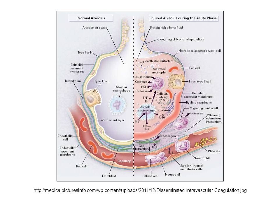
Disorders ranging from obstetric complications to advanced malignancy can be complicated by DIC, the sudden or insidious onset of widespread fibrin thrombi in the microcirculation. Although these thrombi are not grossly visible, they are readily apparent microscopically and can cause diffuse circulatory insufficiency, particularly in the brain, lungs, heart, and kidneys. To complicate matters, the widespread microvascular thrombosis results in platelet and coagulation protein consumption (hence the synonym consumption coagulopathy), and at the same time, fibrinolytic mechanisms are activated. Thus, an initially thrombotic disorder can evolve into a bleeding catastrophe. It should be emphasized that DIC is not a primary disease but rather a potential complication of any condition associated with widespread activation of thrombin. DISSEMINATED INTRAVASCULAR COAGULATION (DIC)
, and at the same time, fibrinolytic mechanisms are activated. Thus, an initially thrombotic disorder can evolve into a bleeding catastrophe. It should be emphasized that DIC is not a primary disease but rather a potential complication of any condition associated with widespread activation of thrombin. DISSEMINATED INTRAVASCULAR COAGULATION (DIC).")
57
http://medicalpicturesinfo.com/wp-content/uploads/2011/12/Disseminated-Intravascular-Coagulation.jpg
58
https://www.meducation.net/encyclopedia/29623 http://upload.wikimedia.org/wikipedia/commons/thumb/0/0b/Acute_thrombotic_microangiopathy_- _pas_-_very_high_mag.jpg/230px-Acute_thrombotic_microangiopathy_-_pas_-_very_high_mag.jpg

59
A 50-year-old woman was admitted to the hospital with fatigue and shortness of breath. Dyspnea after moderate exertion had developed gradually, starting 1 week before presentation, along with profound malaise and a nonproductive cough. In the 48 hours before admission, her shortness of breath had worsened, with dyspnea at rest. She had palpitations, nausea, and bilious emesis without hematemesis but reported no fever, chills, diaphoresis, chest pain, paroxysmal nocturnal dyspnea, orthopnea, platypnea, pleurisy, edema of the legs, or changes in weight or urine output. The Case

62
Extra Stuff

63
Fat and marrow embolism Release of fatty marrow from broken bones Onset of symptoms 1 – 3 days after injury Leads to pulmonary insufficiency Tachypnea, dyspnea, tachycardia Neurological symptoms Irritability, restlessness Thrombocytopenia Platelets adhere to fat globules Diffuse petechial rash

64
Fat and marrow embolism Mechanical obstruction Fat emboli with RBC and platelet aggregates occlude pulmonary and cerebral microvasculature Biochemical injury FFA released from fat globules injure endothelium initiating inflammation Platelet aggregation and granulocyte recruitment result in free radicals, proteases, eicosanoids

65
Fat embolism

66
Air embolisms Iatrogenic consequences of Coronary bypass surgery Neurosurgery Laparoscopic procedures Chest wall injury Decompression sickness Nitrogen bubbles from blood within muscle, lungs, joints Edema or ischemic necrosis in lungs (emphysema), femoral head, tibia, humerus
, femoral head, tibia, humerus")
67
Amniotic fluid embolism Infusion of amniotic fluid containing fetal components into uterine veins via rupture Incidence 1:40K; mortality 80%; morbidity 13% total incidence, 85% survivors Pulmonary microcirculation may contain Fetal cells, vernix caseosa fat, fetal respiratory or GI mucin, lanugo hair Onset characterized by sudden, severe dyspnea, cyanosis, shock, headache, seizures Followed by pulmonary edema Diffuse Intravascular Coagulation (DIC)
")
Similar presentations


: (1)endothelial injury, (2) stasis or turbulent blood flow (3) hypercoagulability.>")
, fibrous material and.>")


>")


>")
