Download presentation
Presentation is loading. Please wait.

1
CMR-Based Differentiation of AL and ATTR Cardiac Amyloidosis February 2014,JACC

2
Systemic amyloidosis is a condition characterized by extracellular deposition of pathologic insoluble fibrillar proteins. Cardiac light chain amyloid (AL) amyloidosis, in which amyloidfibrils are derived from monoclona immunoglobulin light chains, is associated with poor prognosis, with a median survival from diagnosis of <12 months.
 amyloidosis, in which amyloidfibrils are derived from monoclona immunoglobulin light chains, is associated with poor prognosis, with a median survival from diagnosis of <12 months..")
3
The prognosis of transthyretinrelated amyloidosis (ATTR) is typically 3 to 5 years. Transthyretin (TTR) is a plasma protein, predominantly synthesized in the liver, which acts as a transporter for thyroxine and retinol- binding protein.
 is a plasma protein, predominantly synthesized in the liver, which acts as a transporter for thyroxine and retinol- binding protein..")
4
Senile systemic amyloidosis due to nonmutant (wild-type) TTR is a sporadic disease among older people presenting as isolated cardiomyopathy. Hereditary ATTR amyloidosis is an autosomal dominant disease associated with >100 transthyretin gene mutations. Cardiac involvement is now known to occur in association with most hereditary ATTR variants.

5
There has been a marked increase (143%) in new cases of ATTR amyloidosis referred to our center in the past 5 years. The increased detection of ATTR amyloidosis correlates with increased availability of cardiac magnetic resonance (CMR) for investigation of cardiomyopathy.
 for investigation of cardiomyopathy..")
6
Gadolinium-enhanced CMR visualizes expansion of the interstitial space and may be used to demonstrate the accumulation of abundant amyloid in this site.

7
An early CMR study in cardiac amyloidosis described altered gadolinium kinetics, difficulty nulling the myocardium, and a global subendocardial pattern of late gadolinium enhancement (LGE).
.")
8
However, the precise pattern of LGE has been reported to be more variable in recent studies. Our impression has been that LGE is often diffuse in ATTR amyloidosis,but very few ATTR patients have been included in CMR studies, and there are no reports of differences in LGE patterns according to amyloid fibril protein type.

9
We report here the standard CMR measurements and LGE patterns in the largest cohort of cardiac ATTR amyloidosis patients characterized by using CMR at the time of writing.

10
METHODS Patients were identified between January 2007 and June 2012 at the UK National Amyloidosis Centre, a specialist referral center for the analysis of systemic amyloidosis. All patients had a CMR scan for the investigation of suspected amyloidosis.

11
Histological examination was performed in all cases, with immunohistochemistry, to confirm or rule out systemic amyloidosis.

12
Cardiac amyloidosis was confirmed according to results of endomyocardial biopsy, with Congo red staining and immunohistochemistry to determine the amyloid type.

13
In the absence of cardiac histology analysis, patients were diagnosed with cardiac amyloidosis if there were extracardiac biopsy results confirming systemic amyloidosis, immunohistochemistry to determine the amyloidfibril type and echocardiographic criteria for cardiac amyloidosis.

14
Transthyretin gene sequencing to differentiate wild-type and mutant TTR was performed in all cases of ATTR amyloidosis.

15
We obtained the raw CMR DICOM data for all patients. De novo analysis was performed with dedicated software and reported according to international guidelines. Left ventricular (LV) volumes and mass were indexed to body surface area.
 volumes and mass were indexed to body surface area..")
16
Patterns of LGE were described in the left ventricle at 3 levels (base, midventricle, and apex) and in the right ventricular (RV) free wall and atrium (present or absent). The LGE pattern was described as transmural if it involved the subendocardium through to the epicardial layer.

17
Subendocardial LGE was described if there was global subendocardial involvement but no transmural LGE. Nulling was defined as dark, unenhanced areas within the myocardium or skeletal muscle in LGE images. The presence of pericardial and pleural effusion was also recorded.

18
RESULTS A total of 115 CMR studies were obtained. We performed complete analysis of 108 studies; 7 scans with insufficient data were not included in the analyses. Studies were received from 46 different centers.

19
Only 1 center (St. George’s Hospital in South London) performed >10 of the studies analyzed in this series (24 [22%]). Most studies (84%) were performed at 1.5-T field strength; 17 studies (16%) were performed at 3.0-T field strength.
 performed >10 of the studies analyzed in this series (24 [22%]). Most studies (84%) were performed at 1.5-T field strength; 17 studies (16%) were performed at 3.0-T field strength..")
20
Afinal diagnosis of cardiac amyloidosis was made in 100 patients (93%). The cardiac amyloidosis type was AL in 46 patients and ATTR in 51 patients(51%, including 28 patients with senile systemic amyloidosis secondary to wild-type nonmutant ATTR and 23 patients with hereditary amyloidosis secondary to mutant TTR).
..")
21
Non-AL, non-TTR cardiac amyloidosis was diagnosed in 3 patients (3%; AA amyloidosis (causative protein serum amyloid A [SAA]) in 2 patients and apolipoprotein A1 in 1 patient).
![ Non-AL, non-TTR cardiac amyloidosis was diagnosed in 3 patients (3%; AA amyloidosis (causative protein serum amyloid A [SAA]) in 2 patients and apolipoprotein A1 in 1 patient).](http://images.slideplayer.com/21/6329084/slides/slide_21.jpg " Non-AL, non-TTR cardiac amyloidosis was diagnosed in 3 patients (3%; AA amyloidosis (causative protein serum amyloid A [SAA]) in 2 patients and apolipoprotein A1 in 1 patient).")
22
Cardiac histology results were available in 50 patients overall (46.3%), but the remaining patients all had extracardiac biopsy confirmation of systemic amyloidosis, and cardiac involvement was defined according to international guidelines.
, but the remaining patients all had extracardiac biopsy confirmation of systemic amyloidosis, and cardiac involvement was defined according to international guidelines.")
23
Cardiac amyloidosis was ruled out on the basis of endomyocardial biopsy results in 6 patients after a CMR report with amyloid in the differential diagnosis.

24
Thefinal diagnosis in patients without cardiac amyloidosis was as follows: hypertensive cardiomyopathy in 3 patients, hypertrophic cardiomyopathy in 2 patients, and alcoholic cardiomyopathy in 1 patient.

25
Results of CMR were normal in 2 patients with systemic AL amyloidosis in whom cardiac involvement was ruled out according to conventional echocardiographic criteria.

26
Differentiating cardiac ATTR from cardiac AL amyloidosis. We focused on the differences between cardiac AL and ATTR amyloidosis, the most common causes of cardiac involvement in systemic amyloidosis.

27
BASELINE CHARACTERISTICS Baseline differences between AL and ATTR amyloidosis are presented in Table 1.Table 1 Patients with cardiac ATTR amyloidosis were older (74 vs. 63 years; p<0.001) compared with cardiac AL amyloidosis patients and predominantly male (88% vs. 59%; p ¼0.009).
 compared with cardiac AL amyloidosis patients and predominantly male (88% vs. 59%; p ¼0.009)..")
28
There was no significant difference in New York Heart Association functional class at presentation or N-terminal pro–B-type natriuretic peptide (NT-proBNP) concentration according to amyloid fibril type in this cohort.
 concentration according to amyloid fibril type in this cohort.")
29
table1

30
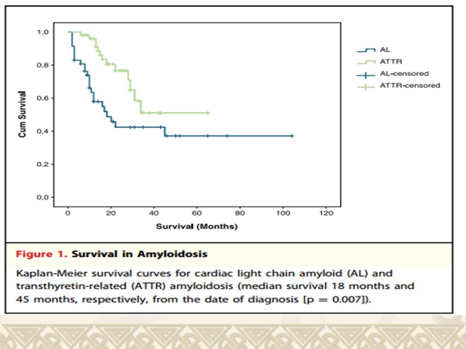
SURVIVAL Median survival from the date of diagnosis in patients with cardiac ATTR amyloidosis was better (45 months) compared with cardiac AL amyloidosis (18 months) (p¼0.007) (Fig. 1).Fig. 1
.Fig. 1.")
32
CMR: CARDIAC DIMENSIONS AND FUNCTION LV volumes and left ventricular ejection fraction (LVEF) were within the normal range, when indexed for body surface area, in cardiac AL and ATTR amyloidosis (Table 2).Table 2
 were within the normal range, when indexed for body surface area, in cardiac AL and ATTR amyloidosis (Table 2).Table 2")
33
LV end-diastolic and end-systolic volumes were significantly lower in AL compared with ATTR amyloidosis, but LV stroke volume was similar in both amyloid types. Thus, LVEF was significantly lower in cardiac ATTR compared with AL amyloidosis, albeit with absolute and indexed values within the normal range for both amyloidosis subtypes.

34
Table 2

35
LV mass index was only mildly increased in cardiac AL amyloidosis but was markedly increased in cardiac ATTR compared with AL amyloidosis.

36
Interventricular septum and RV free wall thickness were increased in both amyloidosis types, with significantly increased wall thickness in cardiac ATTR (left ventricle 18±2 mm; right ventricle 8±2 mm) compared with cardiac AL amyloidosis (left ventricle 14±3 mm; right ventricle 6±2 mm) (p<0.001).
 compared with cardiac AL amyloidosis (left ventricle 14±3 mm; right ventricle 6±2 mm) (p<0.001).")
37
Pleural effusions were common despite preserved LVEF (observed in 57% of cardiac AL patients and 41% of ATTR amyloidosis patients. There was no significant difference in the prevalence of pericardial effusions according to amyloid type.

38
CMR: LGE LGE was present in all patients with cardiac ATTR amyloidosis and 45 (98%) of 46 patients with cardiac AL amyloidosis (Table 3).Table 3 One male patient with cardiac AL amyloidosis according to conventional echocardiographic criteria did not demonstrate any LGE in the context of increased LV thickness (13 mm), increased LV mass index, and preserved systolic function (LVEF75%).
 of 46 patients with cardiac AL amyloidosis (Table 3).Table 3 One male patient with cardiac AL amyloidosis according to conventional echocardiographic criteria did not demonstrate any LGE in the context of increased LV thickness (13 mm), increased LV mass index, and preserved systolic function (LVEF75%).")
39
RV free wall LGE was detectable in all patients with ATTR amyloidosis but in only 33 patients (72%) with cardiac AL amyloidosis (p <0.001), possibly due to a lesser degree of RV thickening in AL amyloidosis.
 with cardiac AL amyloidosis (p <0.001), possibly due to a lesser degree of RV thickening in AL amyloidosis.")
40
Table 3

41
RV wall thickness was significantly higher in patients with detectable RV LGE (7±2 mm) compared with patients without RV LGE (5±2 mm) (p<0.001). AtrialLGE was detectable in 47 ATTR patients (92%) and 34 cardiac AL amyloidosis patients (74%) (p=0.02). The previously reported global subendocardial pattern of LGE (8) was present in 18 AL patients (39%) and only 6 ATTR patients (12%) (p=0.002).
 and 34 cardiac AL amyloidosis patients (74%) (p=0.02). The previously reported global subendocardial pattern of LGE (8) was present in 18 AL patients (39%) and only 6 ATTR patients (12%) (p=0.002)..")
42
The prevalence of any transmural LGE was significantly higher in patients with cardiac ATTR amyloidosis (n=46 [90%]) than cardiac AL amyloidosis (n=17 [37%]) (p<0.001).
![ The prevalence of any transmural LGE was significantly higher in patients with cardiac ATTR amyloidosis (n=46 [90%]) than cardiac AL amyloidosis (n=17 [37%]) (p<0.001).](http://images.slideplayer.com/21/6329084/slides/slide_42.jpg " The prevalence of any transmural LGE was significantly higher in patients with cardiac ATTR amyloidosis (n=46 [90%]) than cardiac AL amyloidosis (n=17 [37%]) (p<0.001).")
43
A baseapex gradient (more extensive uptake at the base compared with the apex) (Fig. 2) was present in 36 cardiac ATTR patients (71%) and 19 cardiac AL patients (41%) (p=0.004).Fig. 2 Global transmural LGE throughout the left ventricle was evident in 11 ATTR patients (22%) but in only 2 patients with cardiac AL amyloidosis (4%) (p=0.01).
 was present in 36 cardiac ATTR patients (71%) and 19 cardiac AL patients (41%) (p=0.004).Fig. 2 Global transmural LGE throughout the left ventricle was evident in 11 ATTR patients (22%) but in only 2 patients with cardiac AL amyloidosis (4%) (p=0.01)..")
44
Fig. 2

45
Quantification of LGE in cardiac amyloidosis The extent of LGE in ATTR amyloidosis was strikingly greater than that observed in AL amyloidosis. We devised a new retrospective analysis tool to semi-quantitatively assess the degree of LGE in patients with suspected cardiac amyloidosis (Fig. 3).(Fig. 3).
.(Fig. 3)..")
46
The Query Amyloid Late Enhancement (QALE) score was performed on LGE images at the base, mid-ventricle, and apex. The maximum LV LGE score at each level is 4 (maximum LV LGE score 12), plus 6 if RV LGE is present. The QALE score range is 0 (no LGE in the left or right ventricle) to 18 (global transmural LV LGE with RV involvement).
, plus 6 if RV LGE is present. The QALE score range is 0 (no LGE in the left or right ventricle) to 18 (global transmural LV LGE with RV involvement)..")
47
Fig. 3

48
A significant, positive correlation was demonstrated between QALE score and interventricular septal thickness (Spearman’s rho =0.344, p <0.001), LV mass index (Spearman’s rho = 0.348, p<0.001), LV end- diastolic volume index (Spearman’s rho = 0.206, p =0.033), and LV end-systolic volume index (Spearman’srh=0.269, p =0.005).
, LV mass index (Spearman’s rho = 0.348, p<0.001), LV end- diastolic volume index (Spearman’s rho = 0.206, p =0.033), and LV end-systolic volume index (Spearman’srh=0.269, p =0.005).")
49
A negative correlation was demonstrated between QALE score and LVEF (Spearman’srho=–0.267, p=0.005). No significant correlation was observed between QALE score and LV stroke volume index (Spearman’s rho=0.011, p=0.910), NT- proBNP (Spearman’srho=0.068, p=0.487), or duration of symptoms (Spearman’s rho=–0.054, p=0.579).
, NT- proBNP (Spearman’srho=0.068, p=0.487), or duration of symptoms (Spearman’s rho=–0.054, p=0.579)..")
50
Multivariate regression analysis was performed by using clinical and CMR variables (age, sex, interventricular septum thickness, LV mass, and QALE score). The QALE score was identified as an independent predictor of amyloid type.

51
A score model for the detection of ATTR amyloidosis wasdeveloped as a logistic probability unit by using the most significant variables: age and interventricular septum thickness with or without QALE score.

52
A receiver-operating characteristic curve was developed for the scores (Fig. 4). A QALE score ≧ 13 differentiated ATTR from AL type with 82% sensitivity and 76% specificity.Fig. 4 The logistic score with QALE included was the most accurate, with 87% sensitivity and 96% specificity to detect ATTR amyloidosis.
. A QALE score ≧ 13 differentiated ATTR from AL type with 82% sensitivity and 76% specificity.Fig. 4 The logistic score with QALE included was the most accurate, with 87% sensitivity and 96% specificity to detect ATTR amyloidosis..")
53
Fig. 4

54
REPRODUCIBILITY OF QALE SCORE. All CMR scans were reported by a second blinded observer to determine the interoperator variability of QALE scoring. The second observer reviewed the entire study and recorded the QALE score in the left ventricle (3 levels) and the right ventricle.
 and the right ventricle..")
55
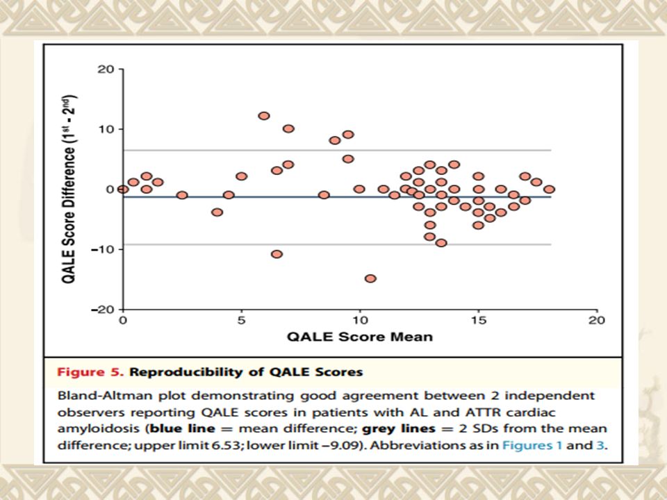
The intraclass correlation coefficient was 0.66 (95% CI: 0.53 to 0.76), confirming fair agreement between the 2 independent observers. Bland-Altman analysis demonstrated a mean difference of–1.28 (95% CI: for mean difference– 0.48 to–2.09) (Fig. 5).Fig. 5
 (Fig. 5).Fig. 5.")
57
FALSE-POSITIVE CMR REPORTS Cardiac amyloidosis was ruled out on the basis of endomyocardial biopsy results in 6 patients after CMR reported findings suggestive of amyloid cardiomyopathy. All 6 patients were male, with Afro-Caribbean ethnicity in 4 cases, and all showed LGE on CMR imaging, but extensive myocardial fibrosis was demonstrated according to histology in each case with negative Congo red staining.

58
The pattern of LGE was variable, but diffuse mid- wall LGE was seen in 2 hypertensive Afro- Caribbean patients (Fig. 6).Fig. 6 None of the patients with falsepositive reports demonstrated transmural LGE or a base-apex gradient.
.Fig. 6 None of the patients with falsepositive reports demonstrated transmural LGE or a base-apex gradient..")
59
Fig. 6

60
DISCUSSION Cardiac amyloidosis of all types is associated with LV volumes and LVEF within the normal range at presentation, but LV wall thickness and mass index are increased. In our experience, preserved LVEF by echocardiography is uncommon as the disease progresses and it should be noted that the CMR scans in this series were performed early, often before a formal diagnosis had been made, and not repeated.

61
Cardiac ATTR amyloidosis is associated with significantly higher LV mass index, higher LV volumes, and lower LVEF compared with cardiac AL amyloidosis. Pleural and pericardial effusions were more common in cardiac AL amyloidosis, despite better LVEF and smaller LV volumes, although not statistically significant.

62
LGE was evident in all but 1 patient with cardiac AL amyloidosis in this series. Two patients with systemic AL amyloidosis and no evidence of cardiac involvement according to conventional echocardiographic criteria had completely normal CMR scans.

63
The pattern of LGE in cardiac ATTR amyloidosis was more extensive, with RV enhancement detected in all cases and a higher prevalence of transmural LV LGE.

64
The absence of detectable RV free wall LGE in more than one-quarter of patients with cardiac AL amyloidosis may distinguish the 2 subtypes; however, the difference in detection of RV LGE may be explained by increased RV wall thickness in ATTR amyloidosis.

65
Suboptimal myocardial nulling was a frequent finding in this series and in itself should raise suspicion of amyloid infiltration. T1-weighted scouting sequences (Look-Locker) incorporating a range of inversion times are helpful to differentiate extensive LGE from poor myocardial nulling.
 incorporating a range of inversion times are helpful to differentiate extensive LGE from poor myocardial nulling..")
66
It may also be helpful to cross-check skeletal muscle signal intensity while choosing the optimal inversion time, particularly in difficult cases; very bright skeletal muscle signal should prompt a reconsideration of the inversion time (Fig. 7).Fig. 7
.Fig. 7.")
68
Gadolinium is an extracellular tracer, and previous studies have confirmed good correlation between LGE and increased extracellular space on histological examination in aortic stenosis and cardiac amyloidosis.

69
A base-apex gradient, common in ATTR amyloidosis in this study, correlates with echocardiographic studies using strain analysis, which have reported reduced basal systolic longitudinal and radial strain (“apical sparing”)in cardiac amyloidosis. The base-apical gradientalso reflects underlying myocardial wall stress in the left ventricle.

70
It remains unclear why survival in cardiac AL amyloidosis is inferior to cardiac ATTR amyloidosis. LGE has been reported as a marker of poor prognosis in ischemic, dilated, and hypertrophic cardiomyopathies. Although Syed et al. reported increased B- natriuretic peptide concentration in amyloidosis patients (predominantlyn AL type) with LGE, there was no correlation between NT-proBNP and LGE when quantified with the QALE score in this study.
 with LGE, there was no correlation between NT-proBNP and LGE when quantified with the QALE score in this study..")
71
Patients with cardiac ATTR amyloidosis in this series had a better prognosis despite more extensive LGE. The previously reported subendocardial LGE pattern is characteristic of AL amyloidosis, but it is very often not global as described and incorporates a degree of transmural enhancement, typically in the basal lateral wall.

72
We have therefore refined the description of global subendocardial LGE resulting in the pattern presenting infrequently in this serie. It is possible that the precise anatomical pattern of LGE (rather than merely the extent of LGE) in amyloidosis has a major influence on causing cardiac dysfunction, with subendocardial deposition potentially causing more marked restrictivephysiology.
 in amyloidosis has a major influence on causing cardiac dysfunction, with subendocardial deposition potentially causing more marked restrictivephysiology..")
73
Diastolic function is not formally assessed with routine CMR, and correlation of the findings in this study with echocardiography is warranted.

74
Direct toxicity of AL amyloid fibrils on myocardial cells has been proposed as an alternative explanation, but it is also possible that the likely much slower accumulation of ATTR amyloid is better tolerated. No significant difference was demonstrated between nonmutant (n=23) and mutant ATTR (n=28) type for any CMR variable in this series.
 and mutant ATTR (n=28) type for any CMR variable in this series..")
75
The newly devised CMR LGE scoring outlined in this study was an independent predictor of amyloid type on multivariate analysis. The logistic regression scoring model differentiated ATTR and AL type with high sensitivity and specificity, and we have confirmed an improvement in accuracy by adding the QALE score.

76
Echocardiography has poor specificity (74% to 82%) to detect amyloid from nonamyloid cardiomyopathy, even using modern speckle tracking strain analysis techniques, and it thus seems inferior to CMR in differentiating between amyloid types (although this has not been formally assessed).
 to detect amyloid from nonamyloid cardiomyopathy, even using modern speckle tracking strain analysis techniques, and it thus seems inferior to CMR in differentiating between amyloid types (although this has not been formally assessed).")
77
99m Tc-3,3-diphos- phono-1,2- propanodicarboxylic acid (DPD) scintigraphy seems to have high specificity to differentiate amyloid types, but it has been studied in much smaller numbers. CMR is currently more widely available than 99m Tc-DPD scintigraphy and also provides additional functional and structural information

78
CMR LGE scoring seems to differentiate between amyloid types but the technique requires validation in additional prospective studies. Novel T1 mapping techniques have been shown to provide quantitative assessment of diffuse extracellular expansion in other cardiomyopathies.

79
T1 mapping may become the gold standard for quantifying the extent of interstitial expansion by amyloid deposition, but it may not be possible to implement the technique into standard clinical practice for all patients due to limited availability (CMR scanning is often performed locally before specialist center referral).
.")
80
Given the retrospective nature of the current study, and the lack of evaluation in a prospective cohort, we accept that treatment decisions should not be based on CMR findings alone, and histological diagnosis (including amyloid type differentiation) remains the gold standard. LGE is not specific to cardiac amyloidosis, and false-positive reporting occurs in the clinical setting.

81
Extensive fibrosis, for example secondary to hypertrophic cardiomyopathy or hypertensive heart disease, may result in diffuse LGE and false-positive CMR findings. The absence of transmural LGE or a base-apex gradient in all patients with false-positive CMR reports in this series may help to identify patients requiring further noninvasive investigation (e.g.,99mTc-DPD scintigraphy) before proceeding to endomyocardial biopsy.
 before proceeding to endomyocardial biopsy..")
82
CONCLUSIONS CMR is often performed to assess patients with suspected amyloidosis or it triggers the differential after an abnormal scan result. The increased availability of CMR will ultimately lead to increased detection of cardiac amyloidosis due to superior tissue characterization, using the LGE technique, compared with echocardiography.

83
This study was a real-world analysis of scans performed in 46 centers without specialist amyloid protocols, and we acknowledge all of the difficulties of the LGE technique. Despite these limitations, the analysis was robust in identifying the amyloidfibril type with high accuracy. CMR LGE scoring has the potential to enhance the management of many patients but cannot replace biopsy as the gold standard.

84
Cardiac amyloidosis should be considered in any patient with LGE and LV hypertrophy, particularly in the presence of preserved ejection fraction and pleural effusions. We have attempted to differentiate between the 2 main subtypes of cardiac amyloidosis, a crucial differentiation due to markedly different outcomes.

85
Cardiac ATTR amyloidosis is associated with improved survival despite increased LV mass and more extensive LGE, afinding at odds with other cardiomyopathies. The explanation for thisfinding needs further investigation.

86
Correct identification of amyloidosis type is imperative to differentiate patients with cardiac AL amyloidosis, a condition potentially amenable to chemotherapy, from cardiac ATTR amyloidosis patients who may benefit from novel TTR-specific treatments now entering Phase III clinical trial assessment.

Similar presentations


 is characterized by.>")


 are characterized by platelet size abnormalities and it has been suggested that this parameter.>")


 and Restrictive cardiomyopathy (RCM) share several clinical, ultrasonographic and hemodynamic.>")


 History: A man in his 40s presenting to respiratory medicine with 6 months breathlessness.>")
