Download presentation
Presentation is loading. Please wait.

1
بسم اللّه الرحمن الرحیم

2
INFECTIOUS SPONDILODISCITIS

3
Definition Infection of intervertebral disc and adjacent vertebrae

4
Microbial Agents Pyogens (Staph -E coli) Acute
Brucella- Salmonella Subacute Tuberculosis Chronic

5
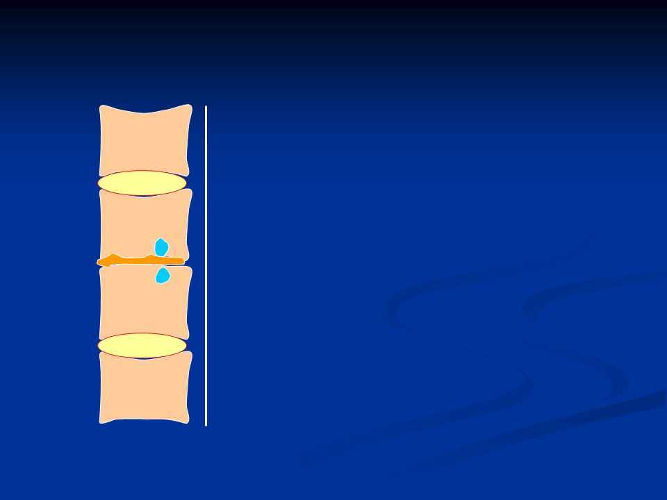
PATHOGENESIS

6
Hematogenous Direct inoculation Adjacent tissue

7
ANT POST Spinal artery

8
ANT POST INITIATION

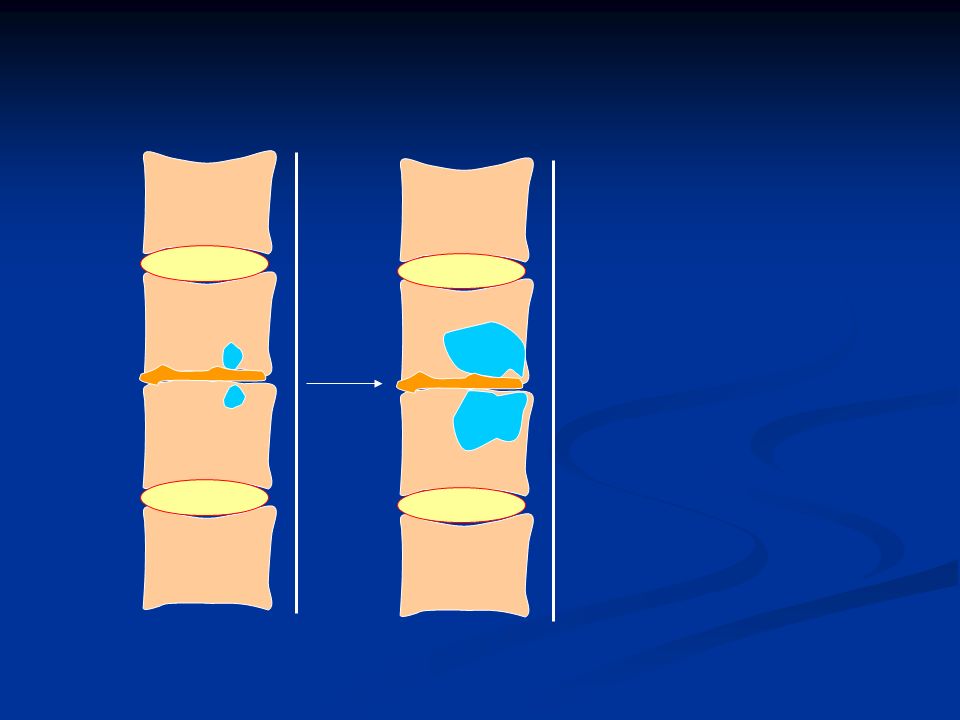
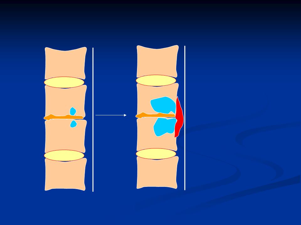
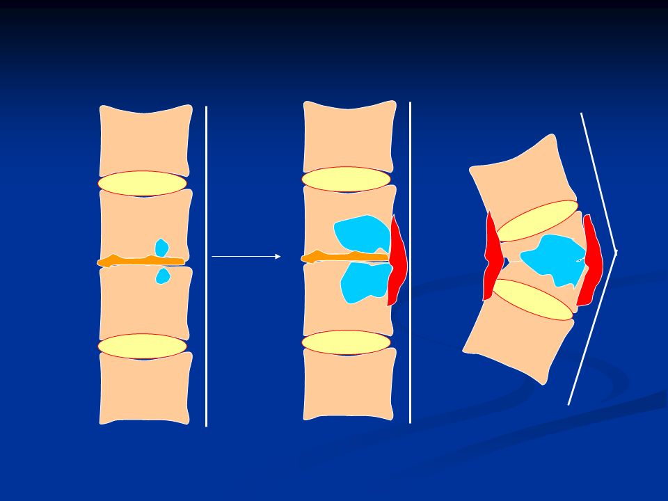
9
ANT POST INITIATION DESTRUCTION

10
TUBERCULOSIS BRUCELOSIS Anterior DESTRUCTION

11
Anterior REPAIR

12
COURSE ANT INITIATION DESTRUCTION REPAIR

13
SPONDYLODISCITIS OF TUBERCULOSIS (POTT’S disease)
")
14
Epidemiology 2 % musculoskeletal Site: Lung involvement: 20-30%
1% spine Site: Upper thoracic: Children Lower thoracic: Adult Lung involvement: 20-30%

19
Clinical Manifestation

20
Clinical Manifestation
STAGE 2 (DESTRUCTION) months 1-2 years STAGE 1 (INITIATION) STAGE 3 (REPAIR)
 months. 1-2 years. STAGE 1. (INITIATION) STAGE 3. (REPAIR)")
21
Clinical Manifestation
STAGE 1 (INITIATION) Mechanical pain Mild tenderness
 Mechanical pain. Mild tenderness.")
22
Clinical Manifestation
STAGE 2 (DESTRUCTION) STAGE 1 (INITIATION) months Constitutional Severe Back pain Ph/Ex: Severe Tenderness Blockage (erect posture) Gibbous deformity Cold abscess Neurological (paraplegia)
 STAGE 1. (INITIATION) months. Constitutional. Severe Back pain. Ph/Ex: Severe Tenderness. Blockage (erect posture) Gibbous deformity. Cold abscess. Neurological (paraplegia)")
23
Clinical Manifestation
STAGE 2 (DESTRUCTION) STAGE 1 (INITIATION) 1-2 years STAGE 3 (REPAIR) Mechanical pain Reduce symptom Reduce spasm
 STAGE 1. (INITIATION) 1-2 years. STAGE 3. (REPAIR) Mechanical pain. Reduce symptom. Reduce spasm.")
24
DIAGNOSIS

25
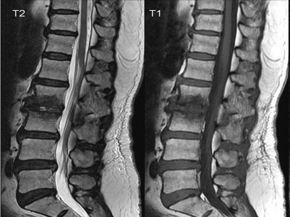
IMAGING

28
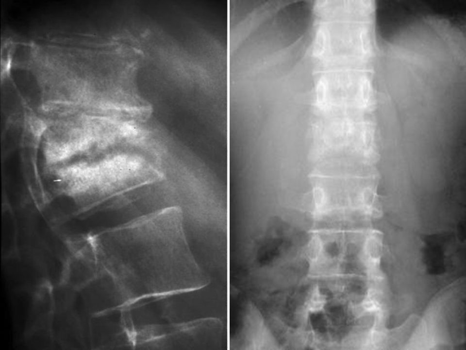
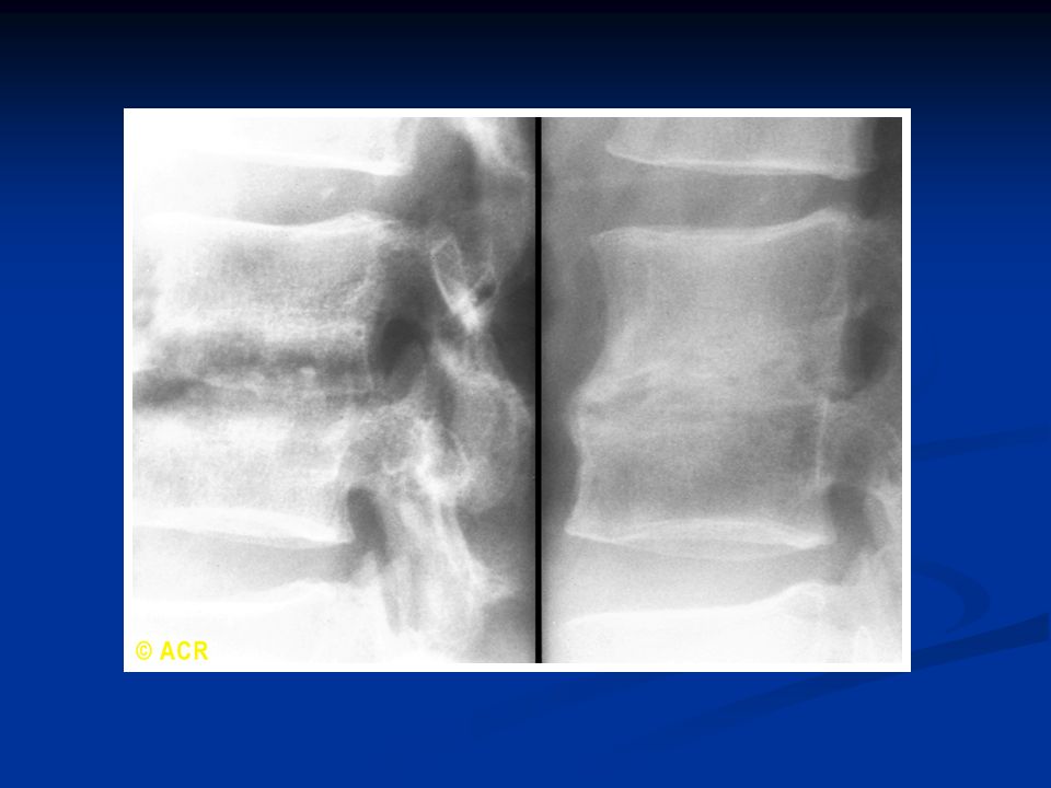
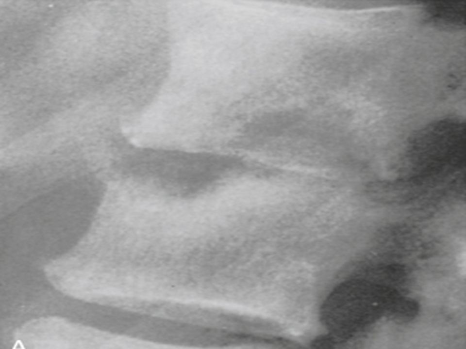
RADIOGRAPHY

31
LAB TESTS

32
LAB TESTS ESR PPD (+) Sputum smear & culture
Abscess or bone biopsy: Culture / Pathology

33
TREATMENT

34
TREATMENT Medical: Mainstay of treatment Alone is successful in 70%
Duration: 18 m (9-12 m ?)
")
35
TREATMENT Surgical: Advanced neurological deficit
Progressive Neurological deficits Kyphosis > 40 degrees

36
Monitoring Clinical Radiology ESR, CRP pain constitutional mobility
neurological signs ESR, CRP Radiology may be progress during first 6 month of treatment

37
BRUCELLOSIS SPONDYLODISCITIS

38
EPIDEMIOLOGY Spine: 7-8% of skeletal Site: Lumbosacral (Anterior)
")
39
BRUCELOSIS

40
CLINICAL MANIFESTATION

41
CLINICAL MANIFESTATION
Constitutional: Fever Night Sweat Malaise Arthralgia Apathetic Then: Severe low back pain

42
CLINICAL MANIFESTATION
Physical examination: Blockage Severe tenderness Fever Splenomegaly Lymphadenopathy

43
DIAGNOSIS

44
DIAGNOSIS Clinical presentation Radiography
Supporting laboratory finding History of potential exposure: Endemic area Microbiology laboratory Unpasteurized milk products Contact with animals History of similar illness in the family (documented in almost 50% of cases).
.")
46
Laboratory WBC: NL or low ESR, CRP: NL Wright; Cooms Wright
B/C or Bone marrow culture (7-35 d) ELISA
 ELISA.")
47
Treatment 2 Drug: 3 months Rifampin 600-900 mg/d
Doxycycline 100 mg twice daily 3 months

48
SALMONELLA SPONDYLODISCITIS

49
SALMONELLA SPONDYLODISCITIS
Clinical sign & radiologic future is similar to Brucellosis. Diarrhea: 60% Positive S/C (most commonly)
")
50
SALMONELLA SPONDYLODISCITIS
Treatment: Quinolones Ceftriaxone For 4 w

51
PYOGENIC SPONDYLODISCITIS

52
PYOGENIC SPONDYLODISCITIS
staphylococcus aureus (50 – 70%) History of recent infection (UTI; septicemia) Acute severe back pain; High fever; Chills Patient is ill. Blockage; Severe tenderness Lumbar spine (45%) Thoracic (35%)
 History of recent infection (UTI; septicemia) Acute severe back pain; High fever; Chills. Patient is ill. Blockage; Severe tenderness. Lumbar spine (45%) Thoracic (35%)")
53
PYOGENIC SPONDYLODISCITIS
Radiography: severe destructive rapidly progressive lesion Large bone bridge

54
PYOGENIC SPONDYLODISCITIS
MRI:

55
PYOGENIC SPONDYLODISCITIS
Laboratory: Leucocytosis ESR (useful in fallow up) Culture B/C: 50% - 70% Biopsy: 70% - 90% UTD 19.3
 Culture. B/C: 50% - 70% Biopsy: 70% - 90% UTD")
56
PYOGENIC SPONDYLODISCITIS
Diagnosis: Clinical presentation Radiology laboratory

57
PYOGENIC SPONDYLODISCITIS
Treatment: Two drugs (Staph & gram negative organisms) Six weeks Surgery: - progressive - cord compression
 Six weeks. Surgery: - progressive. - cord compression.")
59
Paravertebral abscess Psoas abscess
Brucellosis Tuberculosis Site Lumbar and others Dorsolumbar Vertebrae Multiple or contiguous Contiguous Diskitis Late Early Body Intact until late Morphology lost early Canal compression Rare Common Epiphysitis Anterosuperior (Pom's sign) General: upper and lower disk regions, central, subperiosteal Osteophyte Anterolateral (parrot beak) Unusual Deformity Wedging uncommon Anterior wedge, gibbus Recovery Sclerosis, whole body Variable Paravertebral abscess Small, well-localized Common and discrete loss, transverse process Psoas abscess More likely
 General: upper and lower disk regions, central, subperiosteal. Osteophyte. Anterolateral (parrot beak) Unusual. Deformity. Wedging uncommon. Anterior wedge, gibbus. Recovery. Sclerosis, whole body. Variable. Paravertebral abscess. Small, well-localized. Common and discrete loss, transverse process. Psoas abscess. More likely.")
Similar presentations


>")


 in the tissues, especially the lungs.>")


>")


