Download presentation
Presentation is loading. Please wait.

1
Case Conference 指導老師 陳昭文 醫師 Intern 張 盛皇 指導老師 陳昭文 醫師 Intern 張 盛皇 2007.8.7 2007.8.7

2
Patient’s profile Chart NO. : 23803235 Chart NO. : 23803235 Name : 翁 XX Name : 翁 XX Age : 40 y/o Age : 40 y/o Sex : male Sex : male Date of ER arrival : 2007.7.31 (18:30) Date of ER arrival : 2007.7.31 (18:30)
 Date of ER arrival : (18:30).")
3
Condition at ER Chief complaint : falling down accidentally from second floor height this afternoon. Chief complaint : falling down accidentally from second floor height this afternoon. Vital sign : HR= 93 bpm, BT= 36.4 ’ C Vital sign : HR= 93 bpm, BT= 36.4 ’ C RR= 16 /min, BP=251/138 RR= 16 /min, BP=251/138 GCS : E4V5M6 GCS : E4V5M6 Pupil size : L ’ t(3.0) R ’ t(2.5) Pupil size : L ’ t(3.0) R ’ t(2.5)
 R ’ t(2.5) Pupil size : L ’ t(3.0) R ’ t(2.5).")
4
Physical examination Head : not being recorded Head : not being recorded Neck : np Neck : np Chest : np Chest : np Abdomen : np Abdomen : np

5
Multiple laceration wounds over left face 12.5cm 3cm

6
3cm L/W with deformity of left thigh 1cm L/W of left foot

7
L/W 2cm of right forearmL/W 1cm of left forearm

8
Abrasion wound of left elbow Bruise of right knee

9
Past history DM(-) DM(-) HTN(-) HTN(-) Denied any other systemic disease Denied any other systemic disease Operation history:GU history post op about 10+ years ago at 台南市立 hospital Operation history:GU history post op about 10+ years ago at 台南市立 hospital
 DM(-) HTN(-) HTN(-) Denied any other systemic disease Denied any other systemic disease Operation history:GU history post op about 10+ years ago at 台南市立 hospital Operation history:GU history post op about 10+ years ago at 台南市立 hospital")
10
Management (7/31) 18:30 -Arrival ER of KMUH Transferred from 國軍岡山 hospital Brain CT; Abd CT; symptomatic care Imp: R/O ICH with multiple L/W over face Fracture of right wrist Internal bleedinng 18:50 - CBC.GOT.GPT.BUN.Cr.Sugar.Na.K.PT.PTT- CBC.GOT.GPT.BUN.Cr.Sugar.Na.K.PT.PTT - Chest x-ray. Bil wrist x-ray (AP+Lat). Left foot x-ray (AP+Lat)Chest x-ray. Bil wrist x-ray (AP+Lat). Left foot x-ray (AP+Lat) Left femoral x-ray (AP+Lat). Pelvic x-ray (AP).( - Facial CT (non-enhanced)Facial CT (non-enhanced) - L/R 1BOT IVD. Ice package. O2 4L/min. on Foley. 12-lead EKG. EKG Sp02 monitor. Air splint fixation 19:00 -Consult 骨科 & 整外 19:20 -Blood PH/gas analysis & Ethanol(quantitative)Blood PH/gas analysis & Ethanol(quantitative) -Urine routineUrine routine
. Left foot x-ray (AP+Lat)Chest x-ray. Bil wrist x-ray (AP+Lat). Left foot x-ray (AP+Lat) Left femoral x-ray (AP+Lat). Pelvic x-ray (AP).( - Facial CT (non-enhanced)Facial CT (non-enhanced) - L/R 1BOT IVD. Ice package. O2 4L/min. on Foley. 12-lead EKG. EKG Sp02 monitor. Air splint fixation 19:00 -Consult 骨科 & 整外 19:20 -Blood PH/gas analysis & Ethanol(quantitative)Blood PH/gas analysis & Ethanol(quantitative) -Urine routineUrine routine.")
11
Management (7/31~8/1) 21:40 -L/W suture 22:20 -N/S 1BOT IVD. Gastor 1 Amp IV. Laston 1Amp IM -Debridement of the open fracture wounds - C spine collar, C spine x-ray (AP+Lat) C spine x-ray (AP+Lat) 22:35 - PRBC 4U 23:05 -On CVP with N/S 1BOT and Chest x-ray -Gelfusine 1BOT IVD 23:15 -Consult 泌尿科 -Abd. CTAbd. CT -KUBKUB 23:30 -N/S 5000ml irrigation -Right knee x-ray (AP+Lat)Right knee x-ray (AP+Lat) 00:10 -Arm splint (bil. Forearm) -Skeletal tracture ( 骨骼牽引 ) (left femorus) 01:35 -ER to 15ES SICU
 C spine x-ray (AP+Lat) 22:35 - PRBC 4U 23:05 -On CVP with N/S 1BOT and Chest x-ray -Gelfusine 1BOT IVD 23:15 -Consult 泌尿科 -Abd. CTAbd. CT -KUBKUB 23:30 -N/S 5000ml irrigation -Right knee x-ray (AP+Lat)Right knee x-ray (AP+Lat) 00:10 -Arm splint (bil. Forearm) -Skeletal tracture ( 骨骼牽引 ) (left femorus) 01:35 -ER to 15ES SICU.")
12
Management (8/2~8/6) 8/2 -Operation of left femoral neck and shaft fracture -Open reduction and internal fixation + external fixation of femoral shaft -SICU care 8/4 -Transferred to 7C 8/6 -Operation of facial bone fracture -Operation of bilateral wrist fracture → ORIF
 8/2 -Operation of left femoral neck and shaft fracture -Open reduction and internal fixation + external fixation of femoral shaft -SICU care 8/4 -Transferred to 7C 8/6 -Operation of facial bone fracture -Operation of bilateral wrist fracture → ORIF")
13
Free fall trauma Trauma 2006; 8: 157 – 167

14
Epidemiology Falls rank only 2nd to motor vehicle accidents among the causes of trauma deaths in the U.K. and U.S. U.S. Approximately 50% of free falls are accidental 20% are suicide attempts Another 20% are crime related The remainder are from undetermined causes Men predominate over women

15
Risk of dying The distance of a fall Perhaps the strongest single predictor of mortality Falls from 3 storeys(45 feet; 13~14m)carrying a 50% risk of death Falls from 5 storeys(75 feet; 22~23m) or more rarely being compatible with survival The impact surface, attitude of the body at impact, victim ’ s age and location of fall modify chances of survival Immediate death from free fall is usually a result of massive brain damage, thoracic trauma or intraabdominal bleeding alone or in combination.
carrying a 50% risk of death Falls from 5 storeys(75 feet; 22~23m) or more rarely being compatible with survival The impact surface, attitude of the body at impact, victim ’ s age and location of fall modify chances of survival Immediate death from free fall is usually a result of massive brain damage, thoracic trauma or intraabdominal bleeding alone or in combination.")
16
Regional injury Musculoskeletal injuries The lower limbs are the most frequently encountered injuries in feet first landings. The calcaneus is the most frequently fractured bone but complex metaphyseal and epiphyseal fracture patterns of distal joints are often seen. Spinal injury especially to the thoraco- lumbar junction must always be suspected.

17
The association of calcaneus fracture and spinal injury following a fall from height may be as high as 75%. In addition, spinal injuries are commonly unstable, burst or compression type configurations and carry a high incidence of neurological injury. Whole spine imaging is therefore warranted in any patient with calcaneus fracture following free fall.

18
Common sequelae following falls (massive decelerations) Ruptures of the aorta, pericardium and heart Major hepatic and splenic injuries However, such injuries are usually not survivable. Always being borne in mind In the instance of haemodynamic instability in a free fall survivor → The possibility of retroperitoneal haemorrhage especially from vertebral or pelvic fracture.

19
Thank you for your attention!!!

20
Back

25
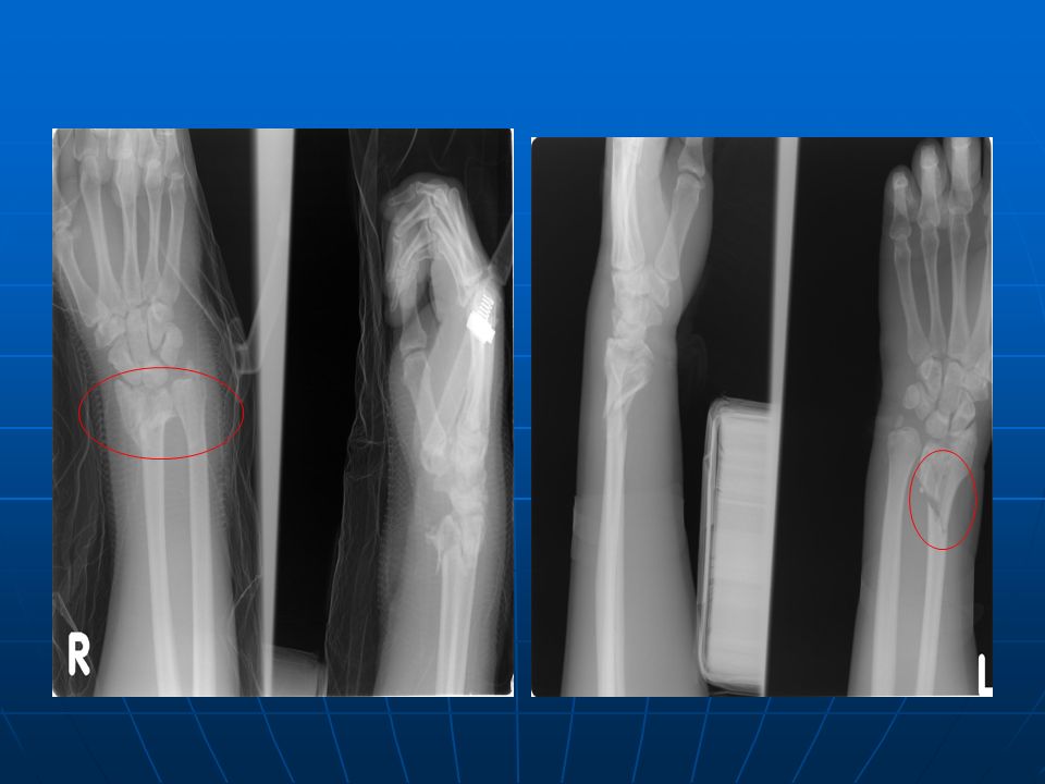
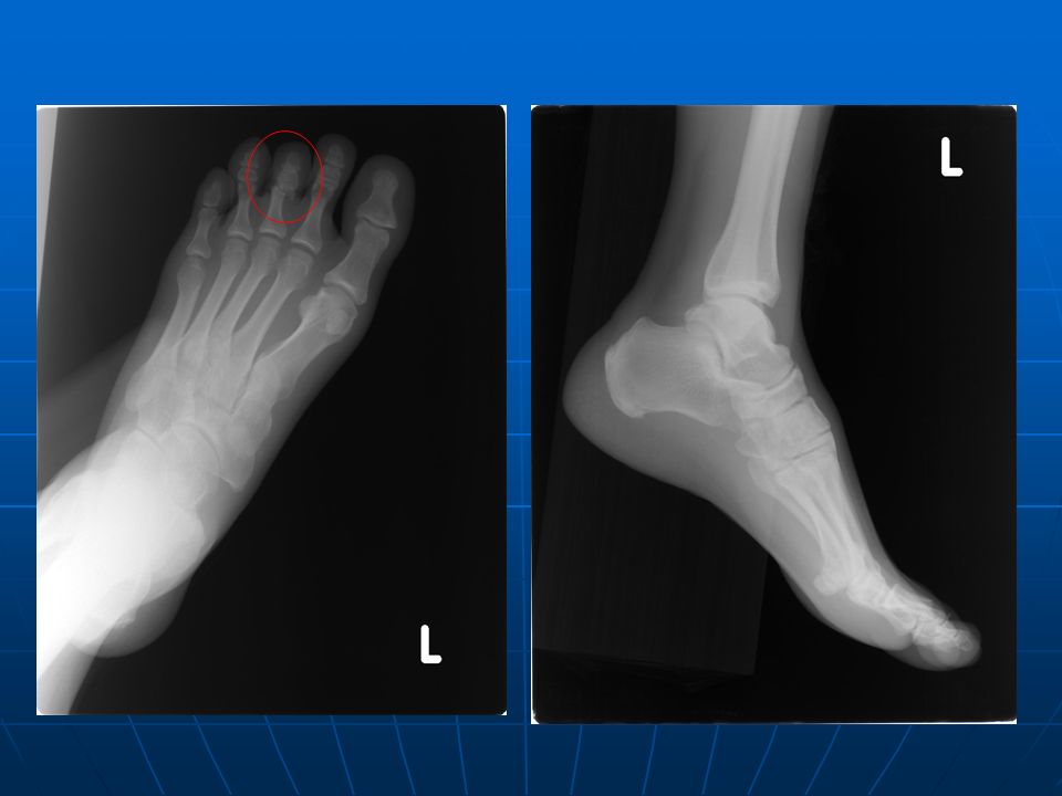
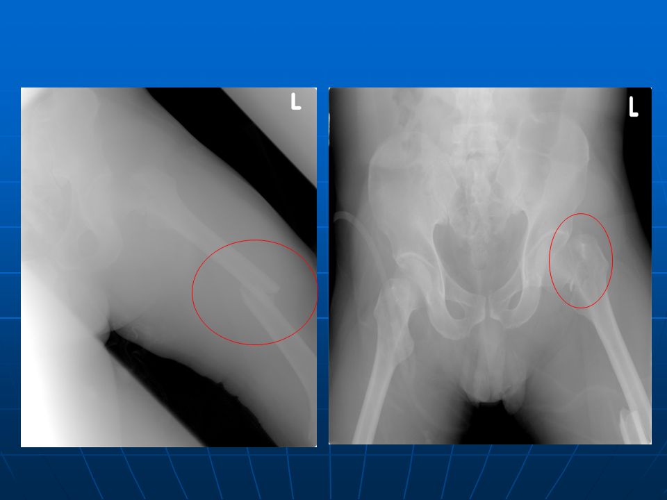
Left radial fracture; Right radial and ulnar fracture Left radial fracture; Right radial and ulnar fracture Left 3rd toe fracture Left 3rd toe fracture Left femoral neck fracture; Left femoral shaft open fracture Left femoral neck fracture; Left femoral shaft open fracture Back

59
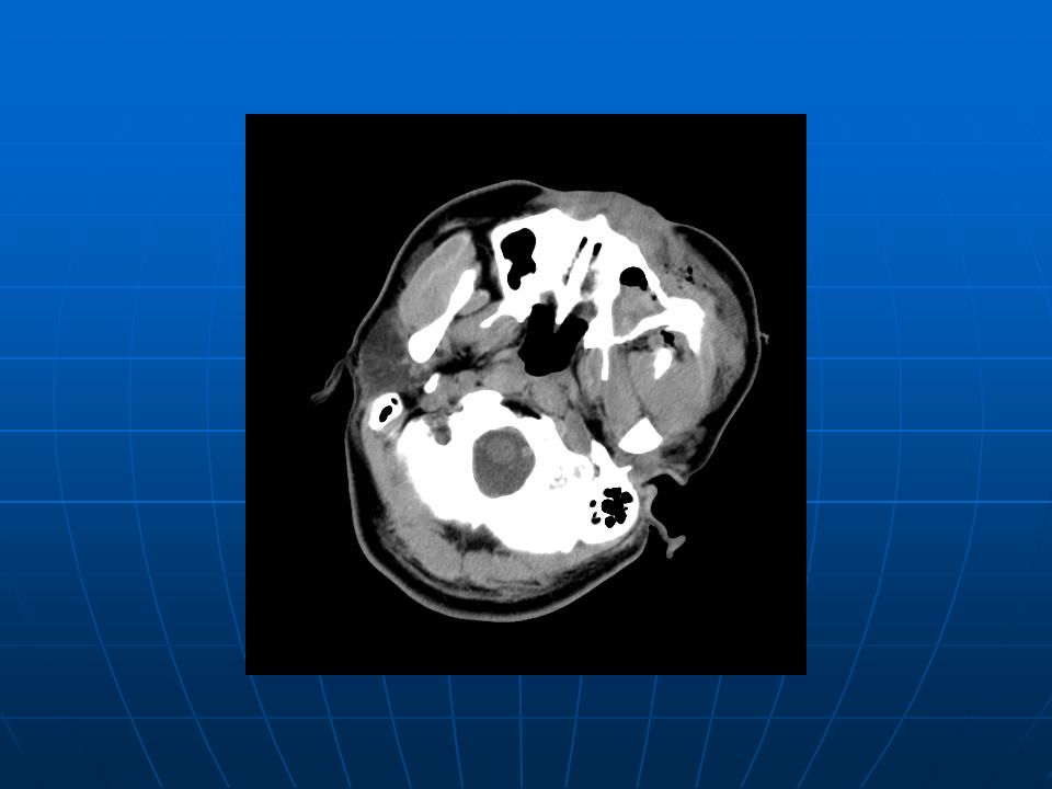
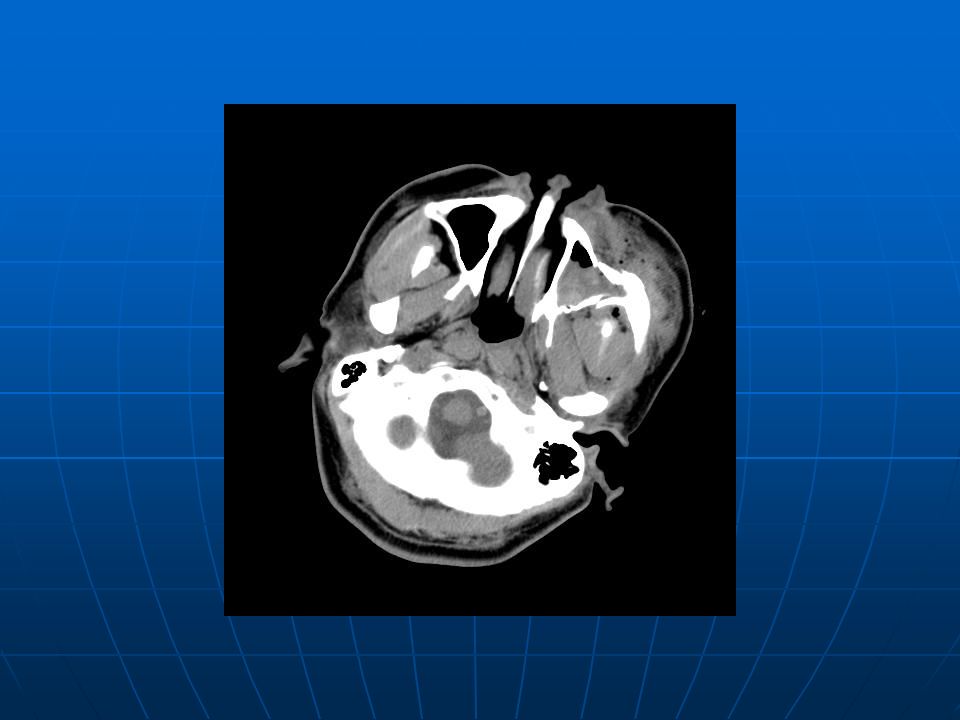
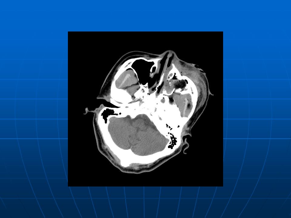
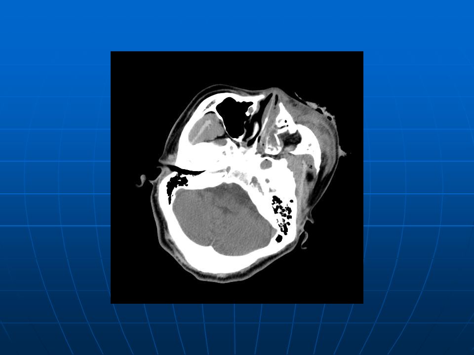
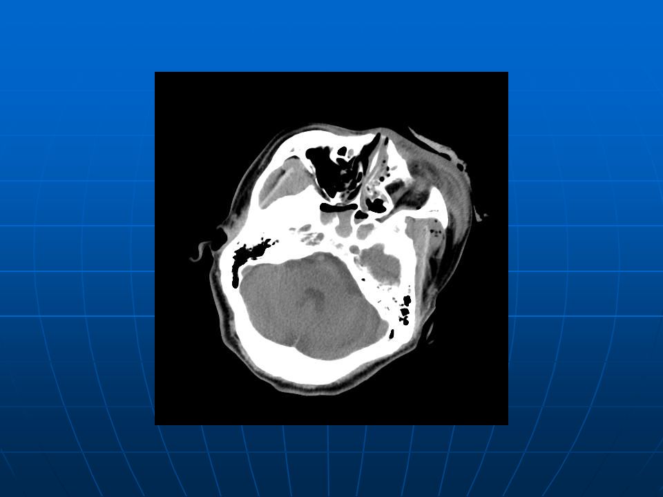
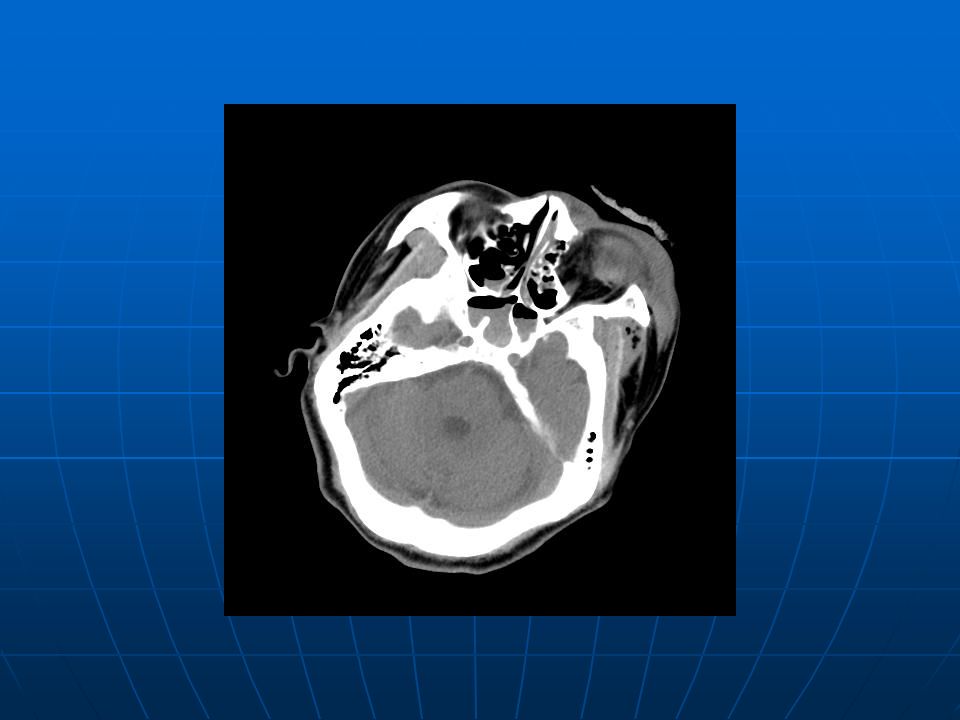
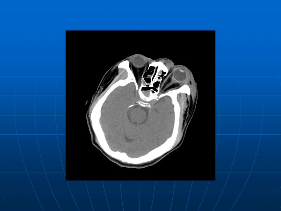
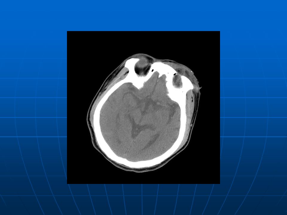
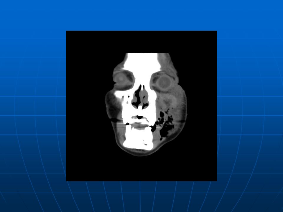
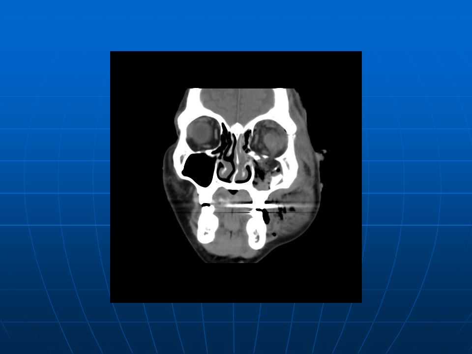
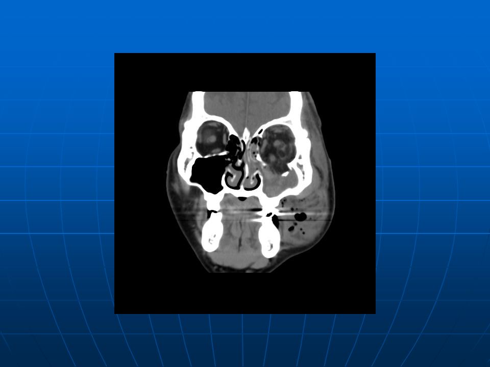
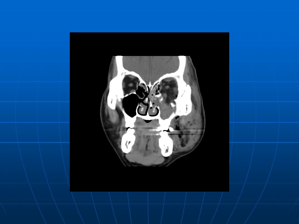
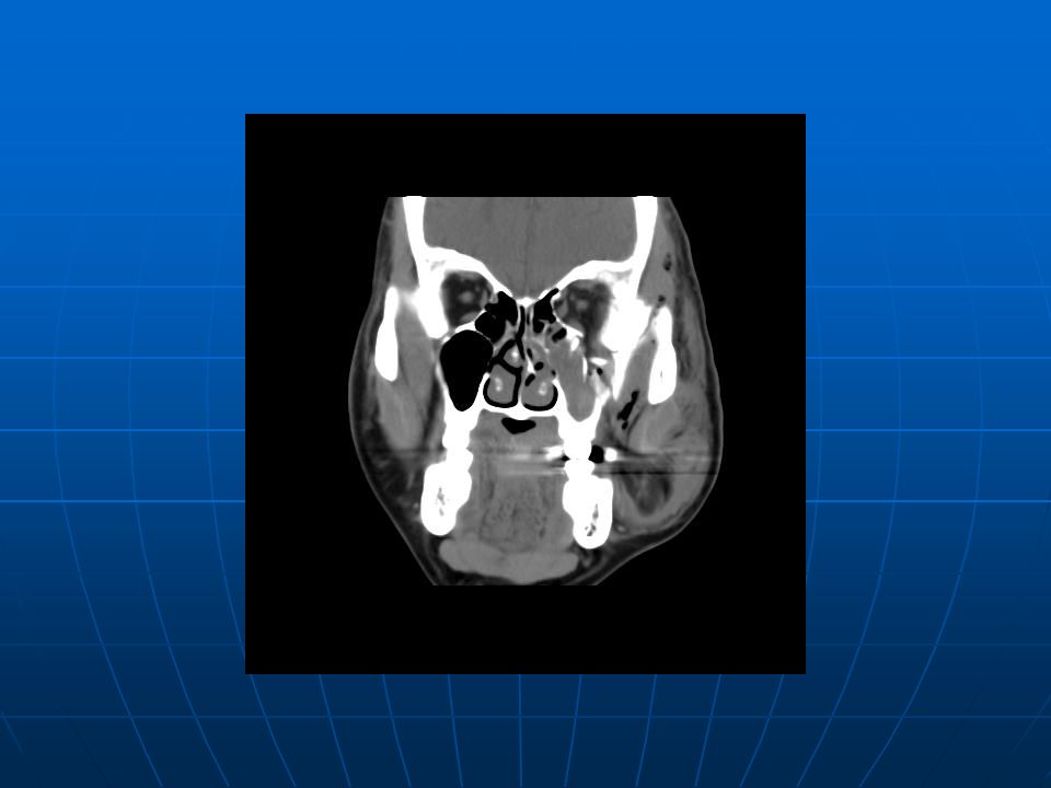
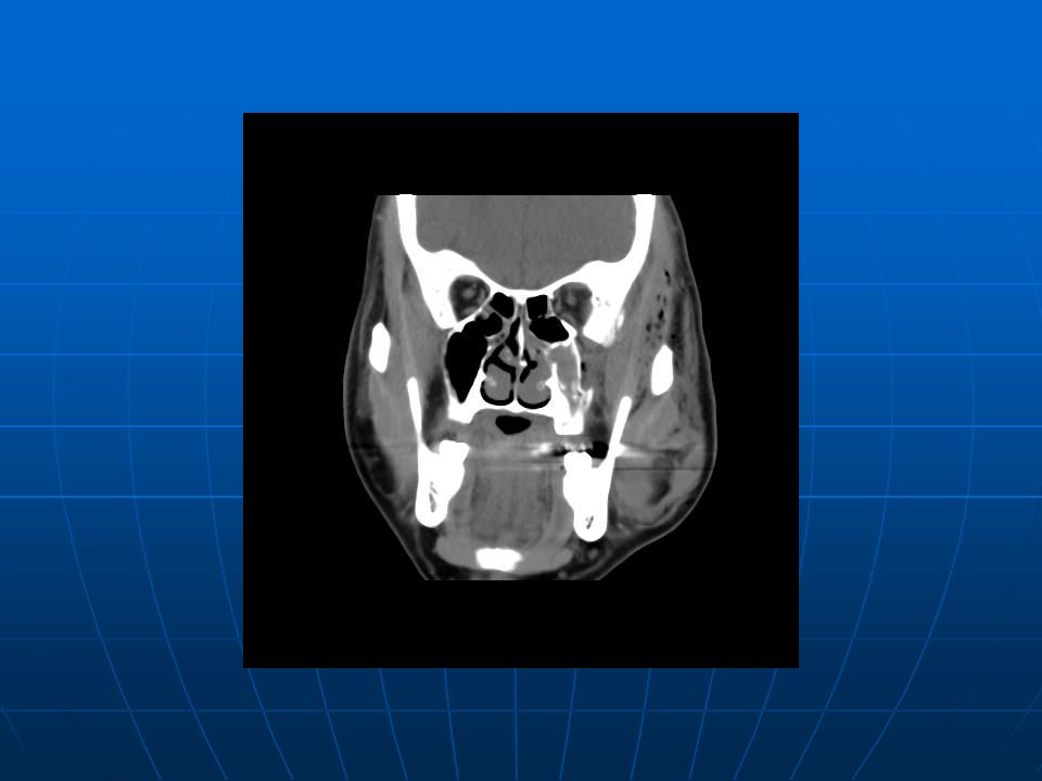
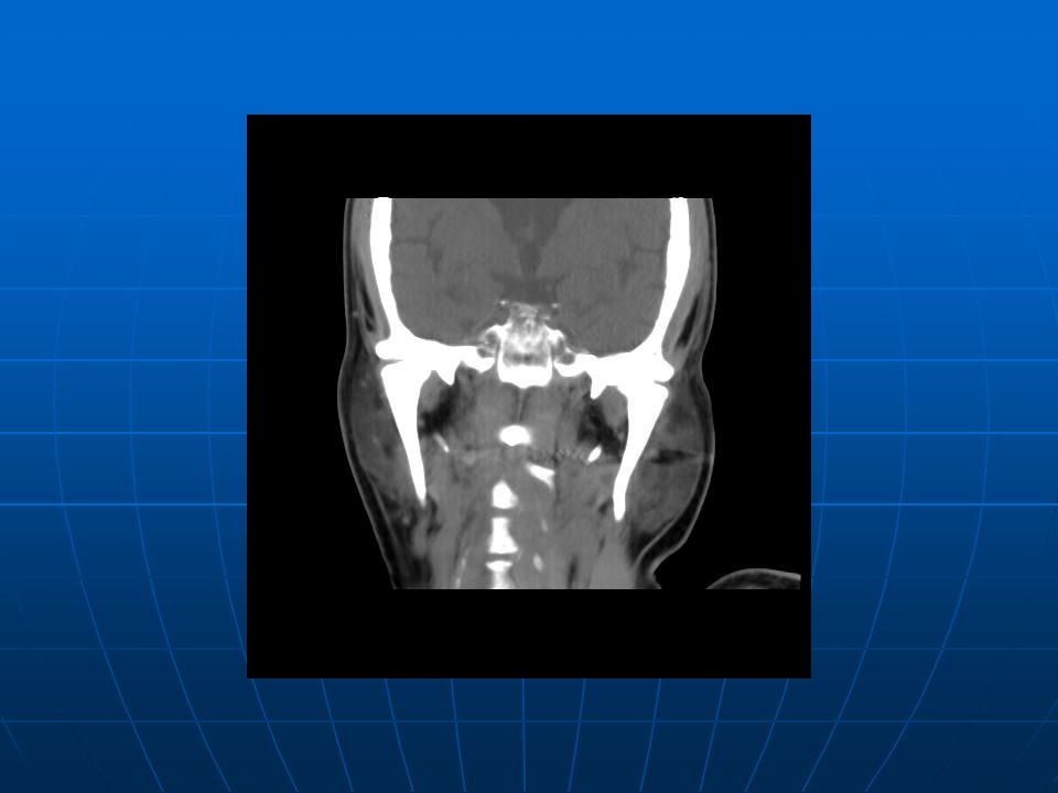
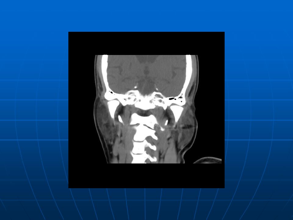
CT of Facial Bones Multiple fracture involving left maxilla, orbital floor, lamina papyracia, zygoma, left zygomatic process of frontal bone, mastoid process of left temporal bone, pterygoid plates, and skull base. Multiple fracture involving left maxilla, orbital floor, lamina papyracia, zygoma, left zygomatic process of frontal bone, mastoid process of left temporal bone, pterygoid plates, and skull base. Hematoma around left eyeball and left periorbital area. Hematoma around left eyeball and left periorbital area. Hemosinus involving left maxillary, ethmoid, and sphenoid sinuses. Hemosinus involving left maxillary, ethmoid, and sphenoid sinuses. Left hemomastoid and bilateral hemotypani. Left hemomastoid and bilateral hemotypani. Associated soft tissue swelling with emphysema in the left zygomatic, matiscator, infraorbital and periorbital areas. Associated soft tissue swelling with emphysema in the left zygomatic, matiscator, infraorbital and periorbital areas. Old infarcts in the right lentiform nucleus. Old infarcts in the right lentiform nucleus. Encephalomalacia in the right frontal lobe. Encephalomalacia in the right frontal lobe. Back

60
Blood PH/gas analysis & Ethanol (quantitative) Back
 Back")
61
Urine routine Back

129
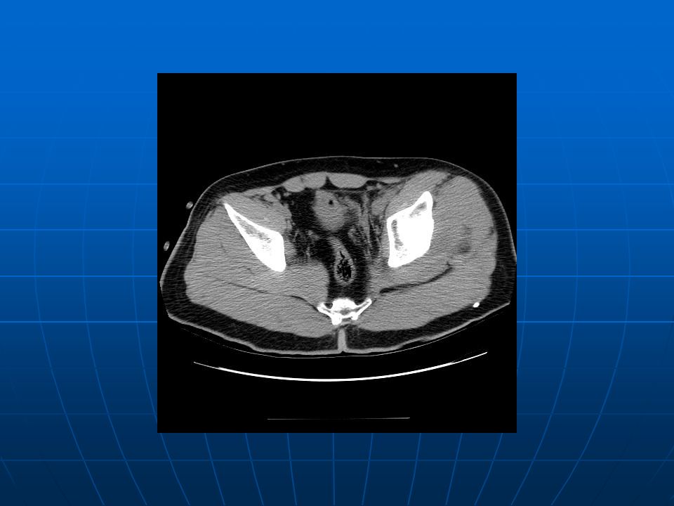
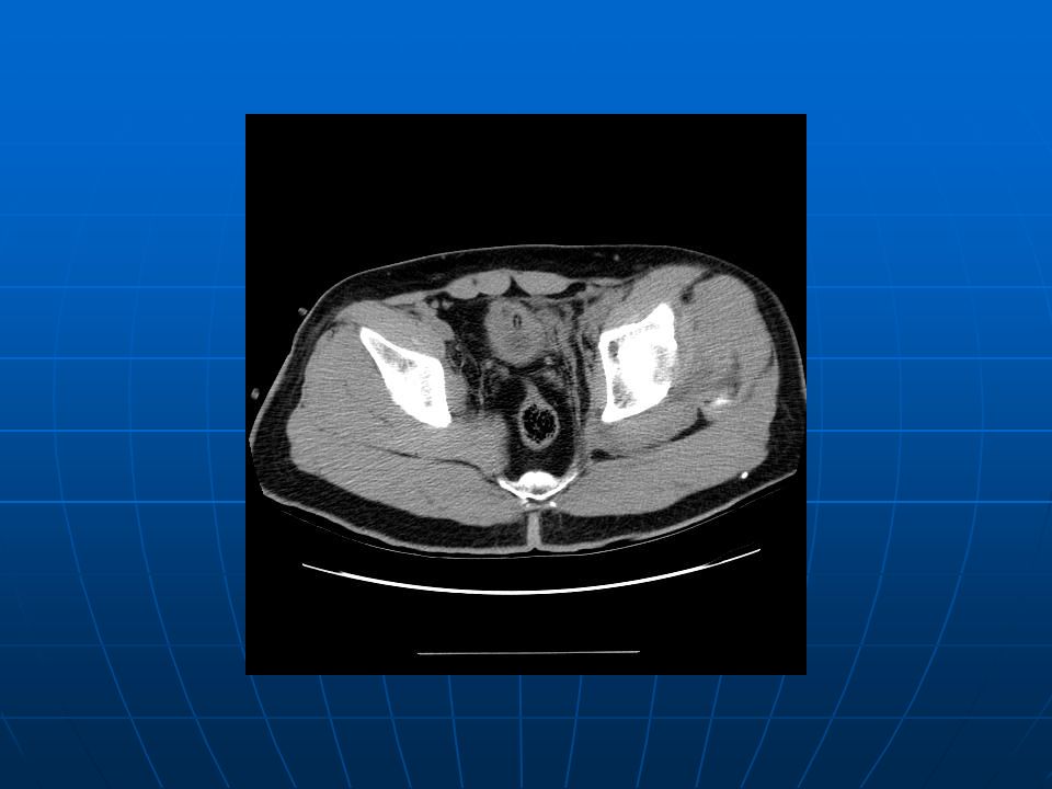
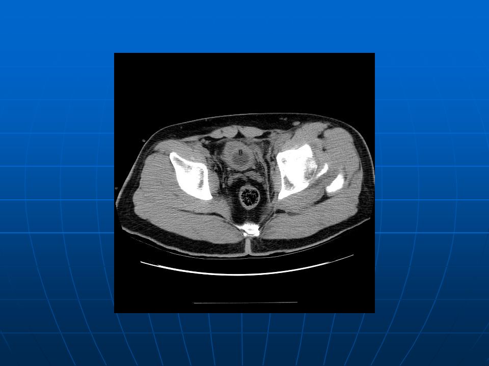
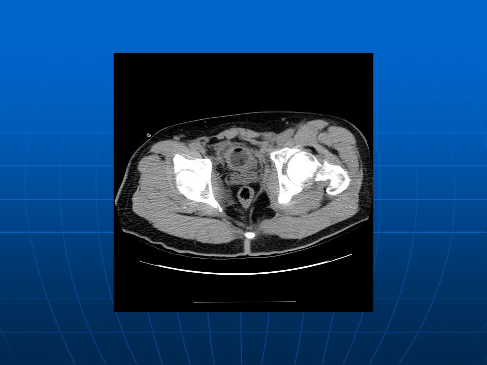
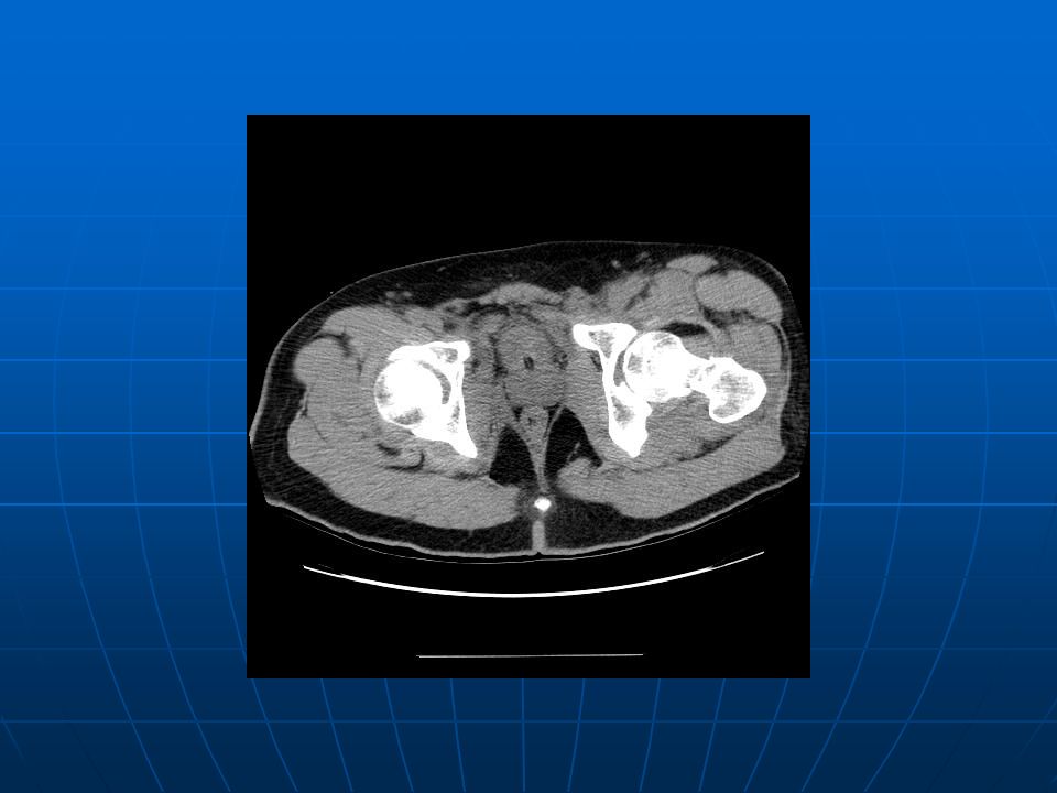
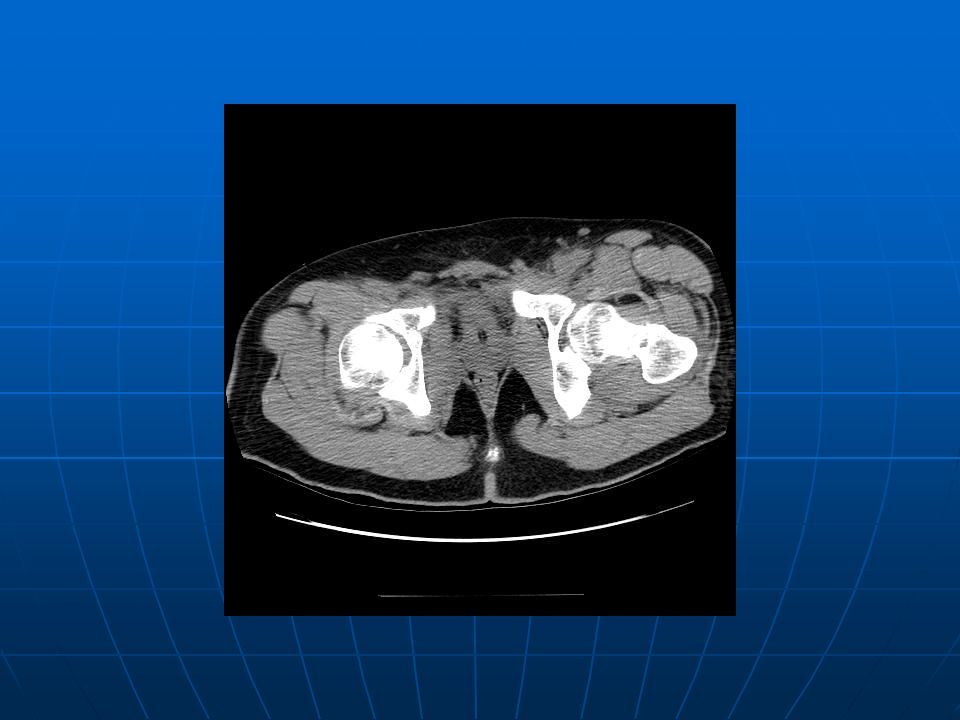
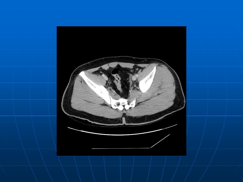
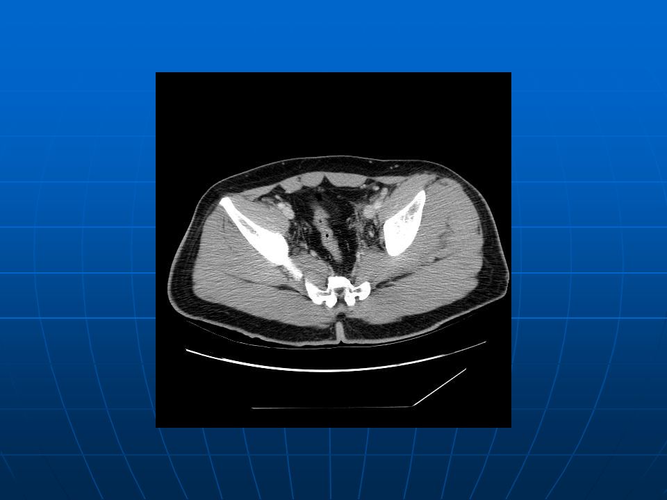
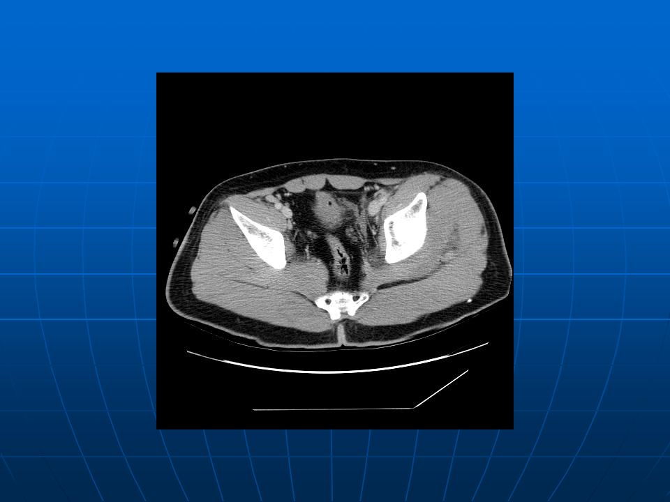
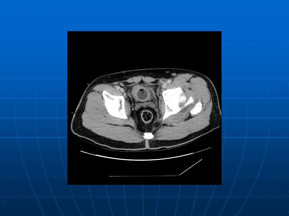
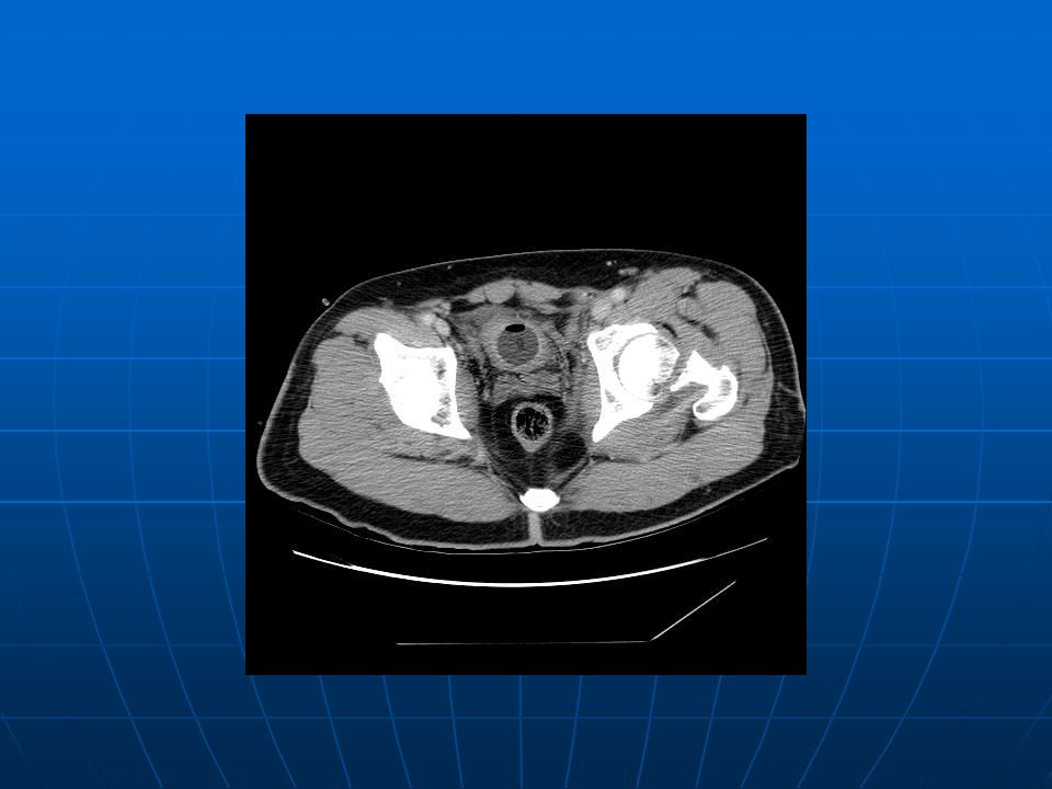
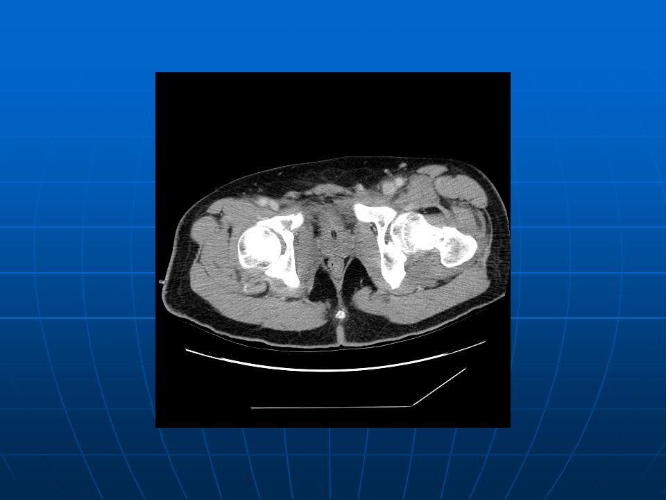
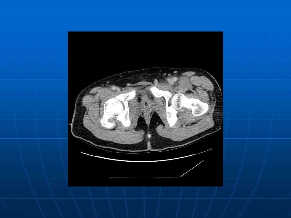
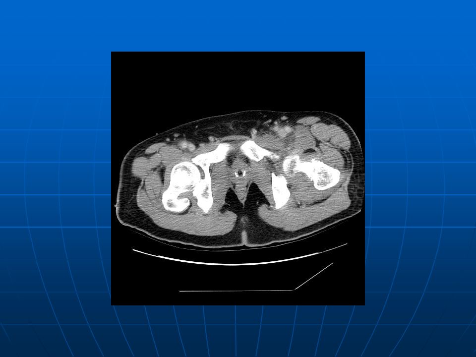
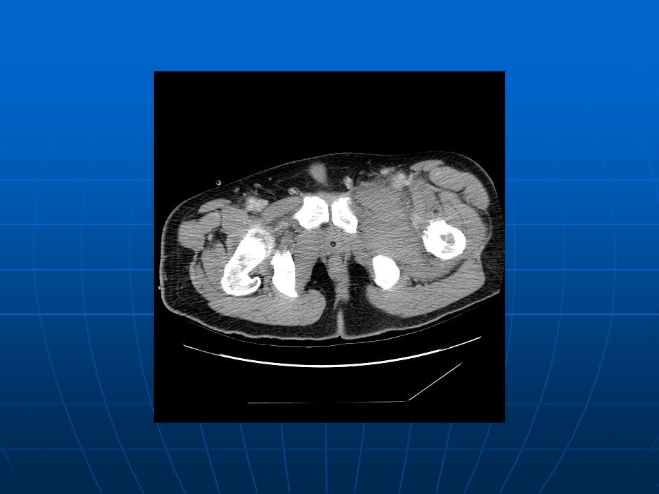
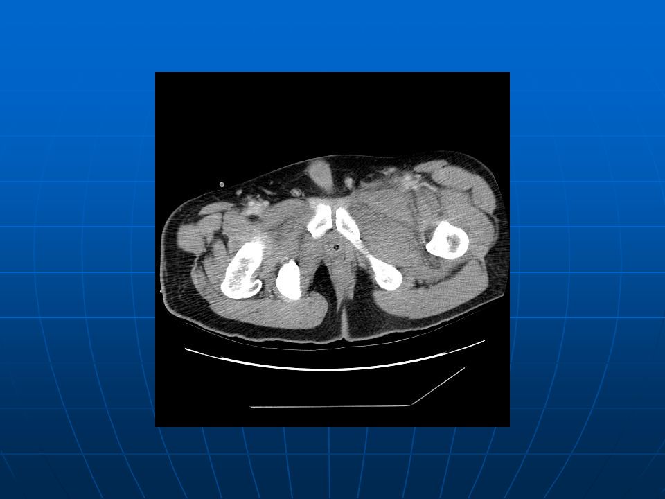
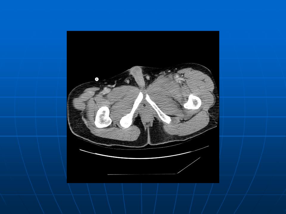
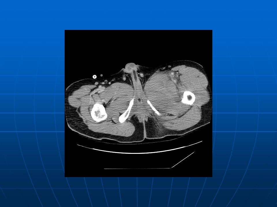
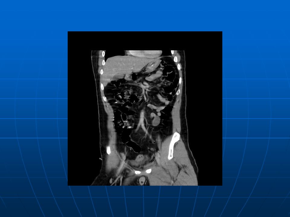
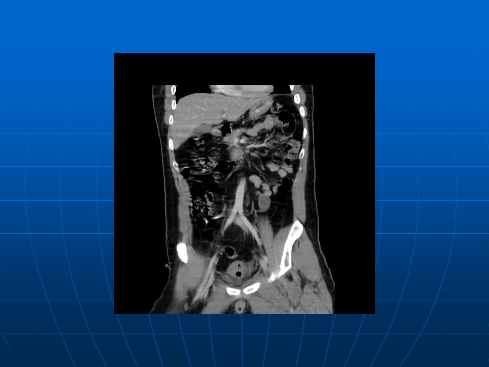
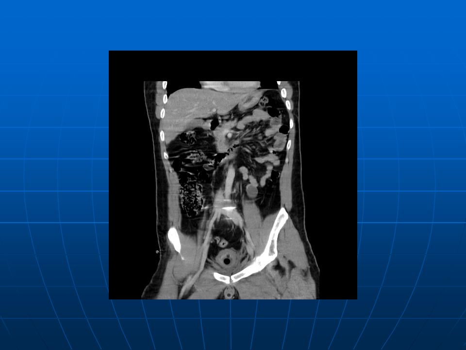
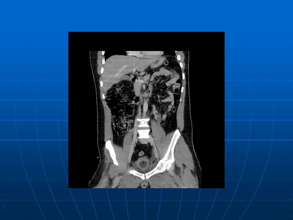
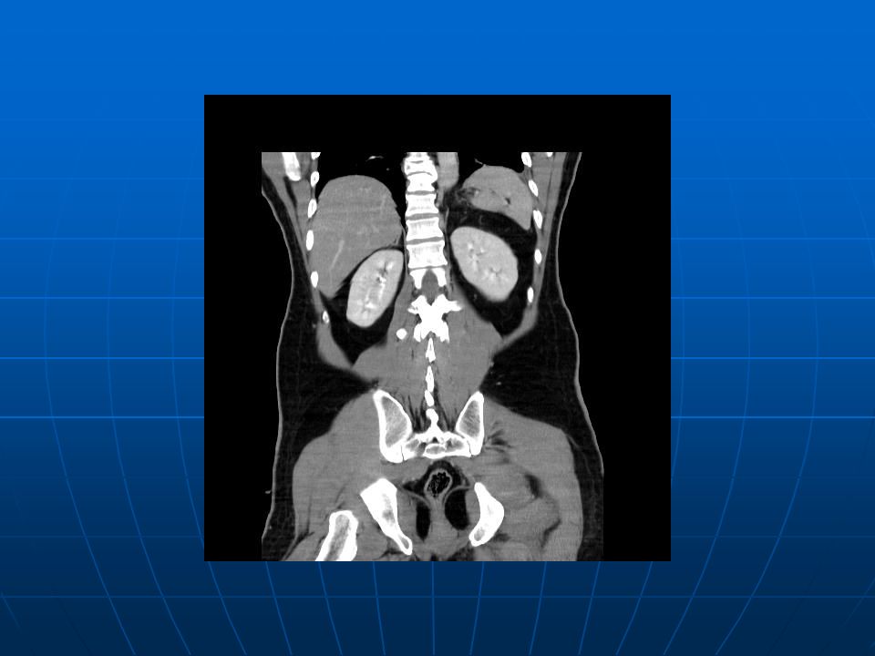
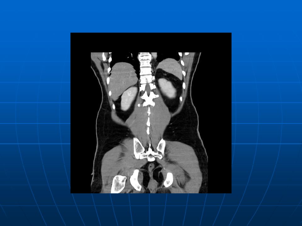
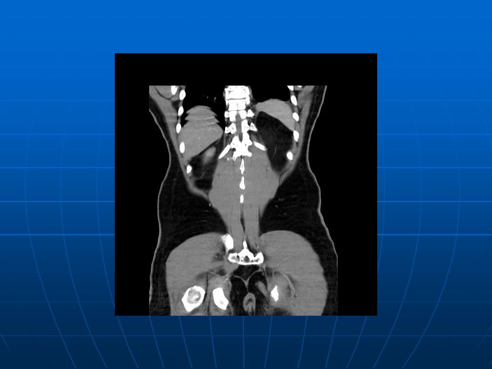
CT of the Abdomen and Pelvis A defect at the wall of the urinary bladder without CT evidence of pericystic or intraperitoneal hemorrhage. (Se/Im=2/70,71) The possibility of laceration of the urinary bladder can not be excluded. A defect at the wall of the urinary bladder without CT evidence of pericystic or intraperitoneal hemorrhage. (Se/Im=2/70,71) The possibility of laceration of the urinary bladder can not be excluded. Fracture of the left femoral neck. Fracture of the left femoral neck. Compression fracture of the T8 vertebral body. Compression fracture of the T8 vertebral body. Prostatic calcification. Prostatic calcification. On Foley catheter. On Foley catheter. Retained surgical clips in the anterior wall of gastric body due to previous surgery. Retained surgical clips in the anterior wall of gastric body due to previous surgery. Consider a calcified lymph node in the left aspect of the pelvic mesentery. (Se/Im=2/70) Consider a calcified lymph node in the left aspect of the pelvic mesentery. (Se/Im=2/70) Back
 The possibility of laceration of the urinary bladder can not be excluded. A defect at the wall of the urinary bladder without CT evidence of pericystic or intraperitoneal hemorrhage. (Se/Im=2/70,71) The possibility of laceration of the urinary bladder can not be excluded. Fracture of the left femoral neck. Fracture of the left femoral neck. Compression fracture of the T8 vertebral body. Compression fracture of the T8 vertebral body. Prostatic calcification. Prostatic calcification. On Foley catheter. On Foley catheter. Retained surgical clips in the anterior wall of gastric body due to previous surgery. Retained surgical clips in the anterior wall of gastric body due to previous surgery. Consider a calcified lymph node in the left aspect of the pelvic mesentery. (Se/Im=2/70) Consider a calcified lymph node in the left aspect of the pelvic mesentery. (Se/Im=2/70) Back.")
130
KUB

131
Right knee x-ray (AP+Lat) Back
 Back")
Similar presentations


-Score: Probability of Mass Transfusion as Surrogate for Life Threatening Hemorrhage after Multiple Trauma The.>")


 Waleed M. Awwad, MD. FRCSC Assistant professor and Consultant Orthopedic Surgery department.>")

.>")


