Download presentation
Presentation is loading. Please wait.

1
Zhang, Lei MS Otolaryngology and Head & Neck Department Sir Run Run Shaw Hospital Email:dr_zhanglei@hotmail.com

2
Acute epiglottis Acute laryngotracheobranchitis in children Chronic laryngitis Vocal fold polyps Vocal fold nodules Leukoplakia of larynx Laryngeal papillomas Laryngeal cancer Squamous cell carcinoma of pharynx(cancer of hypopharynx)
")
3
Verrucous Carcinoma

4
11,000 new cases of laryngeal cancer per year in the U.S. Accounts for 25% of head and neck cancer and 1% of all cancers One-third of these patients eventually die of their disease Most prevalent in the 6 th and 7 th decades of life

5
4:1 male predilection Downward shift from 15:1 post WWII Due to increasing public acceptance of female smoking More prevalent among lower socioeconomic class, in which it is diagnosed at more advanced stages

6
Glottic Cancer: 59% Supraglottic Cancer: 40% Subglottic Cancer: 1% Most subglottic masses are extension from glottic carcinomas

7
The first laryngectomy for cancer of the larynx was performed in 1883 by Billroth Patient was successfully fed by mouth and fitted with an artificial larynx In 1886 the Crown Prince Frederick of Germany developed hoarseness as he was due to ascend the throne.

8
Crown Prince Frederick of Germany

9
Was evaluated by Sir Makenzie of London, the inventor of the direct laryngoscope Frederick’s lesion was biopsied and thought to be cancer He refused laryngectomy and later died in 1888

10
Frederick was succeeded by Kaiser Wilhelm II, who along with Otto von Bismark militarized the German Empire and led them into WW I Could an Otolaryngologist have prevented WW I?

12
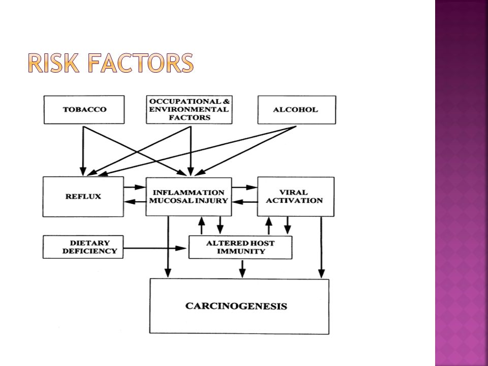
Prolonged use of tobacco and excessive alcohol use primary risk factors The two substances together have a synergistic effect on laryngeal tissues 90% of patients with laryngeal cancer have a history of both

13
Human Papilloma Virus 16 &18 Chronic Gastric Reflux Occupational exposures Asbestos mustard gas petroleum products other risk factors. Prior history of head and neck irradiation

15
85-95% of laryngeal tumors are squamous cell carcinoma Histologic type linked to tobacco and alcohol abuse Characterized by epithelial nests surrounded by inflammatory stroma Keratin Pearls are pathognomonic

16
Verrucous Carcinoma Fibrosarcoma Chondrosarcoma Minor salivary carcinoma Adenocarcinoma Oat cell carcinoma Giant cell and Spindle cell carcinoma

17
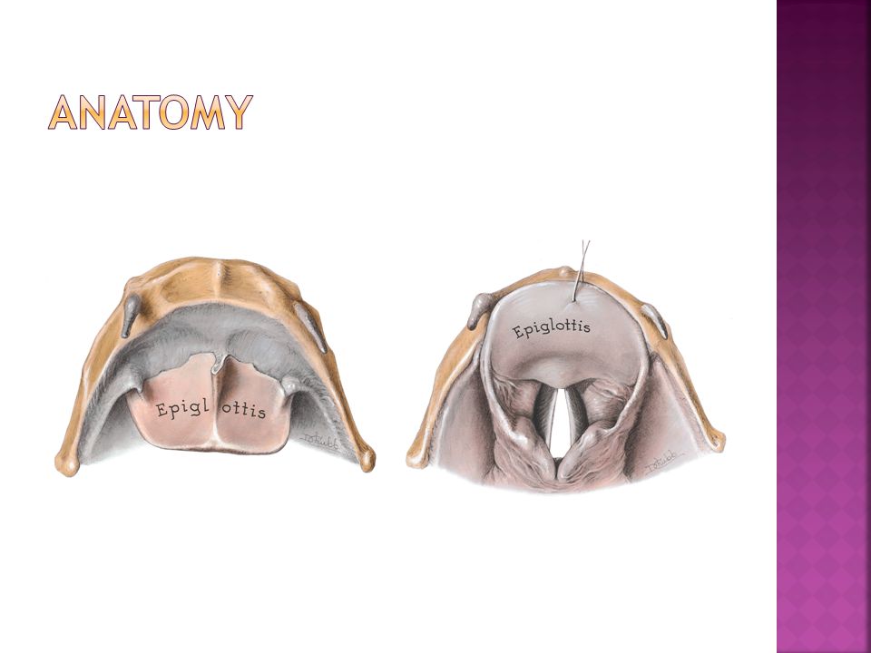
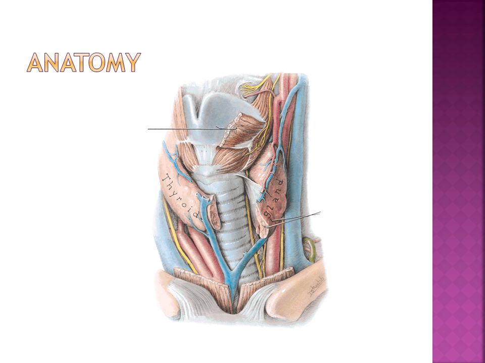
Thyroid cartilage cricoid hyoid epiglottic cricothyroid ligament

19
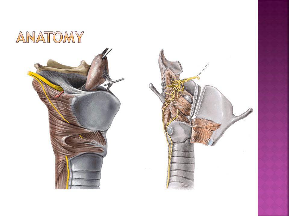
epiglottic Thyroid Arytenoids corniculate, cuneiform cricoid

25
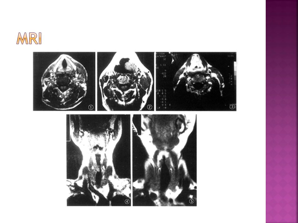
sagittal view coronal view

27
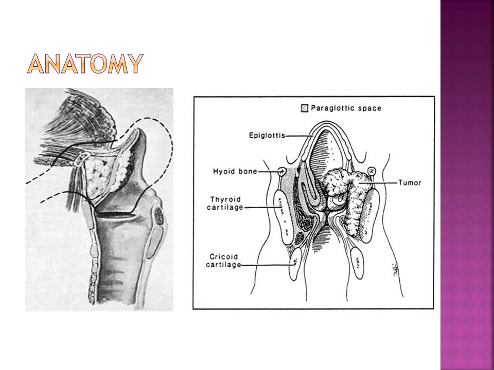
Supraglottic tumors more aggressive: Direct extension into pre-epiglottic space Lymph node metastasis Direct extension into lateral hypopharnyx, glossoepiglottic fold, and tongue base

28
Glottic tumors grow slower and tend to metastasize late owing to a paucity of lymphatic drainage They tend to metastasize after they have invaded adjacent structures with better drainage Extend superiorly into ventricular walls or inferiorly into subglottic space Can cause vocal cord fixation

30
True subglottic tumors are uncommon Glottic spread to the subglottic space is a sign of poor prognosis Increases chance of bilateral disease and mediastinal extension Invasion of the subglottic space associated with high incidence of stomal reoccurrence following total laryngectomy (TL)
")
31
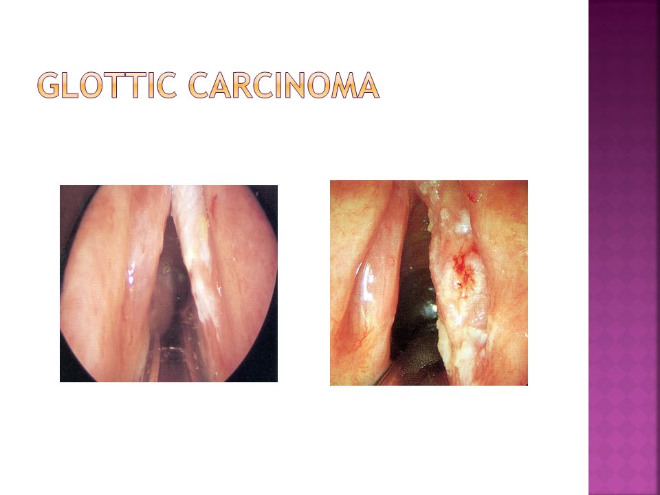
Hoarseness Most common symptom Small irregularities in the vocal fold result in voice changes Changes of voice in patients with chronic hoarseness from tobacco and alcohol can be difficult to appreciate

32
Patients presenting with hoarseness should undergo an indirect mirror exam and/or flexible laryngoscope evaluation Malignant lesions can appear as friable, fungating, ulcerative masses or be as subtle as changes in mucosal color Videostrobe laryngoscopy may be needed to follow up these subtler lesions

33
Good neck exam looking for cervical lymphadenopathy and broadening of the laryngeal prominence is required The base of the tongue should be palpated for masses as well Restricted laryngeal crepitus may be a sign of post cricoid or retropharyngeal invasion

34
Other symptoms include: Dysphagia Hemoptysis Throat pain Ear pain Airway compromise Aspiration Neck mass

35
Biopsy is required for diagnosis Performed in OR with patient under anesthesia Other benign possibilities for laryngeal lesions include: Vocal cord nodules or polyps, papillomatosis, granulomas, granular cell neoplasms, sarcoidosis, Wegner’s granulomatosis

36
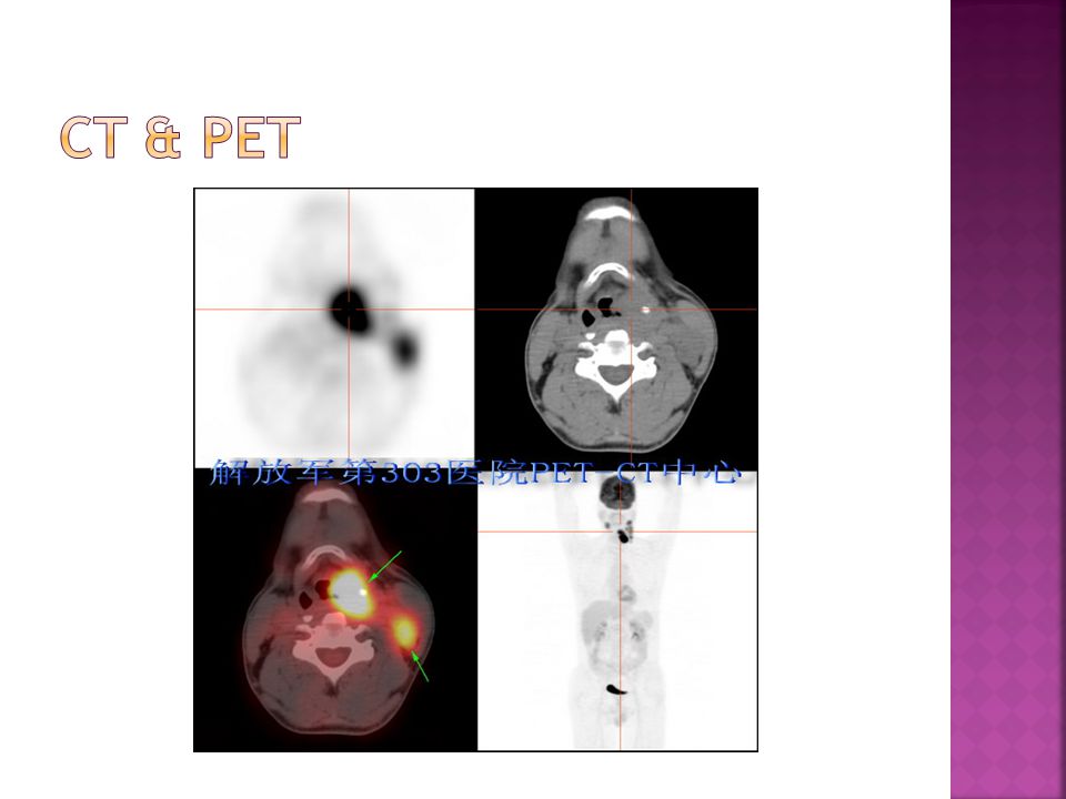
Other potential modalities: Direct laryngoscopy Bronchoscopy Esophagoscopy Chest X-ray CT or MRI Liver function tests with or without US PET ?( Positron emission tomography)
")
39
Malignant lesions can appear as friable, fungating, ulcerative masses or be as subtle as changes in mucosal color

40
TX Minimum requirements to assess primary tumor cannot be met T0 No evidence of primary tumor Tis Carcinoma in situ

41
T1 Tumor limited to one subsite of supraglottis with normal vocal cord mobility T2 Tumor involves mucosa of more than one adjacent subsite of supraglottis or glottis, or region outside the supraglottis (e.g. mucosa of base of the tongue, vallecula, medial wall of piriform sinus) without fixation T3 Tumor limited to larynx with vocal cord fixation and or invades any of the following: postcricoid area, preepiglottic tissue, paraglottic space, and/or minor thyroid cartilage erosion (e.g. inner cortex) T4 a Tumor invades through the thyroid cartilage and/or invades tissue beyond the larynx (e.g. trachea, soft tissues of neck including deep extrinsic muscles of the tongue, strap muscles, thyroid, or esophagus) T4 b Tumor invades prevertebral space, encases carotid artery, or invades mediastinal structures
 without fixation T3 Tumor limited to larynx with vocal cord fixation and or invades any of the following: postcricoid area, preepiglottic tissue, paraglottic space, and/or minor thyroid cartilage erosion (e.g. inner cortex) T4 a Tumor invades through the thyroid cartilage and/or invades tissue beyond the larynx (e.g. trachea, soft tissues of neck including deep extrinsic muscles of the tongue, strap muscles, thyroid, or esophagus) T4 b Tumor invades prevertebral space, encases carotid artery, or invades mediastinal structures.")
42
T1 Tumor limited to the vocal cord (s) (may involve anterior or posterior commissure) with normal mobilty T1a Tumor limited to one vocal cord T1b Tumor involves both vocal cords T2 Tumor extends to supraglottis and/or subglottis, and/or with impaired vocal cord mobility T3 Tumor limited to the larynx with vocal cord fixation and/or invades paraglottic space, and/or minor thyroid cartilage erosion (e.g. inner cortex) T4a Tumor invades through the thyroid cartilage, and/or invades tissues beyond the larynx (e.g. trachea, soft tissues of the neck including deep extrinsic muscles of the tongue, strap muscles, thyroid, or esophagus T4b Tumor invades prevertebral space, encases carotid artery, or invades mediastinal structures
 T4a Tumor invades through the thyroid cartilage, and/or invades tissues beyond the larynx (e.g. trachea, soft tissues of the neck including deep extrinsic muscles of the tongue, strap muscles, thyroid, or esophagus T4b Tumor invades prevertebral space, encases carotid artery, or invades mediastinal structures.")
43
T1 Tumor limited to the subglottis T2 Tumor extends to vocal cord (s) with normal or impaired mobility T3 Tumor limited the larynx with vocal cord fixation T4a Tumor invades cricoid or thyroid cartilage and/or invades tissues beyond larynx (e.g. trachea, soft tissues of the neck including deep extrinsic muscles of the tongue, strap muscles, thyroid, or esophagus) T4b Tumor invades prevertebral space, encases carotid artery, or invades mediastinal structures
 T4b Tumor invades prevertebral space, encases carotid artery, or invades mediastinal structures.")
44
N0 No cervical lymph nodes positive N1 Single ipsilateral lymph node ≤ 3cm N2a Single ipsilateral node > 3cm and ≤6cm N2b Multiple ipsilateral lymph nodes, each ≤ 6cm N2c Bilateral or contralateral lymph nodes, each ≤6cm N3 Single or multiple lymph nodes > 6cm

45
M0 No distant metastases M1 Distant metastases present

46
0TisN0M0IT1N0M0 IIT2N0M0 IIIT3N0M0 T1-3N1M0 IVAT4aN0-2M0 T1-4aN2M0 IVBT4b Any N M0 Any T N3M0 IVC Any N M1

47
Premalignant lesions or Carcinoma in situ can be treated by surgical stripping of the entire lesion CO2 laser can be used to accomplish this but makes accurate review of margins difficult

48
Early stage (T1 and T2) can be treated with radiotherapy or surgery alone, both offer the 85-95% cure rate. Surgery has a shorter treatment period, saves radiation for recurrence, but may have worse voice outcomes Radiotherapy is given for 6-7 weeks, avoids surgical risks but has own complications

49
XRT complications include: Mucositis Odynophagia Laryngeal edema Xerostomia Stricture and fibrosis Radionecrosis Hypothyroidism

50
Advanced stage lesions often receive surgery with adjuvant radiation Most T3 and T4 lesions require a total laryngectomy Some small T3 and lesser sized tumors can be treated with partial larygectomy

51
Adjuvant radiation is started within 6 weeks of surgery and with once daily protocols lasts 6-7 weeks Indications for post-op radiation include: T4 primary, bone/cartilage invasion, extension into neck soft tissue, perineural invasion, vascular invasion, multiple positive nodes, nodal extracapsular extension, margins<5mm, positive margins, CIS margins, subglottic extension of primary tumor.

52
Chemotherapy can be used in addition to irradiation in advanced stage cancers Two agents used are Cisplatinum and 5- flourouracil Cisplatin thought to sensitize cancer cells to XRT enhancing its effectiveness when used concurrently.

53
Induction chemotherapy with definitive radiation therapy for advanced stage cancer is another option Studies have shown similar survival rates as compared to total laryngectomy with adjuvant radiation but with voice preservation. Role in treatment still under investigation

54
Modified or radical neck dissections are indicated in the presence of nodal disease Neck dissections may be performed in patients with supra or subglottic T2 tumors even in the absence of nodal disease N0 necks can have a selective dissection sparing the SCM, IJ, and XI N1 necks usually have a modified dissection of levels II-IV

56
No more than 1cm subglottic extension anteriorly or 5mm posteriorly Mobile affected cord Minimal anterior contralateral cord involvement No cartilage invasion No neck soft tissue invasion

57
T1,2, or 3 if only by preepiglottic space invasion Mobile cords No anterior commissure involvement FEV1 >50% No tongue base disease past circumvallate papillae Apex of pyriform sinus not invloved

58
Resection of true vocal cords, supraglottis, thyroid cartilage Leave arytenoids and cricoid ring intact Half of patients remain dependent on tracheostomy

59
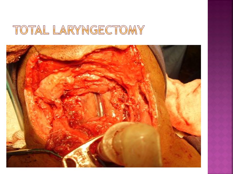
Indications: T3 or T4 unfit for partial Extensive involvement of thyroid and cricoid cartilages Invasion of neck soft tissues Tongue base involvement beyond circumvallate papillae

64
Tracheostomal prosthesis Electrolarynx Pure esophageal speech

65
Inaccurate staging Infection Voice alterations Swallowing difficulties Loss of taste and smell Fistula Tracheostomy dependence Injury to cranial nerves: VII, IX, X, XI, XII Stroke or carotid “blowout” Hypothyroidism Radiation induced fibrosis

66
5 year survival Stage I >95% Stage II 85-90% Stage III 70-80% Stage IV 50-60% After initial treatment patients are followed at 4-6 week intervals. After first year decreases to every 2 months. Third and fourth year every three months, with annual visits after that

67
Patients considered cured after being disease free for five years Most laryngeal cancers reoccur in the first two years Despite advances in detection and treatment options the five year survival has not improved much over the last thirty years

68
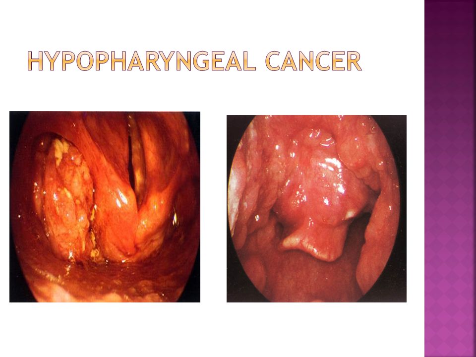
The pyriform sinus is the most common site for hypopharyngeal cancer (65-75%). Cancer may extend from here into the subglottis, thyroid cartilage, postcricoid region, or cricoarytenoid joint. Three of every four patients presenting with hypopharyngeal cancer at this subsite may have regional metastasis with apical primaries, resulting in a poorer prognosis.

71
A life-threatening infection Acute epiglottitis in the Children Acute epiglottitis in the Adult

72
Acute epiglottitis in the Children Organisms non–type B H. influenzae( in vaccinated children) Streptococcus pyogenes, S. pneumoniae S. aureus.
 Streptococcus pyogenes, S. pneumoniae S. aureus..")
73
Diagnosis history and clinical findings Lateral soft tissue radiographs “thumb sign” a dilated hypopharynx. Occasionally, supraglottic region appears hazy In severe cases, treatment should not be delayed to obtain radiographs Differentiating Diagnosis laryngotracheitis is not always easy, but it is of paramount importance

74
The signs and symptoms Signs A toxic appearance is involved, with the child assuming an upright sitting position with the chin up and mouth open, bracing themself on the hands (the "tripod" position). Patients often have difficulty in handling their secretions. Speech is limited due to pain. Stridor is a late finding and signals nearly complete airway obstruction. Symptoms Severe throat pain Fever Irritability and respiratory distress that are rapidly progressive Muffled voice

75
How is acute epiglottitis managed? arrangements for airway endoscopy in the operating room All anxiety-provoking maneuvers should be avoided. endotracheal intubation, and appropriate staff should be prepared to perform a tracheotomy. spontaneous ventilation should be maintained The intubated child should be transferred to the ICU. laryngoscopy to obtain swab cultures from the epiglottis appropriate intravenous antibiotic therapy a second- or third-generation cephalosporin cefuroxime, cefotaxime, or ceftriaxone Ampicillin/ sulbactam trimethoprim/sulfamethoxazole Chloramphenicol

76
Symptom fever, sore throat, a muffled voice, dysphagia, and odynophagia. longer than that seen in children (usually more than 24 hours) Sign swollen, bright-red epiglottis swollen epiglottis and dilated hypopharynx on a lateral neck radiograph infectious etiology Haemophilus group A streptococcus. The clinical course appears less severe Conservative measures include oxygenation, humidification, hydration, corticosteroids, and intravenous antibiotics
 Sign swollen, bright-red epiglottis swollen epiglottis and dilated hypopharynx on a lateral neck radiograph infectious etiology Haemophilus group A streptococcus. The clinical course appears less severe Conservative measures include oxygenation, humidification, hydration, corticosteroids, and intravenous antibiotics.")
78
Acute laryngotracheobronchitis (LTB), or croup Viral laryngotracheitis is the most common laryngeal inflammatory disorder of childhood. Organisms parainfluenza virus respiratory syncytial virus influenza rubeola Adenoviruses Mycoplasma pneumoniae

79
history viral upper respiratory infection with rhinitis, cough, and low-grade fever symptoms hoarseness, dyspnea, stridor, and a barking cough characteristic cough gives its common name, croup airway obstruction is caused by laryngotracheitis, the stridor is characteristically inspiratory, or biphasic.

80
diagnosis based on the history, examination of the larynx erythematous and edematous mucosa with normal vocal fold mobility(although not necessary) Radiographs, reveal a narrowing of the subglottic lumen, the “steeple sign,”
 Radiographs, reveal a narrowing of the subglottic lumen, the steeple sign,")
81
How is LTB managed? Most cases are alleviated by simple home methods, such as humidification most severe cases cause acute airway obstruction Hydration, Humidification supplemental oxygen, fluids, nebulized racemic epinephrine The use of oral and/or intramuscular glucocorticoids ( dexamethasone) Antipyretics, decongestants, Artificial airway support (eg, intubation) is necessary in a relatively small proportion of patients
 Antipyretics, decongestants, Artificial airway support (eg, intubation) is necessary in a relatively small proportion of patients.")
82
Secondary bacterial infection high temperature spikes and exudative, purulent drainage Radiographically the lumen of the upper airway will appear narrowed, shaggy, and irregular Organisms Haemophilus influenzae, Staphylococcus aureus, Streptococcus pneumoniae, Moraxella catarrhalis, hemolytic streptococci Antibiotic therapy is indicated

84
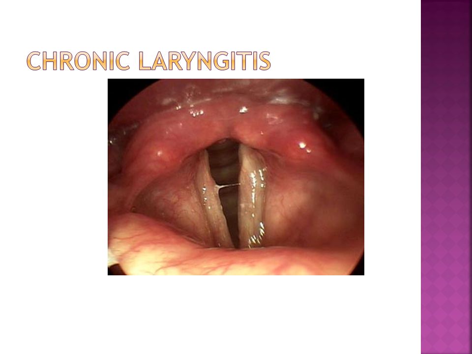
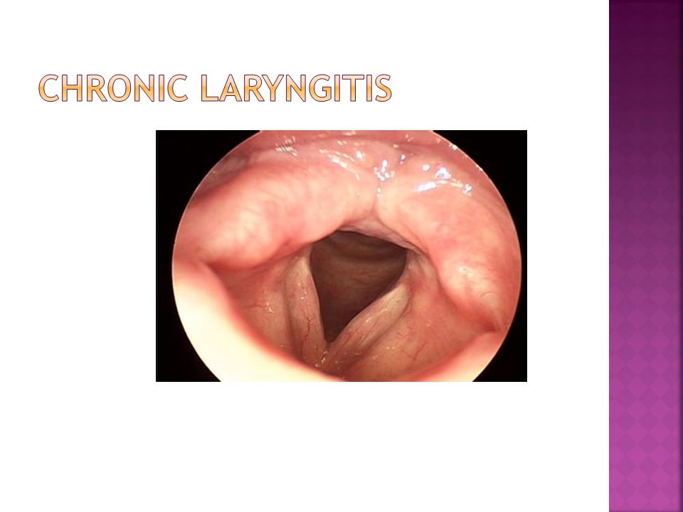
. How is chronic laryngitis treated? general inflammation of the larynx smoking, voice abuse, or laryngopharyngeal reflux symptoms chronic hoarseness, chronic cough, throat irritation, frequent throat clearing, and globus sensation. The voice usually improves if the irritating factors are discontinued. This may involve smoking cessation or voice rest. H 2 blockers and proton pump inhibitors are highly effective in treatment. In addition resting their voice, sleeping with the head of the bed elevated, and waiting 3-4 hours after eating before going to bed.

87
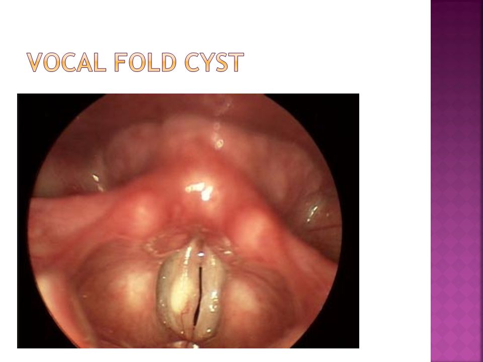
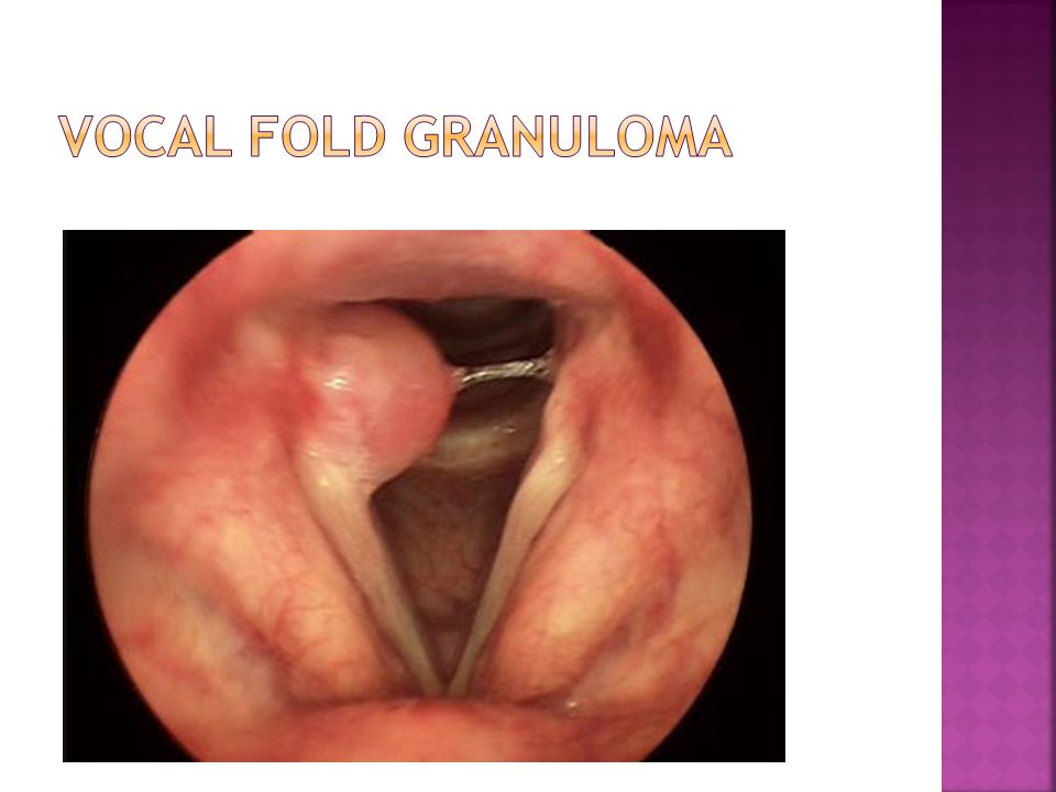
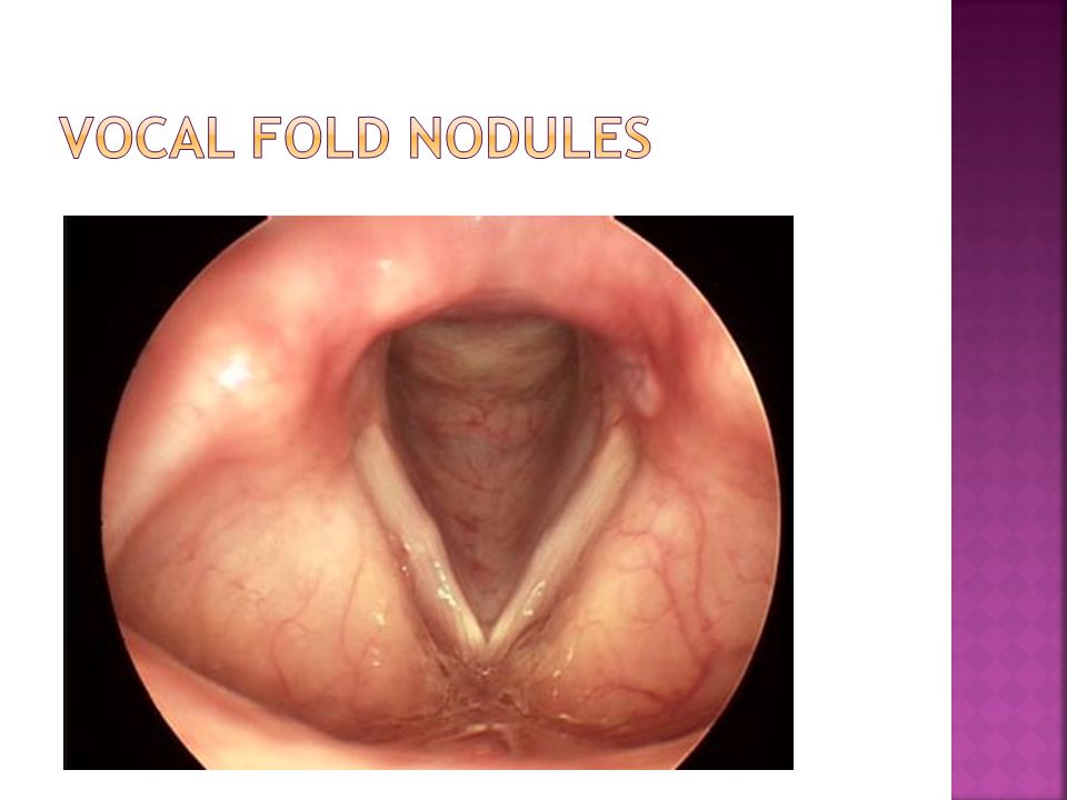
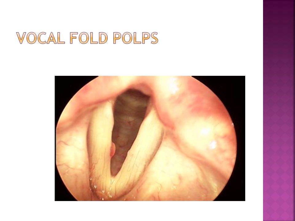
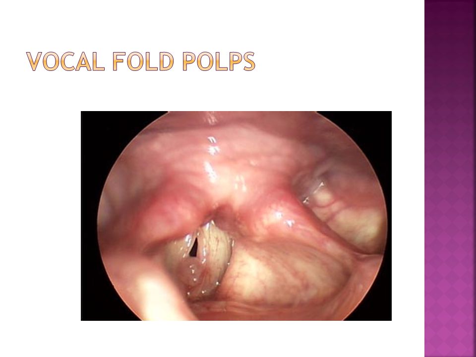
Polyps asymmetric and appear soft and smooth on one or both vocal folds vocal nodules usually paired small and discrete located in the 1/3 the distance from the anterior commissure. Contact granulomas found on the vocal processes of the arytenoid cartilage. Vocal fold cysts mucous retention or epidermoid cysts located in the superficial layer of lamina propria at the middle third of the vocal fold in the medial and superior aspect

96
What are the treatment options for vocal fold nodules? Vocal fold nodules often arise as a result of excessive laryngeal use. Voice therapy is a highly effective method of treatment. In rare cases in which voice therapy does not give satisfactory results, surgical removal of nodules may improve the voice. Generally, surgery will not resolve the hoarseness completely, and it is rarely indicated because vocal coaching is usually curative.

97
How is a laryngeal polyp treated? A laryngeal polyp is a single benign lesion of the larynx. Voice therapy is recommended before and after surgery and could be the only required treatment. Laryngeal polyps can be removed with a standard cold knife, which is preferable, or with a carbon dioxide laser. Microflap technique is used to preserve the mucosal cover and the underlying vocal ligament, when possible. Normal voice usually returns after treatment.

99
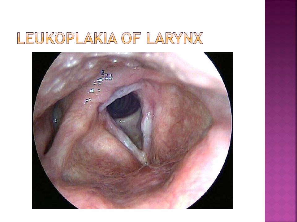
Leukoplakia (precancerous lesions) a characteristic white lesion on the vocal fold exhibit thickening of the epithelial layer abnormal keratinization of the superficial layers solitary or multifocal. benign and malignant Histologically, most of these lesions are benign, but there is thought to be an approximately 3% risk of malignancy for leukoplakia of the vocal fold

101
Laryngeal papillomatosis affects mucous membranse of the larynx characterized by multiple and recurrent squamous papillomatamay more prevalent in children and less common in individuals over 30 years of age. causing hoarseness some degree of respiratory obstruction,particularly in chidren. which is associated with human papilloma virus (HPV) types 6 and 11.
 types 6 and 11..")
102
Papillomatosis in children Papillomatosis in Adult

103
How is laryngeal papillomatosis transmitted? Transmission is multifactorial. Fifty percent of mothers have a history of active or prior HPV infection. The risk of transmission is 1 in 400. Cesarean section is not recommended for mothers with either active or latent infection because transmission has occurred despite cesarean section.

104
gross inspection appear in a multinodular pattern sessile or exophytic. Histologically papillary projections and hypervascular fibroconnective tissus covered by hyperplastic squamous epithelium that shows maturation. Cellular atypia is the rule rather than the exception Histologic differentiation from early carcinoma may sometimes be difficult.

105
What triad is associated with laryngeal papillomatosis? Firstborn child: primigravid mothers are more likely to have a prolonged second stage of labor, which increases the risk for infection Teenage mother Vaginal delivery

106
How are laryngeal papillomas treated? spontaneous remissions can occur Multiple surgical resections, often with a laser, are required. cidofovir (intraoperative injections), indole 3- carbinol/diindolylmethane, acyclovir, and interferon-α are under investigation.
, indole 3- carbinol/diindolylmethane, acyclovir, and interferon-α are under investigation..")
107
Malignant Tumors of the Larynx and Hypopharynx. Cummings- Otolaryngology- Head and Neck Surgery. 4th ed., Mosby, 2005. Malignant Laryngeal Lesions. Lawani- Current Diagnosis and Treatment in Otolaryngology- Head and Neck Surgery. McGraw-Hill and Lange, 2004. Neck. Moore- Essential Clinical Anatomy. 2nd ed., Lippincott, 2002. Head and Neck. Rohen- Color Atlas of Anatomy. 5th ed., Lippincott, 2002. Surgery for Supraglottic Cancer. Myers- Operative Otolaryngology Head and Neck Surgery Vol. 1. 1st ed., Saunders, 1997. Surgery for Glottic Carcinoma. Myers- Operative Otolaryngology Head and Neck Surgery Vol. 1. 1st ed., Saunders, 1997. The Larynx. Lore and Medina- An Atlas of Head and Neck Surgery. 4th ed., Elsevier, 2005. Hinerman, R, Morris, C, et al. Surgery and Postoperative Radiotherapy for Squamous Cell Carcinoma of the Larynx and Pharynx. Am J Clin Oncol. 2006; 29(6): 613-621. Huang, D, Johnson, C, et al. Postoperative Radiotherapy in Head and Neck Carcinoma with Extracapsular Lymph Node extension and/or Positive Resection Margins: a Comparative Study. Int J Radiat Oncol Biol Phy. 1992; 23:737-742. Bernier, J, Domenge, C, et al. Postoperative Irradiation with or without Concomitant Chemotherapy for Locally Advanced Head and Neck Cancer. N Engl J Med. 2004; 350: 1945-1952. Sessions, D, Lenox, J, et al. Supraglottic Laryngeal Cancer: Analysis of Treatment Results. Laryngoscope. 2005; 115: 1402-1410. Wolf, GT. The Department of Veterans Affairs Laryngeal Cancer Study Group. Induction Chemotherapy Plus Radiation Compared with Surgery Plus Radiation in Patients with Advanced Laryngeal Cancer. New England Journal of Medicine. 1991; 324: 1685-90. Lefebre J, Chevalier D, Luboinski B, Kirkpatrick A, Collette L, Sahmoud T. Larynx Preservation in Pyriform Sinus Cancer: Preliminary Results of a European Organization for Research and Treatment of Cancer Phase III Trial. Journal of the National Cancer Institute. Jul 1996. 88(13): 890-899. Grant’s Atlas 10 th ed. CD-ROM
: Huang, D, Johnson, C, et al. Postoperative Radiotherapy in Head and Neck Carcinoma with Extracapsular Lymph Node extension and/or Positive Resection Margins: a Comparative Study. Int J Radiat Oncol Biol Phy. 1992; 23: Bernier, J, Domenge, C, et al. Postoperative Irradiation with or without Concomitant Chemotherapy for Locally Advanced Head and Neck Cancer. N Engl J Med. 2004; 350: Sessions, D, Lenox, J, et al. Supraglottic Laryngeal Cancer: Analysis of Treatment Results. Laryngoscope. 2005; 115: Wolf, GT. The Department of Veterans Affairs Laryngeal Cancer Study Group. Induction Chemotherapy Plus Radiation Compared with Surgery Plus Radiation in Patients with Advanced Laryngeal Cancer. New England Journal of Medicine. 1991; 324: Lefebre J, Chevalier D, Luboinski B, Kirkpatrick A, Collette L, Sahmoud T. Larynx Preservation in Pyriform Sinus Cancer: Preliminary Results of a European Organization for Research and Treatment of Cancer Phase III Trial. Journal of the National Cancer Institute. Jul (13): Grant’s Atlas 10 th ed. CD-ROM.")
108
Laryngeal carcinoma Etiology: tobacco and excessive alcohol use primary Human Papilloma Virus 16 &18 Chronic Gastric Reflux Occupational exposures Presentation Hoarseness, Dysphagia,Hemoptysis,Throat pain, Ear pain, Airway compromise, Aspiration,Neck mass appear as friable, fungating, ulcerative masses or be as subtle as changes in mucosal color

109
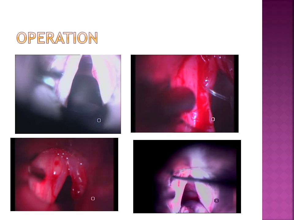
Laryngeal carcinoma Diagnosis History Endoscopy Biopsy Treatment Operation Radiotherapy Chemotherapy

110
Distinguish characteristic between laryngotracheitis and supraglottitis Distinguish characteristic between nodular and polyps Leukoplakia are the precancerous lesions What triad is associated with laryngeal papillomatosis?

Similar presentations


>")


>")

