Download presentation
Presentation is loading. Please wait.

1
THE HEART

2
THE HEART Normal Pathology Heart Failure: L, R Heart Disease
Congenital: LR shunts, RL shunts, Obstructive Ischemic: Angina, Infarction, Chronic Ischemia, Sudden Death Hypertensive: Left sided, Right sided Valvular: AS, MVP, Rheumatic, Infective, Non-Infective, Carcinoid, Artificial Valves Cardiomyopathy: Dilated, Hypertrophic, Restrictive, Myocarditis, Other Pericardium: Effusions, Pericarditis Tumors: Primary, Effects of Other Primaries Transplants This is the chapter outline, fairly logical

3
NORMAL Features 6000 L/day Normal weight – females; grams and Males; gms LV= 1.3 to 1.5 cm RV= 0.30 to0.5 cm Hypertrophy-Increase in the size or thickness Dilation-Enlarged chamber Cardiomegaly-Increase in weight or size(hypertrophy or dilation)
")
4
Whichever artery winds up supplying the posterior interventricular septum is said to be “DOMINANT”.
A thrombosis of WHICH coronary artery would usually result in sudden death? Ans: MAIN left coronary artery.

7
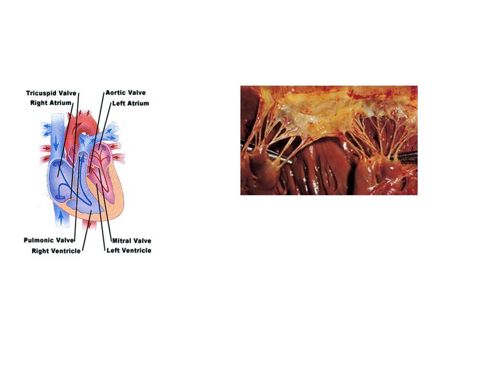
Epicardial Coronary Arteries Chambers
CARDIAC AGING Atherosclerotic plaque Calcific deposits Increased cross-sectional luminal area Tortuosity Epicardial Coronary Arteries Sigmoid-shaped ventricular septum Decreased left ventricular cavity size Increased left atrial cavity size Chambers Amyloid deposits Lipofuscin deposition Brown atrophy Increased subepicardial fat Increased mass Myocardium Buckling of mitral leaflets toward the left atrium Fibrous thickening of leaflets Mitral valve annular calcific deposits Aortic valve calcific deposits Valves These features are seen so commonly in autopsies of elderly people no matter what they died from. Also keep in mind that most people who do not die ACUTELY, die in cardiac failure.

8
CARDIAC AGING Aorta Dilated ascending aorta with rightward shift
Atherosclerotic plaque Elastic fragmentation and collagen accumulation Sinotubular junction calcific deposits Elongated (tortuous) thoracic aorta Dilated ascending aorta with rightward shift Aorta One very key philosophical question is whether atherosclerosis is part of aging or not. We can leave that for the philosophers.
 thoracic aorta. Dilated ascending aorta with rightward shift. Aorta. One very key philosophical question is whether atherosclerosis is part of aging or not. We can leave that for the philosophers.")
9
BROWN LIPOFUCSIN ATROPHY, HEART
The pigment which accumulates with age is called lipofucsin, and caused the heart to appear “browner” than normal. This is called “brown” atrophy of the heart. Lipofucsin is another typical example of a golden brown, slightly refractile, INtrinsic pigment, which looks like hemosiderin, melanin, or bile, but accumulates, as a rule, on opposite poles of the myocyte nucleus. It is also called, appropriately, AGING pigmernt. LIPOFUCSIN

10
Pathology of the Heart All problems are eventually expressed as inadequate cardiac output. Leaks in the system Electrical Conduction, irregular rhythm Obstruction to flow Valve problems, obstruction or incompetence Cardiac muscle weakness and failure

11
HEART FAILURE Heart Failure Definition--The heart is unable to pump blood at a rate that meets the requirements of the metabolizing tissues, or can only do so only with filling pressures that are higher than normal. Onset may be insidious or acute.

12
Congestive Heart Failure
Diminished pumping ability of left ventricle. Back up of blood in pulmonary vasculature. Pulmonary edema Peripheral edema

13
5 million people/year in USA are affected with HF.
300,000deaths /year due to HF and 1 million hospitalization for HF/year. More common after the age of 65.

14
TYPES Left heart failure Right heart failure Biventricular failure
High output failure (least common HF)
")
15
Back Pressure

16
Develops slowly and insidiously Backward failure,
Acute Heart failure Rapidly failing heart .Forward failure Reduced blood flow to the tissues,-reduced renal flow-salt and water retention to increase the blood volume-and venous pressure---edema Chronic heart failure Develops slowly and insidiously Backward failure, Blood backs up to veins –increase venous pressure and –congestion, edema.

17
Right heart failure (Most of the time as consequence of left ventricle failure) The inefficient pumping of the right side of the heart causing fluid build up in lung, legs and abdomen. Left heart failure Inability of the left ventricle to pump enough blood, causing fluid to back up in to the lungs,

18
A 29-year-old woman complains of a 3-month history of nervousness and weakness. She feels hot and sweaty and has experienced a 9-kg (20-lb) weight loss over the past 2 months, despite increased caloric intake. She frequently finds her heart racing and can feel it pounding in her chest. Physical examination reveals an enlarged thyroid, warm hands, and bulging eyes.
 weight loss over the past 2 months, despite increased caloric intake. She frequently finds her heart racing and can feel it pounding in her chest. Physical examination reveals an enlarged thyroid, warm hands, and bulging eyes..")
19
High-output failure heart failure results from greatly increased tissue demands for blood Thyrotoxicosis, Anemia Beri Beri A.V fistula, Paget disease of the bone. pregnancy

20
Low out put failure. IHD, Systemic hypertension A.R and M.R. A.S.
Hypertrophic cardiomyopathy Infiltrative disorders like Amyloidosis, Anti arrhythmic drugs.

21
Compensated heart failure
If the dilated ventricle is able to maintain cardiac output at a level that meets the needs of the body, is called as compensated failure. Decompensate heart failure The failing myocardium is no longer able to propel sufficient blood to meet the needs of the body, even at rest.

22
Systolic failure occurs due to inability of the heart to provide adequate output. IHD, Aortic stenosis disease in the myocardium Ejection fraction falls from 65% to as low as 20%. cardiac remodeling.

23
Diastolic failure The left ventricle is abnormally stiff. Hypertrophic cardiomyopathy, Amyloidosis Ejection fraction remains near normal Occurs over the age of 65. More in women Hypertension is most common cause, Diabetes mellitus Obesity Bilateral renal artery stenosis Also in elderly person of unknown (aging)
")
24
1.The frank starlings mechanism
Physiological mechanism try to compensate heart failure 1.The frank starlings mechanism Increased filling volume dilate the ventricle Increases the contractility 2.Ventricular remodeling with or with out dilation The structural molecular and cellular changes occur in ventricles due to overload is called ventricular remodeling.

25
3.Activation of neurohumoral system
The release of nor epinephrine increases the heart rate and increases the contractility Activation of renin and angiotesnogen aldosterone mechanism Release of atrial natriuritic peptides With all these the heart will be in a state of compensatory failure

26
DECOMPENSATE FAILURE Decompensate failure due to worsening abnormalities state is when heart is unable to bear Myocardial structural changes, including augmented muscle mass (hypertrophy)
")
27
A good general diagram.

28
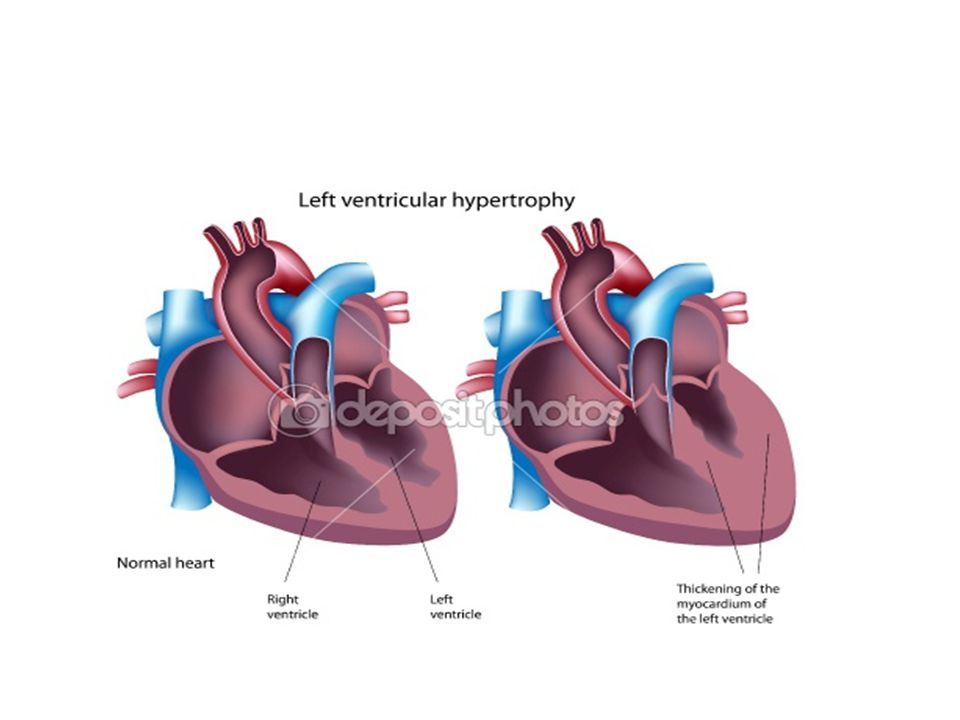
Concentric hypertrophy, In pressure overload states the hypertrophy is characterized by increased diameter of individual muscle fibers. The thickness increases may or may not the size of the ventricle. In hypertension, Aortic valve stenosis,

31
Eccentric hypertrophy
characterized by an increase in heart size as well as an increase in wall thickness. In volume overload states valvular regurgitation or abnormal shunts.

32
Heart is growing outward ,
Shifting the apical impulse displacment. S3 murmur (due to rapid ventricular filling)
")
33
cytoskeletal alterations,
structural and functional disturbances; such degenerative changes include Myocyte apoptosis, cytoskeletal alterations, and altered extracellular matrix synthesis and remodeling. Even hypertrophy comes at a significant.

34
Oxygen requirements of the hypertrophic myocardium are increases as a result of increased myocardial cell mass and increased tension of the ventricular wall. The myocardium becomes vulnerable to ischemic injury.

35
LEFT VENTRICULAR FAILURE
LVF is divided on clinical grounds into Systolic dysfunction Diastolic dysfunction There is stiffening of the left ventricle but output is maintained at rest. but during exercise there is increase in filling pressure leads to backflow of pressure into pulmonary circulation causing Pulmonary edema.

36
The most common causes of left-sided cardiac failure are
(1) IHD, (2) systemic hypertension, (3) mitral or aortic valve disease, (4) primary diseases of the myocardium. (e.g., amyloidosis).
 IHD, (2) systemic hypertension, (3) mitral or aortic valve disease, (4) primary diseases of the myocardium. (e.g., amyloidosis).")
37
SYSTOLIC DYSFUNCTION IHD, due to atherosclerosis
Aortic or mitral valvular disease Hypertension, Dilated cardio myopathy Post myocardial infarction Myocarditis. Low EF<40%

38
DIASTOLIC DYSFUNTION It is commonly seen after 65yers and more common in women. Constrictive pericardiits Amyloidosis Restrictive cardiomyopathy Myocardial fibrosis Hypertropic cardiomyopathy And essential hypertension Also in elderly persons due to age stiffening common. EF normal

39
Left ventricle cannot efficiently eject blood in to the aorta due to
increase in LVEDV and LVEDP (Increase in the hydrostatic pressure). leads to back up of of blood into the lungs PULMONARY EDEMA
. leads to back up of of blood into the lungs PULMONARY EDEMA.")
40
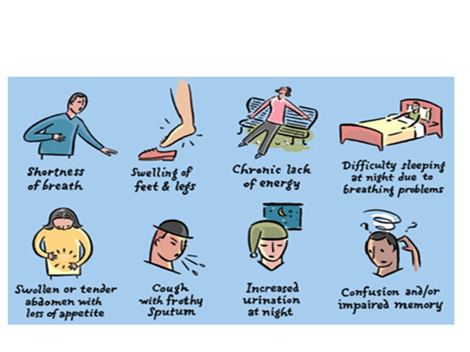
Martha Wilmington, a 74-year-old woman with a history of rheumatic fever while in her twenties, presented to her physician with complaints of increasing shortness of breath ("dyspnea") upon exertion. She also noted that the typical swelling she's had in her ankles for years has started to get worse over the past two months, making it especially difficult to get her shoes on toward the end of the day. In the past week, she's had a decreased appetite, some nausea and vomiting, and tenderness in the right upper quadrant of the abdomen.

41
physical examination jugular veins were noticeably distended. Auscultation of the heart revealed a low-pitched, rumbling systolic murmur, heard best over the left upper sternal border. "S3" heart soundpresent.

42
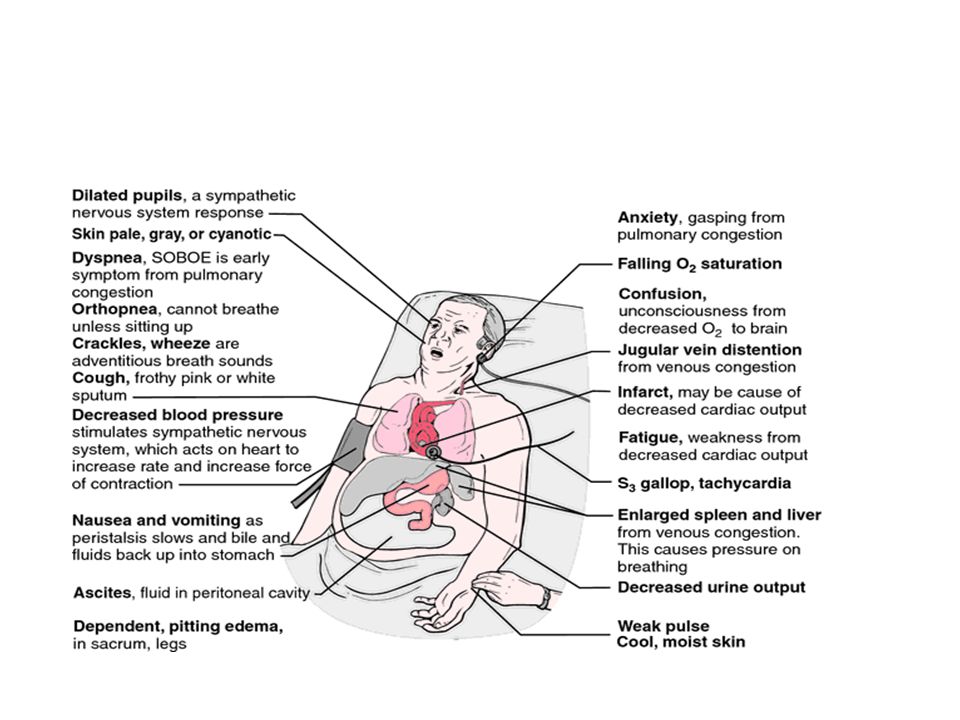
LEFT Heart Failure Dyspnea Orthopnea
PND (Paroxysmal Nocturnal Dyspnea) Blood tinged sputum Cyanosis Can you understand why all of these findings can be related to LEFT sided heart failure? Ans: YES, primarily PULMONARY.
 Blood tinged sputum. Cyanosis. Can you understand why all of these findings can be related to LEFT sided heart failure Ans: YES, primarily PULMONARY.")
43
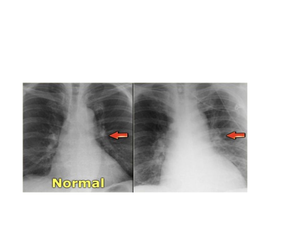
X-ray findings, Appearance of Kerely B lines Straightening of the left border of the heart, Globular heart

45
Pulmonary Edema

47
Mitral regurgitation With progressive ventricular dilation, the papillary muscles are displaced laterally, causing mitral regurgitation and a systolic murmur.

48
Atrial fibrillation Clinical findings
Chronic dilation of the left atrium Clinical findings "irregularly irregular" heartbeat.

49
can reduce stroke volume or lead to blood stasis and thrombus formation a fibrillating left atrium carries a substantially increased risk of embolic stroke

50
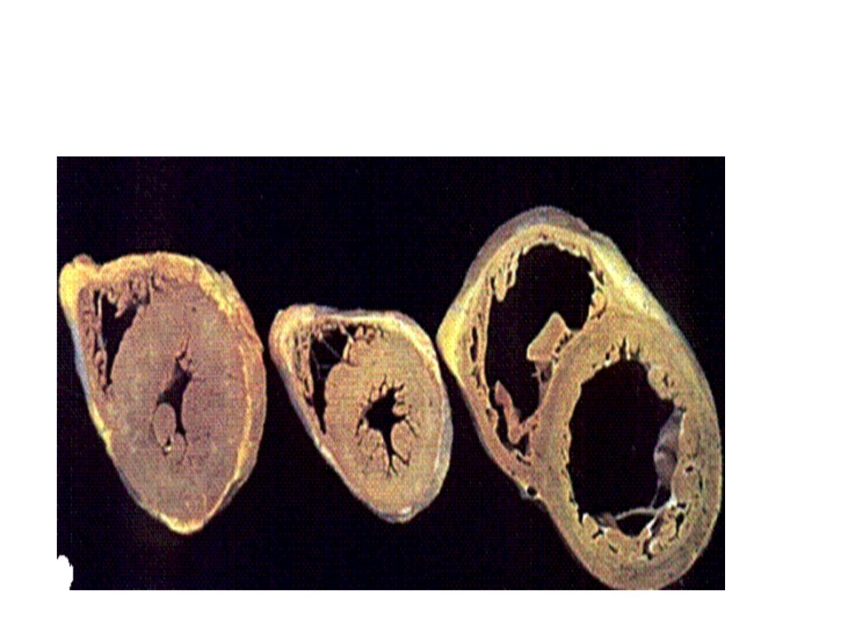
Morphology of LVF The left ventricle is usually hypertrophied and often dilated, sometimes quite massively. There are usually nonspecific changes of hypertrophy and fibrosis in the myocardium. Secondary enlargement of the left atrium with resultant atrial fibrillation. Heart failure cells.

51
LUNGS Rising pressure in the pulmonary veins is ultimately transmitted retrogradely to the capillaries, resulting in pulmonary congestion and edema. The lungs are heavy and boggy, and histologically there are perivascular and interstitial transudate, alveolar septal edema, and intra-alveolar edema .

52
Kidney- Pre renal Azotemia
Kidney- Pre renal Azotemia. Accumulation of nitrogenous waste due to acute tubular necrosis, due to decrease in renal perfusion. Brain-Hypoxic encephalopathy-Restless, irritability and stupor and coma at the end stage of CCF.

53
RHF CAUSES The most common cause of right-sided heart failure is
1. Left ventricular failure, 2.Diseases of the lung parenchyma and/or pulmonary vasculature (cor pulmonale) 3.Primary pulmonic or tricuspid valve disease.
 3.Primary pulmonic or tricuspid valve disease.")
54
Right-Sided Heart Failure
Liver and Portal System. The liver is usually increased in size and weight (congestive hepatomegaly), nutmeg liver congested red centers of the liver lobules are surrounded by paler, sometimes fatty, peripheral regions. the central areas can become fibrotic, creating so-called cardiac cirrhosis.
, nutmeg liver congested red centers of the liver lobules are surrounded by paler, sometimes fatty, peripheral regions. the central areas can become fibrotic, creating so-called cardiac cirrhosis.")
55
Pleural and Pericardial Spaces
Fluid may accumulate in the pleural space (particularly right) and pericardial space (effusions). Pleural effusions (typically serous) can range from 100 mL to well over 1 L and can cause partial atelectasis of the affected lung.
 and pericardial space (effusions). Pleural effusions (typically serous) can range from 100 mL to well over 1 L and can cause partial atelectasis of the affected lung.")
56
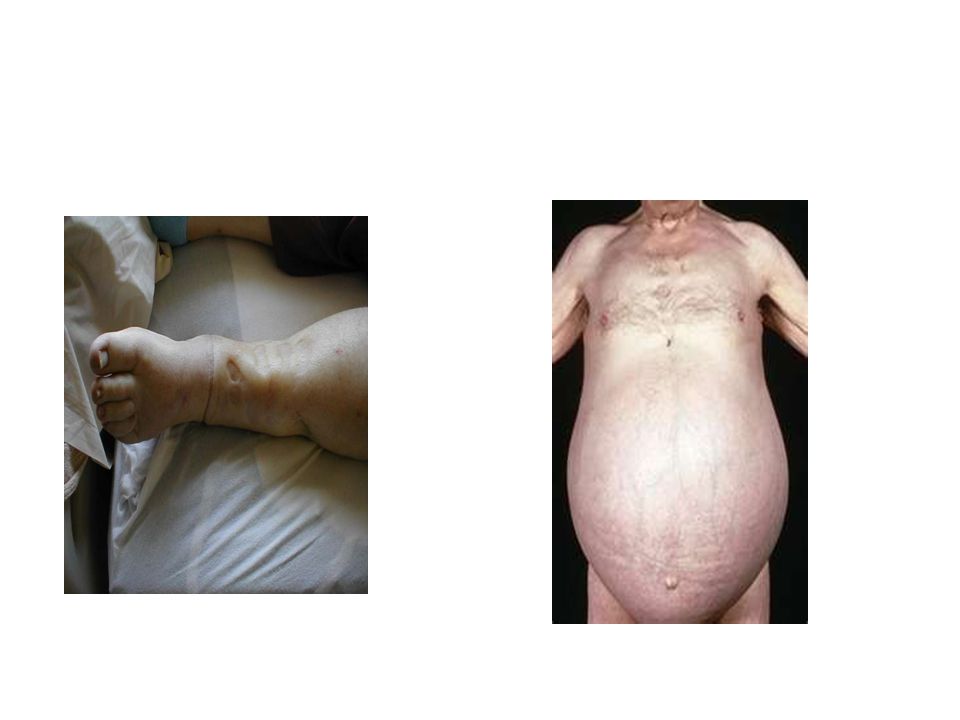
Subcutaneous Tissues. Peripheral edema of dependent portions of the body, especially ankle (pedal) and pretibial edema, is a hallmark of right-sided heart failure. In chronically bedridden patients, the edema may be primarily pre sacral. Generalized massive edema is called anasarca.

57
Pleural effusion pericardial effusion

59
RIGHT Heart Failure FATIGUE “Dependent” edema JVP
Hepatomegaly (congestion) ASCITES, PLEURAL EFFUSION GI Cyanosis Increased peripheral venous pressure (CVP) (nl = 2-6 mm Hg) Can you understand why all of these findings can be related to RIGHT sided heart failure? Ans: YES, primarily STSTEMIC.
 ASCITES, PLEURAL EFFUSION. GI. Cyanosis. Increased peripheral venous pressure (CVP) (nl = 2-6 mm Hg) Can you understand why all of these findings can be related to RIGHT sided heart failure Ans: YES, primarily STSTEMIC.")
60
Pitting Edema

61
Liver Chronic Passive Congestion

62
Note that not only is the FIBER thick, but so are the nuclei
Note that not only is the FIBER thick, but so are the nuclei. Note squaring off of the nuclei, so called “BOXCAR” effect.

Similar presentations


 Occurs when the right ventricle fails as an effective forward pump, causing back-pressure of blood into the systemic.>")


