Download presentation
Presentation is loading. Please wait.

1
Thrombosis and embolism
By Dr S Homathy

2
Thrombosis Thrombosis is the formation of a solid mass (blood clot) from the constituents of blood Platelets Fibrin Entrapped red cells and white cells Within the heart or vascular system in a living organism

3
The development of a clot is life-saving when a large vessel ruptures or is severed.
However, when a thrombus develops within the vascular system, it may be life-threatening

4
Thrombosis is the consequence of inappropriate activation ( pathological) of the processes of normal haemostasis
 of the processes of normal haemostasis")
5
Normal Haemostasis Maintain blood in a fluid, clot-free state in normal vessels Also inducing the rapid formation of a localized haemostatic plug at the site of injury. Both are influenced by components of the blood vessel wall, platelets the clotting sequence.

6
The integrity of the blood vessel wall is crucial in normal haemostasis as well as in thrombosis.

7
Normal Haemostasis Vessel injury –
brief period of arteriolar vasoconstriction (neurogenic reflex ,endothelin) Endothelial injury exposes ECM (highly thrombogenic material). Platelets adhere to endothelial cells and ECM, and are activated.
 Endothelial injury exposes ECM (highly thrombogenic material). Platelets adhere to endothelial cells and ECM, and are activated.")
8
They release their secretary granules.
Platelet aggregation occurs forming haemostatic plug (Primary haemostasis) Tissue factor (produced by endothelium) activates coagulation – formation of thrombin which act on finbrinogen to form fibrin (secondary Haemostsis)
 Tissue factor (produced by endothelium) activates coagulation – formation of thrombin which act on finbrinogen to form fibrin (secondary Haemostsis)")
9
The process continues to form the permanent plug formed by polymerized fibrin and platelet aggregates. At the same time tissue plasminogen activator (t-PA) is formed and it limits haemostatic plug. Fibrinolysis is also activated to limit haemostatic plug to the site of injury
 is formed and it limits haemostatic plug. Fibrinolysis is also activated to limit haemostatic plug to the site of injury.")
10
Normal Endothelium Endothelial cells are activated by injury, infection, plasma mediators and cytokines. They have pro-thrombotic and anti-thrombotic functions

11
The endothelial cells serve to protect against thrombi formation by
Endothelium The endothelial cells serve to protect against thrombi formation by Anti-thrombotic properties: Anti-platelet effect: Non activated platelets do not adhere to endothelium. PGI2, and NO (produced by endothelium) prevent platelet adhesion Anticoagulant properties: Heparin-like molecule activate anti-thrombin III Thrombomodulin binds thrombin which activate protein C (anticoagulant) Fibrinolytic properties: Endothelium synthesize t-PA (fibrinolysis
 prevent platelet adhesion. Anticoagulant properties: Heparin-like molecule activate anti-thrombin III. Thrombomodulin binds thrombin which activate protein C (anticoagulant) Fibrinolytic properties: Endothelium synthesize t-PA (fibrinolysis.")
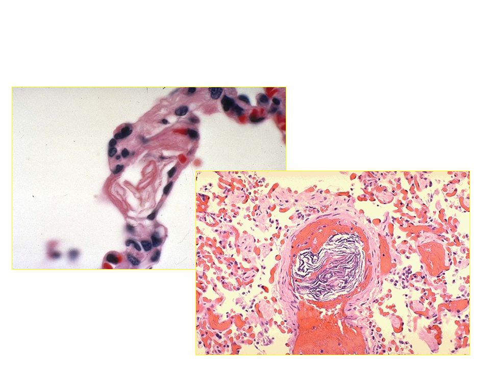
12
Endothelial cells also have the procoagulant properties
Pro-thrombotic properties: Von Willebrand factor: It enhances binding of platelets to ECM. 2.Tissue factor Produced by endothelium, it activates extrinsic clotting pathway Plasminogen activator inhibitors (PAI)
")
13
Platelets Platelets are assigned a central role in normal haemostasis and thrombosis. They adhere to sites of endothelial injury, aggregate to form platelet masses,

14
release granules rich in a variety of secretary products and synthesize several types of prostaglandins. In normal haemostasis, platelets adhere to the severed margins of a vessel within seconds to a few minutes. The most important stimulus to such adherence is the exposure of collagen fibrils

15
Once adhered, platelets release two types of granules:
(1) alpha granules which contain fibrinogen, beta thromboglobulin, cationic protein platelet factor 4 (a heparin neutralizing protein) (2) dense bodies, which are rich in serotonin, ADP, ATP ionized calcium
 alpha granules which contain. fibrinogen, beta thromboglobulin, cationic protein. platelet factor 4 (a heparin neutralizing protein) (2) dense bodies, which are rich in. serotonin, ADP, ATP. ionized calcium.")
16
Initially, the platelet aggregation forms a temporary haemostatic plug
which is friable and easily dislocated in rapidly flowing bloodstreams at this time, the clotting sequence leads to the formation of thrombin which is the most powerful platelet aggregator yet identified

17
Platelets : (1) provide a temporary plug capable of controlling blood flow in small vessels in low pressure systems, (2) initiate the development of a permanent plug composed of aggregated platelets and fibrin, (3) release serotonin which augments vasoconstriction and (4) contributes to the coagulation mechanism.
 initiate the development of a permanent plug composed of aggregated platelets and fibrin, (3) release serotonin which augments vasoconstriction and. (4) contributes to the coagulation mechanism.")
18
Coagulation system The coagulation system plays a major role in normal haemostasis. Maintenance of normal fluidity of blood involves the interplay between procoagulants and anticoagulants. When the procoagulants dominate and clotting is triggered inappropriately in the intact cardiovascular system, thrombi result.

19
The critical events in blood clotting are the conversion of prothrombin to thrombin
the subsequent conversion of soluble fibrinogen into the stable fibrin polymer

20
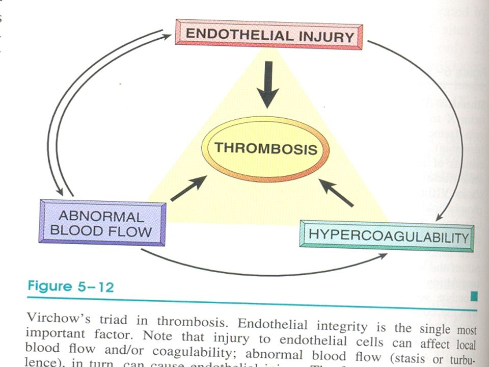
Thrombosis Thrombosis is influenced by three major factors:
VIRCHOW’S TRIAD (1) injury to vascular endothelium, (2) alterations in normal blood flow and (3) alterations in the blood (hypercoagulability).
 injury to vascular endothelium, (2) alterations in normal blood flow and. (3) alterations in the blood (hypercoagulability).")
22
Endothelial injury Endothelial injury plays a dominant role in the formation of thrombi in arteries and in the heart. Once the endothelium is damaged, subendothelial collagen may be exposed and tissue thromboplastin, etc., is released and the sequence of platelet adherence and activation of the clotting sequence follows

23
Endothelial injury occurs in myocardial infarction,
ulcerated atherosclerosis, trauma, and inflammatory disease of vessels. Endothelial dysfunction is also a predisposing factor for thrombosis. Eg: Hypertension, bacterial endotoxins, hypercholestrolemia, radiation, cigarette smoking.

24
Blood Stasis and Turbulence of Flow
Turbulence enhances endothelial injury. Stasis enhances venous thrombosis. Both result in: Bringing platelets close to endothelium Accumulation of clotting factors Prevent clotting factors inhibitors Endothelial activation Eg: aortic aneurysm, MI, valve stenosis, rheumatic heart disease, hyperviscosity, sickle cell disease.

25
Stasis and turbulence Distrupt laminar flow Prevent dilution of activated clotting factors by fresh flowing blood Retard the inflow of clotting factor inhibitors and permits build – up of thrombi Promote endothelial cell activation

26
Hypercoagulability Primary: (genetic)
It is an alteration in coagulation leading to thrombosis. Primary: (genetic) Factor V mutation Prothrombin mutation Antithrombin III deficiency Protein C or S deficiency
 Factor V mutation. Prothrombin mutation. Antithrombin III deficiency. Protein C or S deficiency.")
27
Secondary:( acquired )
High risk for thrombosis Prolonged immobilization Myocardial infarction Tissue damage Cancer Prosthetic cardiac valves DIC Lupus anticoagulant Low risk for thrombosis AF Cardiomyopathy Sickle cell anaemia Nephrotic syndrome Contraceptive pills Smoking

28
Increased numbers of platelets, increased platelet stickiness,
elevated levels of fibrinogen, increased generation of thrombin, etc., have been identified as causing hypercoagulability in various clinical conditions.

29
Special categories among acquired causes
1.Heparin-induced Thrombocytopenia: ( HIT syndrom) When heparin is administered it induces the formation of antibodies that bind platelets and activate them. Occurs when unfractionated heparin is given. Solution – give low-molecular Wt heparin Have anticoagulant activity Do not interact with platelets Prolonged serum half life
 When heparin is administered it induces the formation of antibodies that bind platelets and activate them. Occurs when unfractionated heparin is given. Solution – give low-molecular Wt heparin. Have anticoagulant activity. Do not interact with platelets. Prolonged serum half life.")
30
2.Antiphospholipid syndrome (Lupus anticoagulant):
Antibodies to phospholipid (eg. Cardiolipin) In-vitro: it inhibits coagulation( inhibit assembly of phospholipid cpx) In-vivo: it induces coagulation Approximately 20% of patients with a recent sroke were found to have anticardiolipin antibody
 In-vitro: it inhibits coagulation( inhibit assembly of phospholipid cpx) In-vivo: it induces coagulation. Approximately 20% of patients with a recent sroke were found to have anticardiolipin antibody.")
31
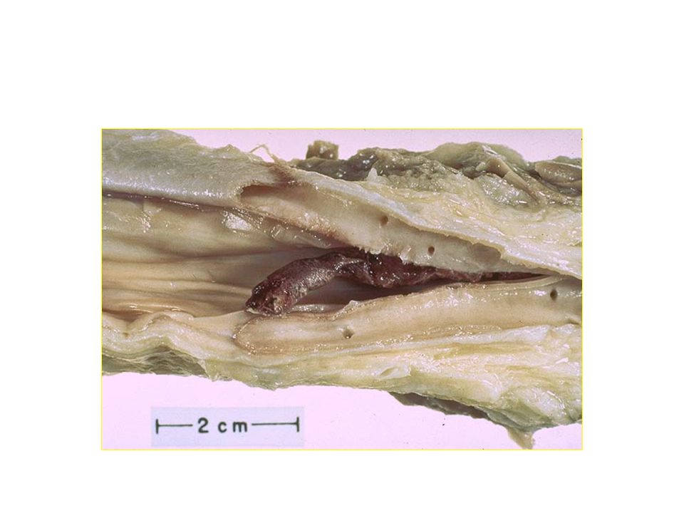
Morphology Thrombi may develop in the heart, arteries, veins and capillaries. Arterial thrombi and cardiac thrombi occur at site of endothelial injury or turbulence of flow. Venous thrombi occur in areas of blood stasis. Thrombi usually are attached to the underlying vessel wall (mural thrombi)
")
32
Arterial thrombi grow back(retrograde direction) to the heart.
Venous thrombi grow toward the heart.

33
Arterial and cardiac thrombi are firmly attached to the wall
Grossly and microscopically show lines of Zahn (layers of fibrin and platelets alternate with layers of RBC and WBC. Implies thrombosis at a site of blood flow Venous thrombi do no show clear lamination. Resemble coagulated blood( like clotted in test tube)
")
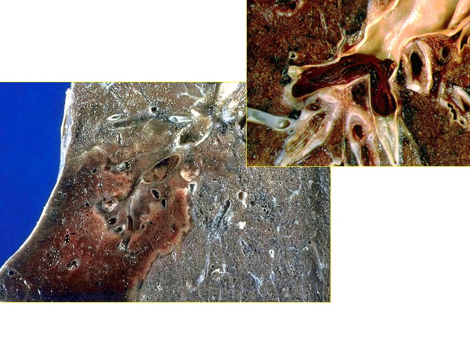
34
Microscopic appearance of thrombi

35
In the heart: common causes: Attached to the underlying structure MI,
Mural thrombi common causes: MI, dilated cardiomyopathy, arrhythmia, myocarditis, valvular disease.

36
Arterial thrombi usually occlude the lumen, common in
In arteries: common causes: atherosclerosis, aneurysm. Arterial thrombi usually occlude the lumen, common in coronary, cerebral femoral arteries.

37
Deep Vein thrombosis (phlebothrombosis)
are almost always occlusive, Red / stasis thrombi, 90% occur in lower extremities. Resemble postmortem clots Firmer , almost always have a point of attachment Transection reveal vague strands of pale gray fibrin

38
Under special circumstances thrombi may form on heart vales.
Bacterial and fungal blood-born infection may lead to valve damage Development of large thrombotic masses/ vegetations ( infective endocarditis)
")
39
Sterile vegetations can also develop on noninfected valves(NBTE)
Hypercoagulable states Libman-sacks endocarditis Occurs in SLE

40
Classification of Thrombus according to
Color Pale, formed of platelets and fibrin, small, grayish white, firm and adherent Red, formed of red cells and fibrin, dark soft and loosely attached to the vessel Mixed, common and has pale and red components Presence or absence of bacteria Infected or septic Non infected or aseptic

41
Sites of Thrombus Formation
1.Thrombus in veins: More common because of thin wall and slow blood flow: Thrombophlebitis ----Septic Phlebothrombosis---- occurs in the veins of the calf Ms and femoral ,iliac veins pulmonary emboli In the varicose veins

42
2.Thrombosis in Arteries
less common than veins because of rapid flow and thick elastic wall but occur in arteries affected by: Atheroma, polyarteritis nodosa and thromboangitis obliterans (roughness of the intima) Aneurysm due to stasis Lead to ischaemia
 Aneurysm due to stasis. Lead to ischaemia.")
43
3.Thrombosis in the heart
more common in the the left side Mural---- occur over infarction Vegetations---- pale over the valve Auricular--- adherent to valve, if detach called ball thrombus Agonal--- red thrombi occurring in Rt. V at the time of death specially lobar pneumonia Arterial and cardiac thrombosis possibly embolise to brain, kidneys, spleen

44
Coronary artery thrombosis

45
4.Thrombosis in capillaries (very rare):
occur in acute inflammation ,sever cold and frost bite

47
Fate of thrombus

48
its element disintegrate and form a pale red structure less mass
1-septic thrombus fragmented by the proteolytic enzymes into septic emboli causes pyaemic abscesses 2-Aseptic thrombus its element disintegrate and form a pale red structure less mass If mass is small it dissolves by 1).fibrinolysis(dissolution) If mass is large it undergoes:
.fibrinolysis(dissolution) If mass is large it undergoes:")
49
2.Propagation (progression)
3.Embolization 4.Organization and recanalization (inflammation and fibrosis)
")
50
If mass is large it undergoes
Organization: the thrombus is invaded by capillaries and fibroblast change to fibrous mass lead to permanent vascular occlusion Organization and Canalization; some time capillaries dilated and allow Passage of blood through the thrombus;

51
Dystrophic calcification
Incorporation : the fibrosed thrombus shrinks from the vascular wall leaving a space which gets lined by endothelium Dystrophic calcification phlebolith Detachment aseptic emboli--- infarctions Propagating thrombus--- due to spread of venous thrombosis

53
Propagation progressive spread of thrombosis distally in arteries proximally in veins

54
Organisation reparative process ingrowth of fibroblasts and capillaries (similar to granulation tissue) lumen remains obstructed

55
Effects of thrombosis Arterial ischaemia infarction Venous congestion
depends on site and collateral circulation Venous congestion oedema ischaemia infarction

56
1-Red or current jelly clot:
Post-mortem clots( occur in cardiac chambers after death) there are two types: 1-Red or current jelly clot: occur when the components of the blood are evenly distributed throughout the clot. This type develops when there is rapid clotting of blood formed of fibrin ,red and white blood cells
 there are two types: 1-Red or current jelly clot: occur when the components of the blood are evenly distributed throughout the clot. This type develops when there is rapid clotting of blood. formed of fibrin ,red and white blood cells.")
57
2-Yellow or chicken fat clot:
result from a settling and separation of erythrocytes from the fluid phase of the blood. Such clots occur when postmortem clotting is delayed which allow sedimentation of red cells with plasma, fibrin and white cells above.

58
Venous Thrombosis Superficial: eg. Saphenous vein Local congesion,
edema, swelling, pain, tenderness, ischemia, risk of infection Rarely embolize

59
Venous thrombosis Thrombosis is commoner in vein than in arteries
The venous system is capacious, and of low pressure and velocity Liable to injury Two types of venous thrombosis Phlebothrombosis Due to stasis of blood in uninflamed veins Usually in the calves of the legs Thrombophlebitis Vein wall is inflamed

60
eg. Popliteal, femoral, iliac veins.
Deep Vein Thrombosis: eg. Popliteal, femoral, iliac veins. There is a lot of collaterals so the congestion and edema are not prominent. 50% are asymptomatic. Most serious as it Can embolize

61
Causes of DVT

62
Blood stasis is common predisposing factor for deep vein thrombosis.
Mostly in leg veins, whenever the cardiac output is reduced Local factors responsible for regional venous stasis

63
Also can occur in variety of hypercoagulable state Eg: pregnancy ,
Eg.1. General conditions a.Heart failure, b.Shock due to bleeding, trauma, burn c.low metabolic rate 2. Local causes a. Lack of muscular activity b. incompetent valves c.pressure from outside Also can occur in variety of hypercoagulable state Eg: pregnancy , cancer (Trousseau syndrome / migratory thrombophlebitis)
")
64
Advanced age Bed rest Immobilization Increase the risk of DVT
Reduced physical activity diminishes the milking action of muscles in the lower leg and slows venous return

65
Commonly starts in the deep veins of the calf
Site affected Commonly starts in the deep veins of the calf Then spread to the posterior tibial vein From here it may extend to involve the Poplitial, femoral and iliac veins Even to the IVC Iliac thrombosis is more common on left side than right ( compression of L iliac vein by the R common iliac artery)
")
66
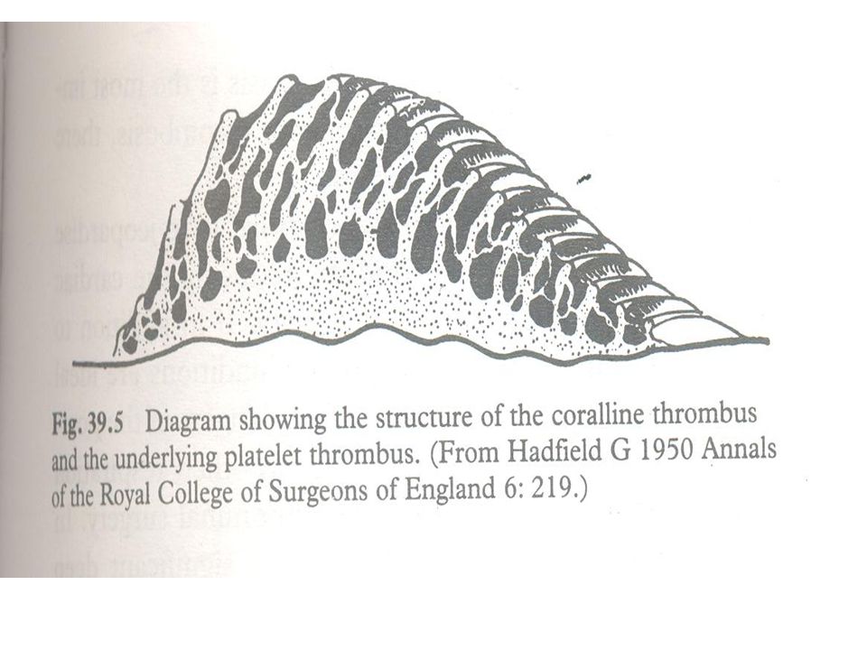
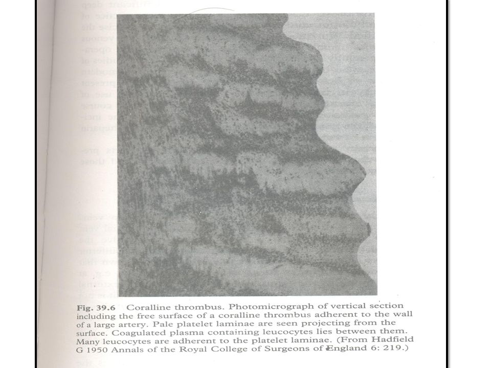
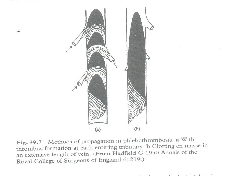
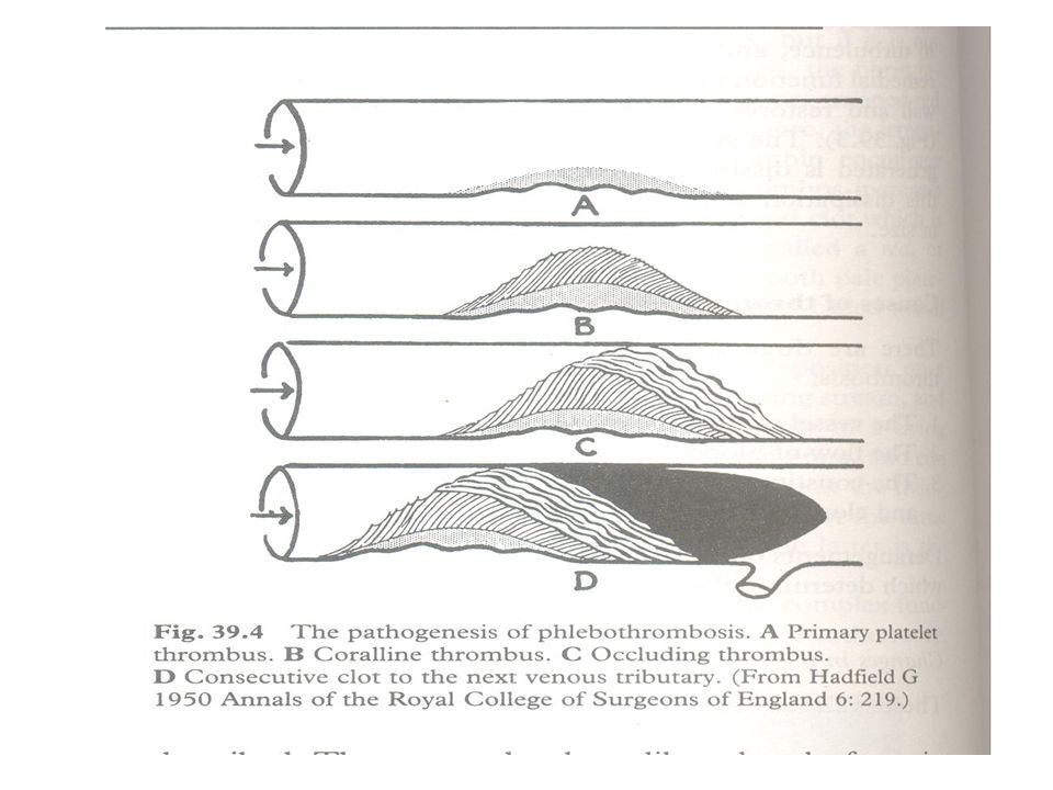
Pathogenesis of DVT Five stages Primary platelet thrombosis
Following trivial intimal damage platelets adhere to the vein wall Aggregate to form a pale thrombus Stasis is paramount important Accumulation of clotting factors and Promote an increase in the Fibrin element- stabilizes the mass of platelets and leads to the next stage

67
2.Coralline thrombus Primary platelet thrombus encourages further platelet accumulation Which take the form of upstanding laminae growing across the stream They are bent in the direction of the blood flow by the force of the stream These laminae anastomose to form an intricate structure Resemble coral

70
This is an example of a mixed thrombus
Up to this stage it is possible for the process to cease

71
3.Occluding thrombus Growth of the coralline thrombus progressively occludes the lumen of the vein. Causes further stasis Formation of more thrombus Which completely occlude the lumen Trails away from the coralline thrombus in the direction of the blood flow This thrombus composed of blood clot with a smaller platelet element. Red thrombus

72
Once the vein is occluded blood flow stops
4. Consecutive clot Once the vein is occluded blood flow stops It stops thrombosis Can occur only in the streaming blood Stationary column of blood beyond the occluding thrombus clots to form a consecutive clot. Which extends up to the entrance of the next venous tributary

73
Can occur by two methods
5. Propagated clot Can occur by two methods Clot when reaches the entrance of the venous tributary Lead to the formation of another platelet and coralline thrombus Occlusion of the ostium of the tributary Then a consecutive clot will form up to the osteum of the next venous tributary

74
2. Sometimes the column of blood above the consecutive clot is so stagnant
Forming one long cord of clotted blood Anchored only at the site of thrombus formation This clot retracts and lies loose in the vein except at its one point of anchorage It can easily break off and be carried to the heart as a massive pulmonary embolus.

77
Morphology Long propagated clot or tail
Red in colour With retraction- thin, pale Loosly attached Head (platelet and coralline thrombus) Is small Firmly attached to the vein wall
 Is small. Firmly attached to the vein wall.")
78
Clinical features Remarkably silent Tenderness
Pain on passive dorsiflexion of the foot (Homans’s sign) Oedema distal to the obstructed veins All the clinical signs are unreliable Frequently the first indication is the occurrence of pulmonary embolism.
 Oedema distal to the obstructed veins. All the clinical signs are unreliable. Frequently the first indication is the occurrence of pulmonary embolism.")
79
Venous thrombosis

80
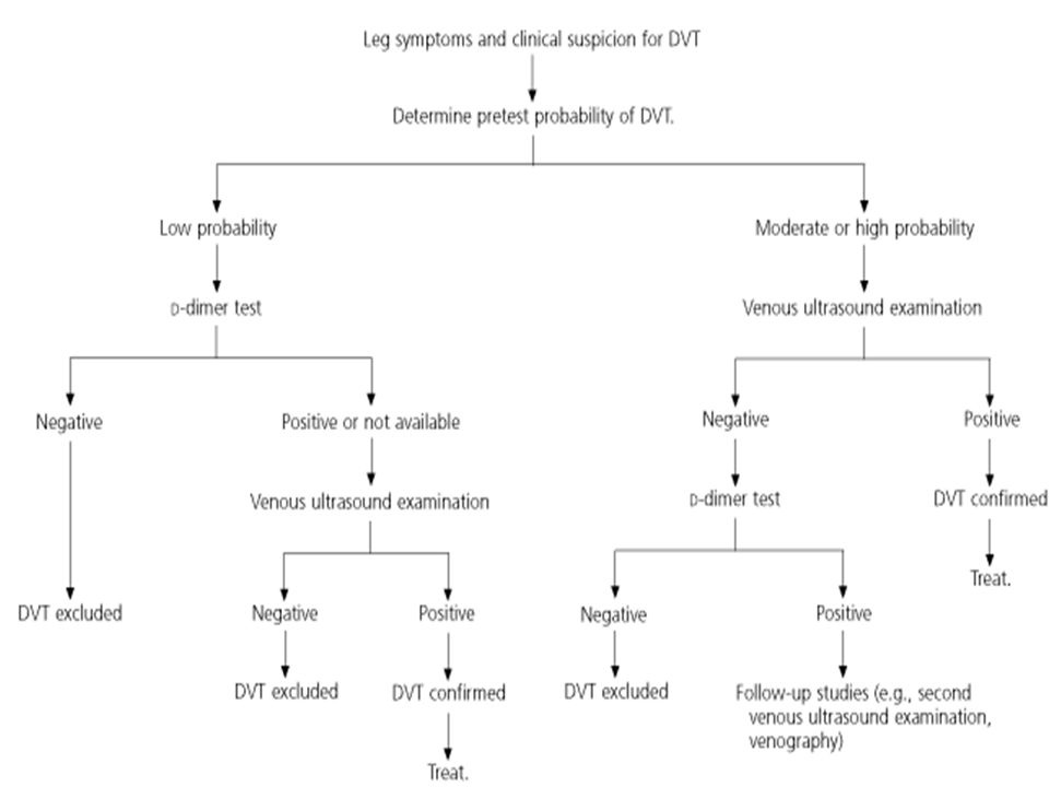
Specialized diagnostic procedure
Phlebography Radioactive iodine- labelled fibrinogen test Doppler ultrasound

82
Complications Massive pulmonary embolism
Smaller pulmonary emboli, with or withoutpulmonary infarction Repeated episodes of silent embolization leading to a syndrome of progressive pulmonary HT

83
Thrombophlebitis Inflammation of a vein wall causes damage to the endothelial lining On this platelets are deposisted Blood flow is either normal or accelerated Thrombosis proceeds to complete obstruction Thrombus is so firmly adherent to the wall Danger of embolism is negligible except pyogenic inflammtion

84
DisseminatedIntravascularCoagulation(DIC)
This condition occurs under two circumstances Which may be found separately or in combination The release of clotting factors into the blood stream Extensive endothelial damage

85
The result is the formation of fibrin in the circulation
This produces vascular obstruction and micro-infarction The extensive intravascular coagulation consumes the coagulation factors Characteristic features are Afibrinogenaemia thrombocytopaenia

86
Disseminated IntravascularCoagulation(DIC)
Refers to widespread microthrombi formation in capillaries, arterioles venules

87
Thrombi are not visible on gross inspection
Leading to circulatory insufficiency: brain , lung, heart, kidneys

88
the development of the multiple thrombi
Although the fibrinolytic system is activated, it cannot effectively deal with the large deposits of fibrin. As a result, there is rapid consumption and eventually a deficiency of clotting factors, including fibrinogen, platelets, prothrombin and factor V, VII, and X

89
Two effects of DIC are a sever bleeding Tendency to infarction
a deficiency of fibrinogen, platelets and prothrombin is required for the diagnosis of DIC. Tendency to infarction Primarily microscopic in extent

90
DIC have bleeding tendencies on hemorrhagic diathesis.
Also the widespread occlusion of the microcirculation may induce signs of shock, acute respiratory distress, central nervous system depression, heart failure or renal failure. Affected tissues may not necessarily disclose the microthrombi because of prompt activation of the fibrinolytic system.

91
Disseminated IntravascularCoagulation(DIC)
A variety of disorders may be complicated by DIC In abruptio placentae with amniotic fluid embolism Intrauterine retention of a dead fetus Incompatible blood transfusion After Sever trauma Fat embolism Open-heart surgery

92
In the newborn after Sever infection Purpura fulminans
Abruptio placenta Birth asphyxia Hypothyroidism Rhesus immunisation Sever infection Purpura fulminans

93
Metastatic cancers Acute leukamia
Usually of prostate Occationally of the lung, pancreas stomach Acute leukamia Certain vascular disorders (giant haemangiomas, aneurysms of aorta and other large vessels, cyanotic congenital heart disease.

94
Clinical features Bleeding ,ecchymosis and haemorrhage from the body’s orifices Thrombocytopaenia Mild haemolytic anaemia It is an emergency condition Is treated with transfusion of fresh blood or fibrinogen

95
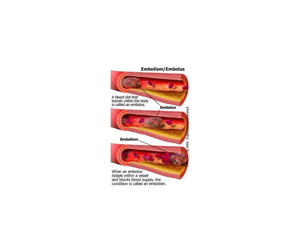
Embolism Definition Embolism is the blockage of a blood vessel by detached intravascular solid, liquid or gaseous mass That is carried by the blood to a site distant from its point of origin. Embolus: An insoluble solid, liquid or gaseous mass circulating in blood stream

96
Virtually 99% of emboli are thrombo-emboli
Arterial (systemic) emboli Venous (pulmonary) emboli
 emboli. Venous (pulmonary) emboli.")
97
Rare forms ( non-thrombotic )
fat Bubbles of air / nitrogen AS debris (cholesterol emboli) Tumour fragments bits of bone marrow Foreign bodies Amniotic fluid embolism
 Tumour fragments. bits of bone marrow. Foreign bodies. Amniotic fluid embolism.")
98
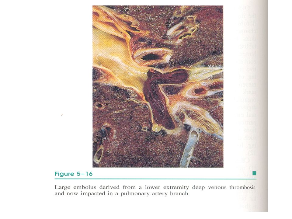
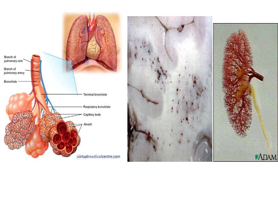
Pulmonary Thromboembolism
20-25/ 100,000 of hospital patients 95% coming from DVT (above knee) Depending on the size of the embolus it may occlude main pulmonary artery (Saddle embolus) in small branches of vessels (multiple) Paradoxical embolus: cardiac embolus passing to the right side through septal defect
 Depending on the size of the embolus it may occlude. main pulmonary artery (Saddle embolus) in small branches of vessels (multiple) Paradoxical embolus: cardiac embolus passing to the right side through septal defect.")
99
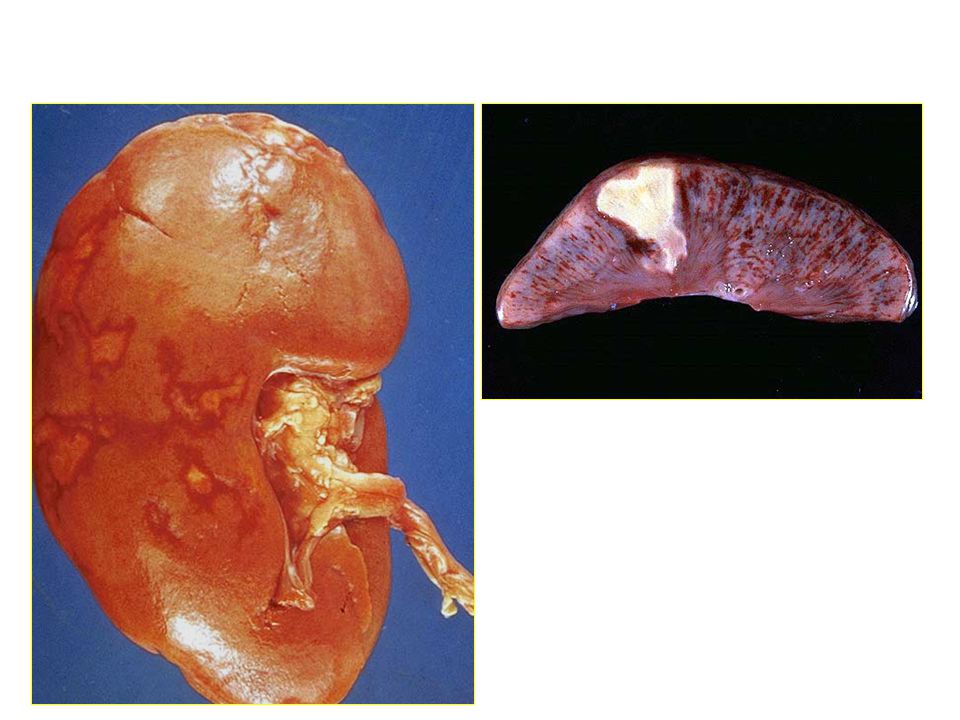
Effects of emboli of Thrombotic origin depends upon
Size and nature (septic or aseptic) State of the collateral circulation in affected organ Aseptic produces transient ischemia if it has good collateral circulation and infarction if poor Septic produces pyaemic abscess at the site of impaction
 State of the collateral circulation in affected organ. Aseptic produces transient ischemia if it has good collateral circulation and infarction if poor. Septic produces pyaemic abscess at the site of impaction.")
100
Size of occluded artery
Number of occluded arteries Release of pro-thrombogenic vs thrombolytic factors locally Background cardiovascular status

101
Pathophysiological consequences of PE
unperfused but ventilated segment respiratory compromise PE ↑ resistance to pulmonary blood flow haemodynamic compromise

102
Pulmonary Thromboembolism
Most pulmonary emboli (60-80%) are asymptomatic because they are small. most organized and incorporated into the vessel wall Can also lead to right ventricular failure (cor pulmonale) / sudden death. Cardiovascular collapse occur when 60% or more of the pulmonary circulation is obstructed with emboli
 are asymptomatic because they are small. most organized and incorporated into the vessel wall. Can also lead to right ventricular failure (cor pulmonale) / sudden death. Cardiovascular collapse occur when 60% or more of the pulmonary circulation is obstructed with emboli.")
103
Obstruction of medium- sized arteries may result
in hemorrhage, and rarely infarction Obstruction of small vessels lead to small infarctions Multiple emboli over time may lead to pulmonary hypertension

104
Pulmonary arterial thrombo-embolism - sequelae
Resorption and resolution (asymptomatic or transient SOB) Organisation → pulmonary hypertension → cor pulmonale Pulmonary infarction (pleuritic chest pain, cough, SOB, haemoptysis, hypoxaemia) Sudden death
 Organisation → pulmonary hypertension → cor pulmonale. Pulmonary infarction (pleuritic chest pain, cough, SOB, haemoptysis, hypoxaemia) Sudden death.")
108
It is important to differentiate embolus from post-mortem clot
Post mortem clot is Moist, shiny and gelatinous Loosely inserted into the pulmonary trunk Shape conforms to that of the situation where it is found Thromboembolus is Dry, friable and granular Already retracted in the leg veins Ripple of platelets may be visible on its surface Shape does not conform to that of its surroundings Tightly inserted into the pulmonary tree Removal is difficult

109
Clinical Features Massive Pulmonary Embolism Shock Dyspnea
Apprehension tachycardia Sweating Chest pain Faintness Cyanosis AF collapse 2/3 die in the first 2 hours. It is a Medical Emergency

110
Dyspnea 73% • Pleuritc Pain 66% • Cough 43% • Leg Swelling 33% • Leg Pain 30% • Hemoptysis 15% • Palpitations 12% • Wheezing 10% • Angina-Like pain 5%

111
Differential Diagnosis
Myocardial Infarction. Dissecting Aortic Aneurysm. Peumothorax. Major Pulmonary Collapse. Shock. Perforating Peptic Ulcer. Acute Pancreatitis

112
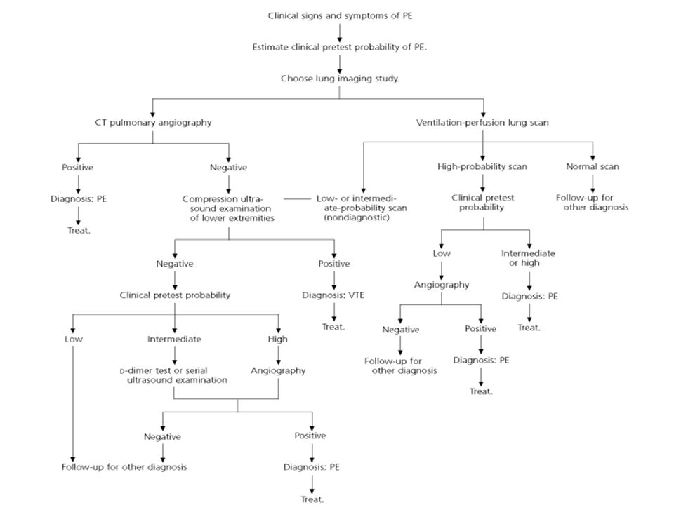
Diagnosis of Pulmonary Embolism (PE)
Clinical picture. Look for risk or predisposing factors for DVT Look for ventilation-perfusion mismatch Testing for PE. chest radiograph ECG Ventilation-perfusion scanning (V/Q scanning). Angiography Spiral CT D-dimer
. Angiography. Spiral CT. D-dimer.")
113
-A marker for thrombosis and fibrinolysis,
D-dimer -A marker for thrombosis and fibrinolysis, can be useful in the exclusion of PE. Specific conditions that will give positive Ddimer tests include trauma, postoperative state, malignancies. -30% with PE will have normal D-dimer

116
Systemic emboli 80% cardiac 20% aortic
2/3rd associated with LV wall infarction 1/4th – dilated left atria- in MS On the mitral or aortic valves- infective endocarditis/ valvular prosthesis Cardiomyopathy 20% aortic AS Aneurysms Valvular vegetations Very small fraction –paradoxical emboli

117
Embolization lodging site is proportional to the degree of flow (cardiac output) that area or organ gets, Lower extremities (75%) brain (10%), kidneys splanchnic liver
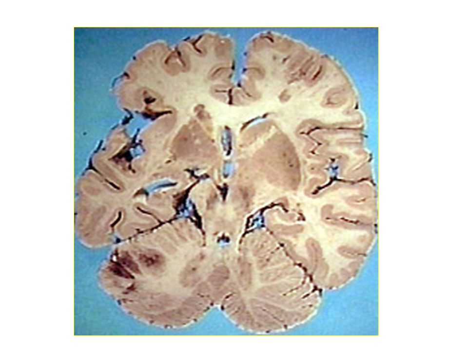
 brain (10%), kidneys. splanchnic. liver.")
118
Consequence depends on the
extent of the collateral supply in the affected tissue Tissue’s vulnerability to ischaemia Caliber of the vessel occluded

119
Effects of systemic emboli
Ischaemia in various organs Septic emboli according to anatomical circumstancesproduce Pyaemic abscesses Septic infarcts mycotic aneurysms Spontaneous embolistion can occur with aortic AS

124
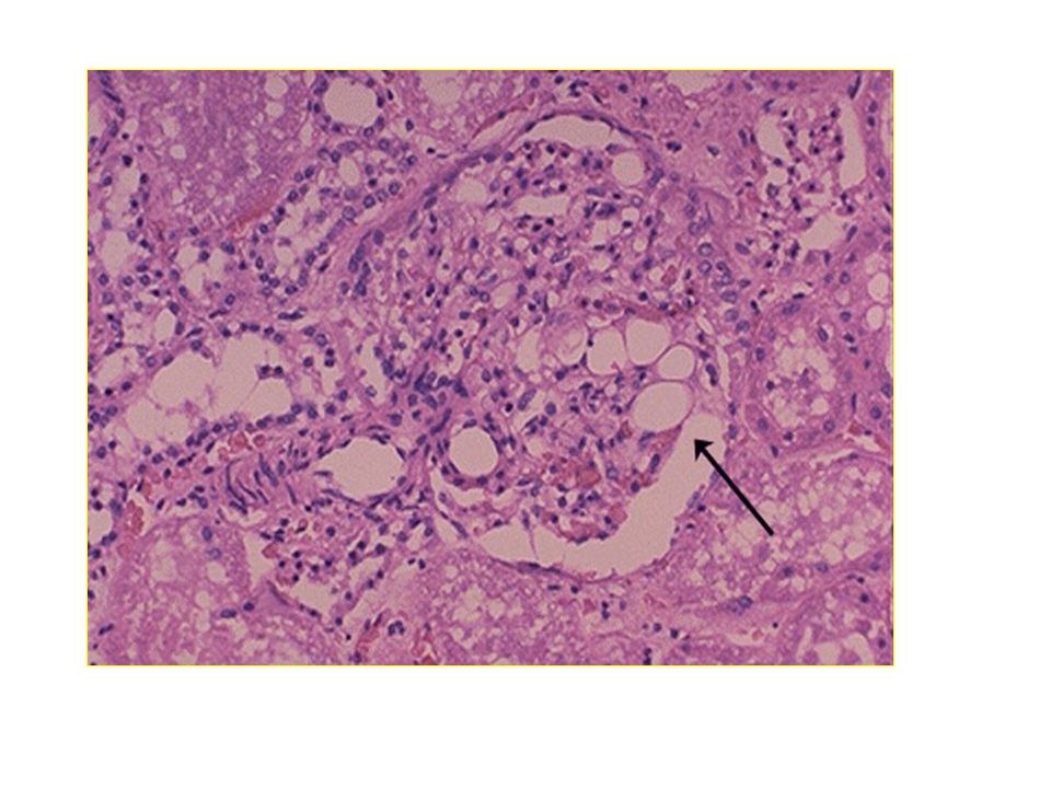
Fat Embolism A process by which fat emboli passes into the bloodstream and lodges within a blood vessel. Fat Embolism Syndrome (FES): serious manifestation of fat embolism occasionally causes multi system dysfunction, the lungs are always involved and next is brain
: serious manifestation of fat embolism occasionally causes multi system dysfunction, the lungs are always involved and next is brain.")
126
Causes of FES Blunt Trauma Long bone (Femur, tibia, pelvic) factures
 factures")
127
Non Trauma: agglutination of chylomicrons and VLDL by high levels of plasma CRP.
disease-related Diabetes, acute pancreatitis, burns, SLE, sickle cell crisis drug-related parenteral lipid infusion procedure-related Orthopedic surgery, liposuction

128
Pathophysiology of FES
Exact mechanism unknown, but two main hypothesis Mechanical Hypothesis Biochemical Hypothesis

129
Mechanical Hypothesis
Obstruction of vessels and capillaries Increase in intermedullary pressure forces fat and marrow into bloodstream Bone marrow contents enter the venous system and lodge in the lungs as emboli

131
Smaller fat droplets may travel through the pulmonary capillaries into the systemic circulation and hence to the brain and other organs. Embolization to cerebral vessels or renal vessels also leads to central nervous system and renal dysfunction

132
Biochemical Hypothesis
Toxicity of free fatty acids circulating free fatty acids directly affect the pneumocytes, producing abnormalities in gas exchange Coexisting shock, hypovolemia and sepsis impair liver function and augment toxic effects of free fatty acids

133
hormonal changes caused by trauma and/or sepsis induce systemic release of free fatty acids as chylomicrons. Acute-phase reactants, such as C-reactive proteins, cause chylomicrons to coalesce and create the physiologic reactions described above. The biochemical theory helps explain nontraumatic forms of fat embolism syndrome and why symptoms take 12 hours to develope.

134
Fat embolism

137
Clinical Manifestations
Pulmonary fat embolism Systemic fat embolism Fat emboli enters the systemic circulation

138
Pulmonary Dysfunction
Asymptomatic for the first hours Pulmonary Dysfunction Respiratory Failure and ARDS (tachypnea, dyspnea, crackles, cyanosis) Hypoxemia systemic arterial hypotension, a decrease in cardiac output, and arrhythmias
 Hypoxemia. systemic arterial hypotension, a decrease in cardiac output, and arrhythmias.")
139
Systemic fat embolism Fat emboli enters the systemic circulation
Lodged in the capillaries of the brain, kidney, skin and other organs Serious event Constitutes the fat embolism syndrome Lungs are certainly always involved

140
Most usually symptoms occur 24-48hours after injury
In sever cases, the patient become comatose within a few hours of injury Dies with 1-2 days Most usually symptoms occur 24-48hours after injury Fever, cyanosis, restlessness, respiratory distress, cerebral symptoms

141
Neurological (nonspecific)
acute confusion, headache, stupor, coma, rigidity or convulsions If brain damage is severe, coma and death results Dermatological Signs A reddish brown petechial rash - helpful diagnostically But it is not manifest until the 2nd or 3rd day distributed to the upper body, chest, neck, upper arm, axilla, shoulder, oral mucous membranes and conjunctivae Subconjunctival and retinal haemorrhages also

142
Laboratory Studies Arterial Blood Gases (ABGs)
Urine and sputum examination Examination of the urine may reveal fat, the test is unreliable Haemotological Tests Platelet count is invariably lowered Biochemical tests

143
Imagining Chest x-ray CT Scan brain Helical CT Scan chest
shows multiple flocculent shadows (snow storm appearance). picture may be complicated by infection or pulmonary edema. CT Scan brain may be normal or may reveal diffuse white-matter petechial haemorrhages Helical CT Scan chest may be normal as the fat droplets are lodged in capillary beds. Can detect lung contusion, acute lung injury, or ARDS may be evident.
. picture may be complicated by infection or pulmonary edema. CT Scan brain. may be normal or may reveal diffuse white-matter petechial haemorrhages. Helical CT Scan chest. may be normal as the fat droplets are lodged in capillary beds. Can detect lung contusion, acute lung injury, or ARDS may be evident.")
144
Risk Factors

145
Gaseous emboli Air may be introduced into a systemic vein in various situations Operations on the head and neck Mismanaged blood transfusion During haemodialysis Insufflations of the uterine tubes

146
Enters the circulation in decompression sickness ( caisson disease)
Nitrogen Enters the circulation in decompression sickness ( caisson disease) Occurs in people who have been exposed to a high pressure Eg : deep sea divers those encased in a diving bell(caisson) under water tunnellers returns to a normal atmospheric pressure too rapidly
 Occurs in people who have been exposed to a high pressure. Eg : deep sea divers. those encased in a diving bell(caisson) under water tunnellers. returns to a normal atmospheric pressure too rapidly.")
147
As the pressure is reduced
Bubbles of air come out of solution from the blood and interstitial fluid O2 and CO2 are readily absorbed and removed Inert nitrogen remains in the tissues for some time Its presence causes mechanical damage

148
Clinical features Prutitus
Severe pain around the joints and muscle(‘the bends’) ARDS(‘the chokes’) Involvement of nervous system Paralysis Confusion, seizure, coma and death Chronic decompression syndrome Multiple foci of bone necrosis arthritis
 ARDS(‘the chokes’) Involvement of nervous system. Paralysis. Confusion, seizure, coma and death. Chronic decompression syndrome. Multiple foci of bone necrosis. arthritis.")
149
Amniotic fluid embolism
Amniotic fluid containing meconium and squamous cells may enter the uterine veins and travel to the lungs Characterized by the sudden onset of respiratory difficulty, cyanosis and shock.

Similar presentations


, fibrous material and.>")


