Download presentation
Presentation is loading. Please wait.

2
In the name of GOD

3
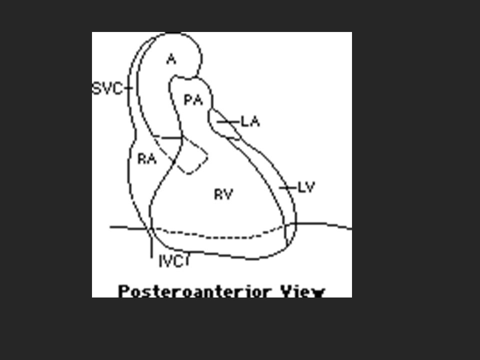
Mediastinum

4
Anatomy

12
Pathology

13
Masses and mass like lesions Inflammatory changes Hematoma

14
Mediastinal masses

15
Imaging strategy Localized to mediastinum Localize within the mediastinum

16
Localize to the mediastinum Unlike lung lesions, a mediastinal mass will not contain air bronchograms. The margins with the lung will be obtuse. Mediastinal lines (azygoesophageal recess, anterior and posterior junction lines) will be disrupted. There can be associated spinal, costal or sternal abnormalities.
 will be disrupted. There can be associated spinal, costal or sternal abnormalities..")
17
LEFT: A lung mass abutts the mediastinal surface and creates acute angles with the lung. RIGHT: A mediastinal mass will sit under the surface of the mediastinum, creating obtuse angles with the lung.

18
The lesion on the left was a pancoast tumor. The lesion on the right was a thymoma, located within the anterior mediastinum.

19
Anterior Mediastinum Thymus Teratoma (germ cell) Thyroid Terrible Lymphoma
 Thyroid Terrible Lymphoma")
20
ThymomaThymoma : most common primary neoplasm of the anterosuperior mediastinum Invasive thymoma Thymic carcinoma ThymolipomaThymolipoma / thymoliposarcomathymoliposarcoma Thymic cyst Benign thymic hyperplasia Thymic carcinoid Thymus

21
Thymic cyst

22
Cystic thymoma

23
Thyroid Thyroid neoplasms Thyroid goiter Parathyroid mass

24
Retrosternal thyroid

25
Retrosternal thyroid mass

27
Cervicothoracic sign

28
Lymphoma Hodgkin Lymphoma Non-Hodgkin lymphoma

29
lymphoma

30
Lymphoma (Hilum Overlay Sign: hilar vessels are seen through a mediastinal mass)
")
31
lymphoma

32
Germ cell tumours Mediastinal teratoma Mature:75% of mediastinal germ cell tumours Immature Teratocarcinoma Mediastinal seminoma Mediastinal emberional cell carcinoma Mediastinal yolk sac tumour Mediastinal choriocarcinoma Mediastinal mixed cell type germ cell tumour

33
Germ cell tumor

34
A fat-containing teratoma

35
Mediastinal teratoma

36
Germ cell tumor

39
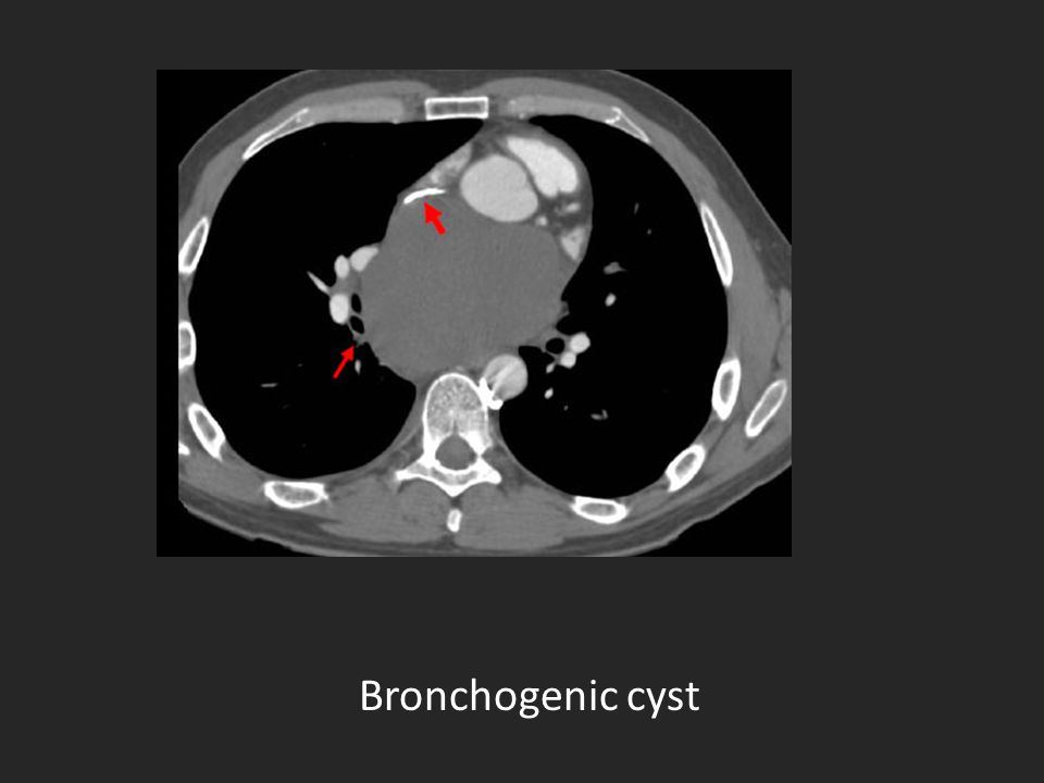
Bronchogenic cyst – A benign growth with respiratory origins. Lymphadenopathy mediastinal – An enlargement of the lymph nodes. Pericardial cyst – A benign growth that results from an "out- pouching" of the pericardium (the heart’s lining). Thyroid mass mediastinal – Usually a benign growth, such as a goiter. These types of tumors can occasionally be cancerous. Tracheal tumors – These include tracheal neoplasms and non- neuplastic masses, such as tracheobronchopathia osteochondroplastica (benign tumors). Vascular abnormalities including aortic aneurysm and aortic dissection. Middle mediastinum
. Thyroid mass mediastinal – Usually a benign growth, such as a goiter. These types of tumors can occasionally be cancerous. Tracheal tumors – These include tracheal neoplasms and non- neuplastic masses, such as tracheobronchopathia osteochondroplastica (benign tumors). Vascular abnormalities including aortic aneurysm and aortic dissection. Middle mediastinum.")
40
Pericardial cyst

41
Aortic aneurysm

42
Bronchogenic cyst

44
Posterior mediastinum Neurogenic tumors Neuroblastic tumors Non-neurogenic tumours Hernias

46
Neurogenic tumor

47
Neurogenic tumors Schwannoma Neurofibroma Malignant peripheral nerve sheath tumor

48
Neuroblastic tumors Neuroblastoma ganglioneuroma

49
neuroblastoma

51
Others Chordoma Pheochromocytoma Praspinal abscess Descending aortic aneurysm Esophageal neoplasm Hiatal hernia Bochdalek hernia Lymphadenopathy Extramedullary hematopoiesis Duplication cysts Thoracic meningocele

53
Extramedullary hematopoiesis

54
Bochdalek hernia

55
Intranthoracic meningocele

56
discitis

57
Foregut duplication cysts occasionally contain milk of calcium

58
4-year-old child with stridor

59
Duplication cyst

61
Hernia hernia

63
Esophageal varicosis

64
Mediastinal widdening >8 cm in the aortic knob depression of the left main-stem bronchus deviation of the naso-gastric tube to the right apical pleural haemoatoma (cap) disruption of the calcium ring in the aortic knob (broken-halo) Aortic injury in blunt trauma
 disruption of the calcium ring in the aortic knob (broken-halo) Aortic injury in blunt trauma")
65
Mediastinal hematoma

66
Some tips in differential diagnosis of mediastinal masses

67
Thymolipoma Teratoma (Germ cell tumors) Esophageal lipoma Fat deposition Lipoma Lipoblastoma Liposarcoma Extramedullary hematopoiesis Fat containing masses
 Esophageal lipoma Fat deposition Lipoma Lipoblastoma Liposarcoma Extramedullary hematopoiesis Fat containing masses")
68
Hyperenhancing lymph nodes Thyroid tissue Paragangliomas Hemangiomas Vascular Etiologies Enhancing masses

69
hemangioma

70
thyroid mass

71
Melanoma Renal cell carcinoma Thyroid carcinoma Castlemann's disease Enhancing lymphomas can be seen in:

72
Castlemann's disease

73
Thymic Cyst Thymoma Teratoma Pericardial Cyst Foregut Duplication Meningocoele Neuroenteric Cyst Cystic Lymphadenopathy Lymphangioma Fluid containing masses

74
Thank you

Similar presentations


disease TB Histoplasmosis Chicken box Sarcoidosis LCH Pneumoconiosis Alveolar microlithiasis Metastasis.>")


: Principal Modality (2): Trauma CT General Radiography.>")


 have two basic components. Proliferating neoplastic cells that constitute.>")


 SHEN JIN The First Affiliated Hospital of Kunming Medical College.>")

