Download presentation
Presentation is loading. Please wait.

1
Mohammad Ali Tahririan Department of Orthopedics Kashani Hospital
Impingement Syndrome Mohammad Ali Tahririan Department of Orthopedics Kashani Hospital

2
History Jarjavavy 1867 Subacromial bursitis
Codman Supraspinatus rupture Neer Impingement syndrome

3
Developmental Stages of Impingement Syndrome
Edema and Hemorrhage Typical age of patient: <25 years old Differential diagnosis: subluxation, acromioclavicular joint arthritis Clinical course: reversible Treatment: conservative

4
Developmental Stages of Impingement Syndrome
Fibrosis and Tendinitis Typical age of patient: 25 to 40 years old Differential diagnosis: frozen shoulder, calcium deposits Clinical course: recurrent pain with activity Treatment: consider bursectomy or division of coracoacromial ligament

5
Developmental Stages of Impingement Syndrome
Bone Spurs and Tendon Rupture Typical age of patient: >40 years old Differential diagnosis: cervical radiculitis, neoplasm Clinical course: progressive disability Treatment: anterior or acromioplasty, rotator cuff repair

6
History Pain, weakness and loss of motion are the most common symptoms reported. Minor pain that is present both with activity and at rest Pain radiating from the front of the shoulder to the side of the arm Pain is exacerbated by overhead or above-the-shoulder activities. A frequent complaint is night pain, often disturbing sleep, particularly when the patient lies on the affected shoulder. The onset of symptoms may be acute, following an injury, or insidious, particularly in older patients, where no specific injury occurs.

7
Impingement Syndrome 1) Primary impingement 2) Secondary impingement
3) Internal impingement 4) Subcoracoid impingement
 Internal impingement. 4) Subcoracoid impingement.")
8
Primary impingement Usually older than 40 years,
complain of anterior shoulder and upper lateral arm pain, with an inability to sleep on the affected side. They have complaints of “shoulder weakness,” and difficulty performing overhead activities.

9
Primary impingement On physical examination, patients may exhibit a loss of motion or weakness of rotator cuff strength secondary to pain. They will usually have a positive Hawkins sign and a positive impingement sign as described by Neer. The impingement test is performed by injecting 10 ml of 1% lidocaine into the subacromial space

10
Primary impingement Intrinsic type Extrinsic type
Tendinopathy(thickening of the RC) Calcific tendinitis Subacromial bursitis Intrinsic type Extrinsic type .Subacromial spur .Acromial fracture .Os acromiale .ACJ arthritis .Exostoses of the GT When the structures passing beneath the coracoacromial arch become enlarged resulting in abutment against the arch, the cause of the impingement is considered to be intrinsic. Examples of this condition include thickening of the rotator cuff, calcium deposits within the rotator cuff, and thickening of the subacromial bursa. Extrinsic impingement occurs when the space available for the rotator cuff is diminished; examples include subacromial spurring, acromial fracture or pathological os acromiale, osteophytes off the undersurface of the acromioclavicular joint, and exostoses at the greater tuberosity.
 Calcific tendinitis. Subacromial bursitis. Intrinsic type. Extrinsic type. .Subacromial spur. .Acromial fracture. .Os acromiale. .ACJ arthritis. .Exostoses of the GT. When the structures passing beneath the coracoacromial arch become enlarged resulting in abutment against the arch, the cause of the impingement is considered to be intrinsic. Examples of this condition include thickening of the rotator cuff, calcium deposits within the rotator cuff, and thickening of the subacromial bursa. Extrinsic impingement occurs when the space available for the rotator cuff is diminished; examples include subacromial spurring, acromial fracture or pathological os acromiale, osteophytes off the undersurface of the acromioclavicular joint, and exostoses at the greater tuberosity.")
12
Left) Coronal PO FSE MR shows increased signal intensity within the supraspinatus critical zone (arrow), consistent with tendinopathy. (Right)Coronal FS PO FSf MR shows thickening and increased signal intensity (arrow) of thp distal supraspinatus tendon, representing tendinopathy (tendinosis).
Coronal FS PO FSf MR shows thickening and increased signal intensity (arrow) of thp distal supraspinatus tendon, representing tendinopathy (tendinosis)..")
14
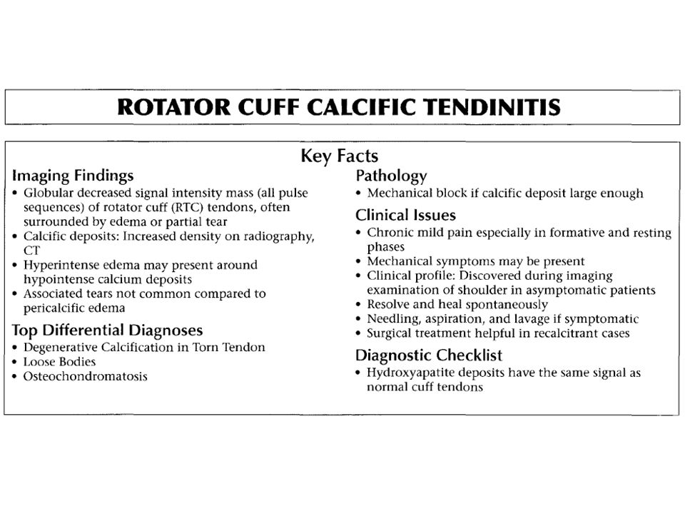
Calcific tendinitis Calcific tendinitis is a painful, largely self-limited disorder of the rotator cuff in which the tendons are infiltrated with calcium deposits.

15
Calcific tendinitis Location
This condition most frequently affects the rotator cuff of the shoulder. supraspinatus - 80% infraspinatus - 15% subscapularis - 5% periarticular soft tissues in addition to tendons ligaments capsule bursae The most common site of occurrence is within the supraspinatus tendon and at a location 1.5 to 2 cm away from the tendon insertion on the greater tuberosity.

16
Calcific tendinitis Mostly asymptomatic Older than 30 y
10% of population 10% bilaterally F ˃ M

17
Calcific tendinitis Painful Phase I—precalcification stage
phase of formation Phase II—calcification stage resting phase Painful resorptive phase pain due to extravasation of calcium hydroxyapatite into adjacent tissues, especially subacromial bursa pain typically lasts 2 weeks Phase III—postcalcification phase

20
(Left) Coronal PO FSE MR shows clump-like calcification (arrow) within the supraspinatus tendon, consistent with the silent phase of calcific tendinitis. (Right) Coronal FS PO FSE MR shows hypointense clumped calcification with subacromial bursal hyperintensity.
 Coronal FS PO FSE MR shows hypointense clumped calcification with subacromial bursal hyperintensity..")
21
Calcific tendinitis Nonoperative treatment
Nonoperative management is the initial treatment of choice. Nonoperative treatment usually includes physical therapy, exercises, anti inflammatory medications, and steroid injections. The efficacy of any of these treatment methods has not been proved, however. Corticosteroids have been suggested to abort the resorptive phase, returning the lesion to dormancy and setting into motion the factors necessary for recurrence. We believe that only patients in the resorptive phase should have treatment directed at the calcium deposit itself. Others should follow treatment protocols directed at the particular pathological condition (e.g., impingement).
.")
22
Calcific tendinitis Operative treatment indications:
symptom progression constant pain that interferes with activities of daily living absence of improvement after conservative therapy.

23
successful in approximately 70% of patients.
ultrasound-guided percutaneous needling + subacromial corticosteroid injection successful in approximately 70% of patients. Long-term follow-up studies have confirmed the benign natural history of this disorder: although treated patients tend to have better results in the short-term (1-year follow-up), at longer-term follow-up there are no differences
, at longer-term follow-up there are no differences.")
24
(A) Type 1 acromion with flat acromial undersurface
(A) Type 1 acromion with flat acromial undersurface. Type 1 acromion with straight inferior margin as seen on a sagittal T2 FSE image.
 Type 1 acromion with flat acromial undersurface. Type 1 acromion with straight inferior margin as seen on a sagittal T2 FSE image.")
25
A) Type 2 acromion with a curved convex inferior surface that parallels the contour of the humeral head. (B) Type 2 curved acromion on a corresponding sagittal PD FSE image.
 Type 2 curved acromion on a corresponding sagittal PD FSE image..")
26
(A) Type 3 acromion with an inferiorly directed beak or hook, which contributes to narrowing of the supraspinatus outlet for the supraspinatus tendon. (B)Sagittal PDFSE image of the type 3 or anterior hooked acromion. The type 3 acromion is assessed at least one to two images lateral to the ACjoint In a cadaver study of 140 shoulders, one third had full-thickness tears of the rotator cuff, 73% of which were in shoulders with type III acromions.

28
Coronal FS PD FSE image showing an acromial "keel" spur associated with a full-thickness rotator cuff tear with retraction.

29
AC joint Arthrosis (A) Although AC arthrosis may be concurrent with impingement, it is the more lateral acromial spurs that are directly associated with symptomatic bursal-side cuff damage. (8) AC joint degenerative disease with hypertrophic inferior acromial side spur.
 Although AC arthrosis may be concurrent with impingement, it is the more lateral acromial spurs that are directly associated with symptomatic bursal-side cuff damage. (8) AC joint degenerative disease with hypertrophic inferior acromial side spur.")
30
Os Acromiale The acromion forms from 4 ossification centers that normally fuse by age 18 years, and acromion fuses to the spine at y.

31
Os Acromiale 1-15% of nl population M˃F B˃W 30-60% Bilat.
Mean age: 50 y

32
The os acromiale may cause impingement because if it is unstable, it may be pulled inferiorly during abduction by the deltoid, which attaches here.

33
Os Acromiale Optimal surgical treatment for symptomatic os acromiale is unclear.

34
The most common type treated mesoacromiale (94%)
")
35
The most common surgical technique:
IF (60%) Excision (27%) Acromioplasty (13%) The most common surgical technique: in patients who are considered for subacromial decompression, the removal of the acromion distal to the synchondrosis may further destabilize the synchondrosis and allow for even greater mobility of the os acromiale after surgery and worsening of the impingement The most common concurrent surgical technique: RC Repair All techniques Improve clinical outcome
 Excision (27%) Acromioplasty (13%) The most common surgical technique: in patients who are considered for subacromial decompression, the removal of the acromion distal to the synchondrosis may further destabilize the synchondrosis and allow for even greater mobility of the os acromiale after surgery and worsening of the impingement. The most common concurrent surgical technique: RC Repair. All techniques Improve clinical outcome.")
36
Subacromial Impingement

38
Subacromial bursitis

39
No response after 3-4 months
Treatment No response after 3-4 months NSAID,s 1-2 subacromial cortisone injections Physical therapy program focusing on stretching for full shoulder motion and strengthening the rotator cuff Operative intervention may be indicated

40
Treatment Principles:
We use arthroscopic and occasionally open techniques. Principles: ■ Release (but not resection) of the coracoacromial ligament ■ Removal of the anterior lip and lateral edge of the acromion ■ Removal of part of the acromion anterior to the anterior border of the clavicle ■ Removal of the distal 1 to 1.5 cm of clavicle if significant degenerative changes are found
 of the coracoacromial. ligament. ■ Removal of the anterior lip and lateral edge of the. acromion. ■ Removal of part of the acromion anterior to the anterior. border of the clavicle. ■ Removal of the distal 1 to 1.5 cm of clavicle if significant. degenerative changes are found.")
41
A, Incision centered on anterolateral corner of acromion, avoiding axillary nerve, and carried medially on superior surface of acromion. B, Deltoid origin elevated from acromion in continuity with acromial periosteum and trapezius insertion. C, Anterior extent of acromion to be removed

42
Complication infection seroma formation hematoma synovial fistula
biceps rupture pulmonary embolus acromial fracture

43
Complication the worst common complication is loss of anterior deltoid function, which is caused by either axillary nerve injury or detachment of the deltoid from the acromion.

44
Secondary impingement
Secondary impingement is a clinical phenomenon that results in a “relative narrowing” of the subacromial space. GH instability Scapulothoracic instability

45
Secondary impingement
Patients with secondary impingement are usually younger and often participate in overhead sporting activities such as baseball, swimming, volleyball, or tennis. They complain of pain and weakness with overhead motions and may even describe a feeling of the arm going “dead.”

46
Secondary impingement
physical examination On physical examination, the examiner should look for possible associated pathology, including GH joint instability with a positive apprehension and relocation test or abnormal scapular function such as scapular winging or asymmetrical scapular motion.

47
Secondary impingement
GH instability Translation of the humeral head, typically anteriorly, resulting contact of the rotator cuff against the coracoacromial arch.

48
Secondary impingement
The loss of the stabilizing function of the rotator cuff muscles also leads to an abnormal superior translation of the humeral head (decreased depression of the humeral head during throwing and less “clearance”) and mechanical impingement of the rotator cuff on the coracoacromial arch .
 and mechanical impingement of the rotator cuff on the coracoacromial arch .")
49
Secondary impingement
Scapulothoracic instability In patients who have scapular instability, impingement results from improper positioning of the scapula with relation to the humerus. The instability leads to insufficient retraction of the scapula, which allows for earlier abutment of the coracoacromial arch on the underlying rotator cuff.

50
Secondary impingement
In patients with secondary impingement, treatment of the underlying problem should result in resolution of the “secondary impingement” symptoms. A subacromial decompression here worsens the symptoms because the shoulder is rendered even more “unstable.”

51
Subcoracoid impingement
Gerber et al. suggested that this painful contact might be caused by a prominent coracoid, for which there may be numerous reasons, including idiopathic and iatrogenic conditions. The iatrogenic form was most common in their series, and it was found in patients who had undergone a Trillat osteotomy of the coracoid for the treatment of anterior instability.

52
Physical findings Gerber Test -Tenderness over the coracoid
-Positive coracoid impingement test. -Subcoracoid injection similar to the Neer impingement test

53
Internal impingement In this condition, internal contact of the rotator cuff occurs with the posterosuperior aspect of the glenoid when the arm is abducted, extended, and externally rotated as in the position of the throwing motion.

54
Internal impingement It often occurs in throwers who have lost internal rotation of the shoulder. This loss causes the center of rotation of the humeral head to move upward so that the contact between the rotator cuff and the biceps tendon attachments increases. Arthroscopic findings include partial rotator cuff tears, posterior and superior labral tears, and anterior shoulder laxity.

55
Internal impingement Early in the course of the condition, aggressive physical therapy with attention to regaining internal rotation and rotator cuff strengthening often is successful.

Similar presentations


 and humerus. Shoulder.>")