Download presentation
Presentation is loading. Please wait.

1
Pacemaker Follow-up Alpay Çeliker MD. Hacettepe University Department of Pediatric Cardiology 3rd International Summer School on Cardiac Arrhythmias, 9-12 September, Eskişehir

3
Organization Regular follow-up schedule Pacemaker record files X-ray ECG Telemetry units

4
Pacemaker Follow-up: Objectives Adjust the pacing system Maximize the benefits of pacing therapy Predict impending pacemaker system failure before the patient is at risk Ascertain the nature of malfunction Look for accompanying complications

5
PATIENT TELEMETRY THRESHOLDS INTRINSIC AMPLITUDES PACEMAKER DEPENDENCY PACING RATIO HISTOGRAMS ECG&TELE PACING EFFICACY LEAD PROBLEMS PHYSICAL EXAMHOLTER, EXERCISE TEST EXERCISE PERFORMANCE MAXIMUM HEART RATE DETECT CAPTURE AND SENSING PROBLEMS ECHO

6
Pacemaker Follow-up Patient evaluation Patient evaluation History Physical examination Chest x-ray Echocardiography Pacing system evaluation Pacing system evaluation Surface ECG Telemetric control of pacemaker Holter monitoring Treadmill testing

7
History Palpitations Rapid ventricular rate, PMT, intrinsic tachycardia Weakness, fatigue, malaise, dyspnea Pacemaker syndrome, capture failure, inappropriate programming, cardiac or pulmonary disease Hiccups Syncope, presyncope Pacemaker syndrome, capture problem, inhibition due to oversensing Cough, chest pain

8
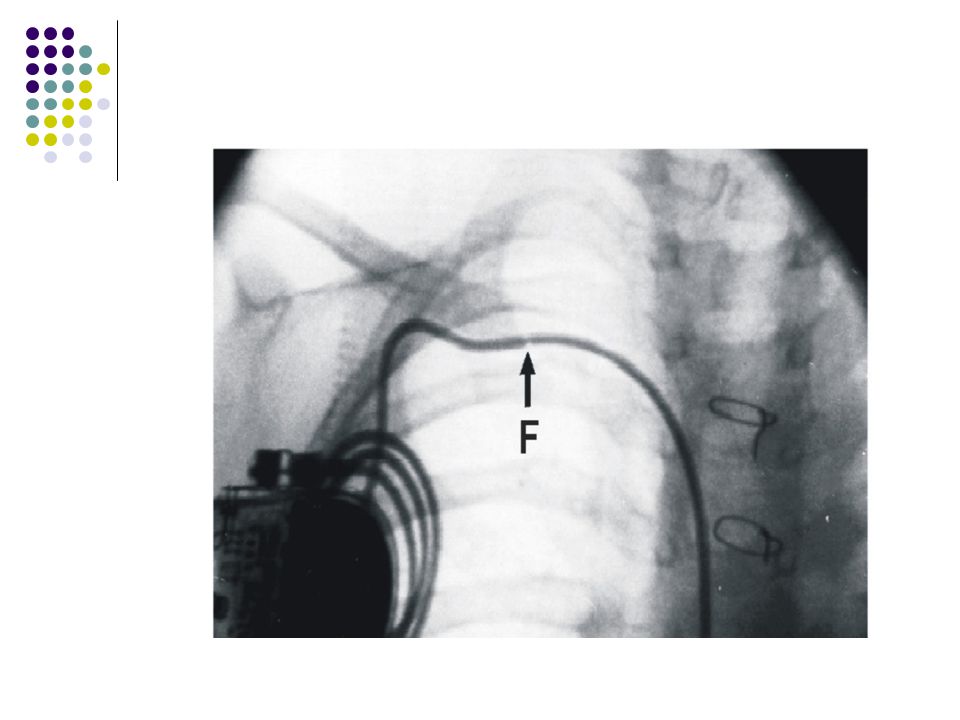
Radiologic Evaluation

9
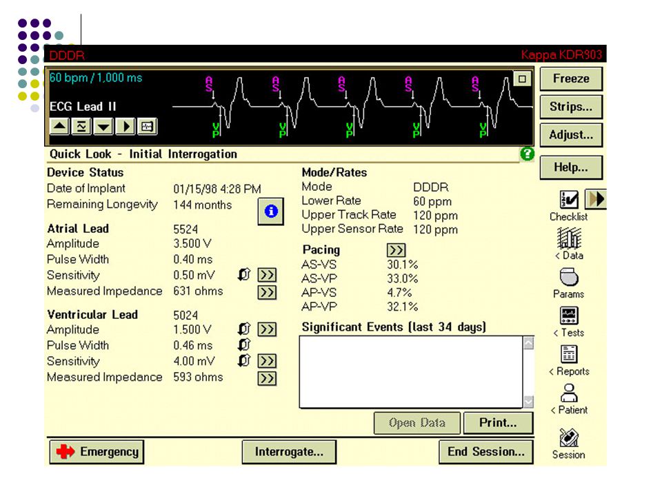
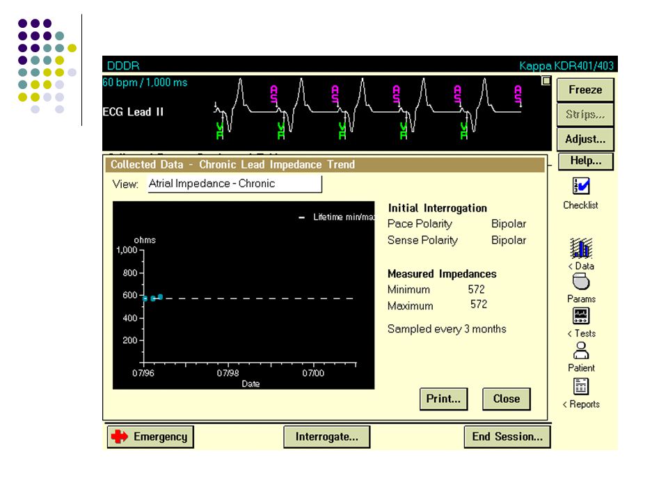
Pacemaker Interrogation Administrative data verification Administrative data verification Name, implant date Programmed data control Programmed data control Examine the pacing&sensing parameters Examine the pacing&sensing parameters Capture threshold Voltage measurements Battery&lead measurements Overview the memorized data Overview the memorized data

11
Capture Thresholds Automatic or manual measurements Voltage or pulse width thresholds Pacing rate > spontan rate during test Test during coughing and deep respiration to detect malfunction

12
Absence of PM Stimuli or Capture Absence of PM Stimuli or Capture Intrinsic rate > pacing rate Hysteresis Very tiny bipolar stimuli Lead problems Fracture, loose connection Pulse generator problems EOL, failure Electromagnetic interference Oversensing

13
Atrial Noncapture

14
Ventricular Noncapture

15
Sensing Thresholds Automatic or manual measurements Print-out of intracardiac electrocardiogram Needs for spontaneous atrial or ventricular rhythm

16
Undersensing

17
Undersensing Low amplitude EGM due to poor lead position Lead dislodgement Lead malfunction Metabolic or toxic causes Development of new bundle branch block Myocardial infarciton near the electrode tip

18
Oversensing

19
Causes of Oversensing Causes of Oversensing Ventricular Ventricular T wave Crosstalk Myopotentials False signals Atrial Atrial Far-field R wave Myopotentials False signals

20
Change in Pacing Rate Battery depletion Runaway pacemaker Component failure Oversensing External effects on battery Phantom or wrong programming

21
Signs of Lead Fracture No stimuli Stimuli without capture Oversensing of false signals Permanent or intermittant high lead impedance Maneuvers X-ray

24
Testing of Specific Functions Check for crosstalk Evaluate the VA interval Examine rate adaptive parameters Hysteresis, sleep rate Automatic mode switching Histogram settings

25
Rate Adaptive Pacemaker

26
Rate Histogram Assess rate response settings Assess high rate events Evaluate percent pacing versus sensing Determine if a change in disease state condition has occurred

27
Atrial Pace/sense Histogram

28
Physical Examination Pacing System Pocket Infection Erosion Migration Twiddler’s syndrome Muscle stimulation Chronic pain Vascular System Venous thrombosis Intracardiac thrombus Lead Endocarditis Tricuspid valve entanglement TR Leads Displacement Perforation Diaphragmatic pacing

29
Lead Endocarditis

30
Venous Obstruction <obstruction

32
Pacemaker Syndrome Dizziness Presyncope Chest tightness Shortness of breath Neck pulsations Apprehension/malaise Fatigue

33
PMT

35
Conclusions I Long rhythm strips with markers and IEGM’s may needed for correct diagnosis 12 lead paced ECG is very valuable Know the timing cycles Do not attribute patient symptoms to age,sex or underlying heart disease Do not leave the pacemaker at factory settings, since every patient has different necessities.

36
Conclusions II Make every effort to prolong battery life The other purpose of pacing is optimization of quality of life Optimal AV delay can not be predicted Test the retrograde VA conduction Keep the records carefully Be obsessive in pacemaker dependent patient

Similar presentations


 Position I IIIII Category Chamber(s) Chamber(s) Response to paced sensed sensing.>")


