Download presentation
Presentation is loading. Please wait.

1
Ahmad Hormati Assistant Professor of Gastroenterology Qom University of Medical Sciences

2
MALABSORPTION SYNDROME

3
Epidemiology, and clinical manifestations of celiac disease in adults

4
Celiac disease occurs primarily in whites of northern European ancestry.
Although classically a disease of infants, celiac disease now often presents later, between the ages of 10 and 40 years

5
Formerly, epidemiological studies using serologic assays for IgA antibodies to gliadin and endomysium with biopsy verification established prevalences of 1:300 to 1:500 in most countries where celiac disease is prevalent. The identification of tissue transglutaminase (TG2) as endomysial autoantigen allowed the development of a highly sensitive and specific serological test and population based screening. Anti-TG2-based screening programs revealed a higher prevalence of celiac disease, reaching 1 percent or more, thus demonstrating a large number of undiagnosed celiac patients in the "normal" populations of affected countries.
 as endomysial autoantigen allowed the development of a highly sensitive and specific serological test and population based screening. Anti-TG2-based screening programs revealed a higher prevalence of celiac disease, reaching 1 percent or more, thus demonstrating a large number of undiagnosed celiac patients in the normal populations of affected countries.")
6
Patients with celiac disease may present with classic symptoms related to malabsorption, including diarrhea, steatorrhea, weight loss, and nutrient or vitamin deficiencies. They may also exhibit only minor gastrointestinal complaints, have nongastrointestinal manifestations, or be asymptomatic. Numerous conditions have been associated with celiac disease including dermatitis herpetiformis, diabetes mellitus, and selective IgA deficiency.

7
CLASSIFICATION Classic disease: includes the following three features:
villous atrophy symptoms of malabsorption such as steatorrhea, weight loss, or other signs of nutrient or vitamin deficiency resolution of the mucosal lesions and symptoms upon withdrawal of gluten-containing foods, usually within a few weeks to months. Patients with classic disease present with diarrhea, weight loss, or malabsorption, and possess antibodies against gliadin and especially tissue transglutaminase.

8
CLASSIFICATION Atypical celiac disease:
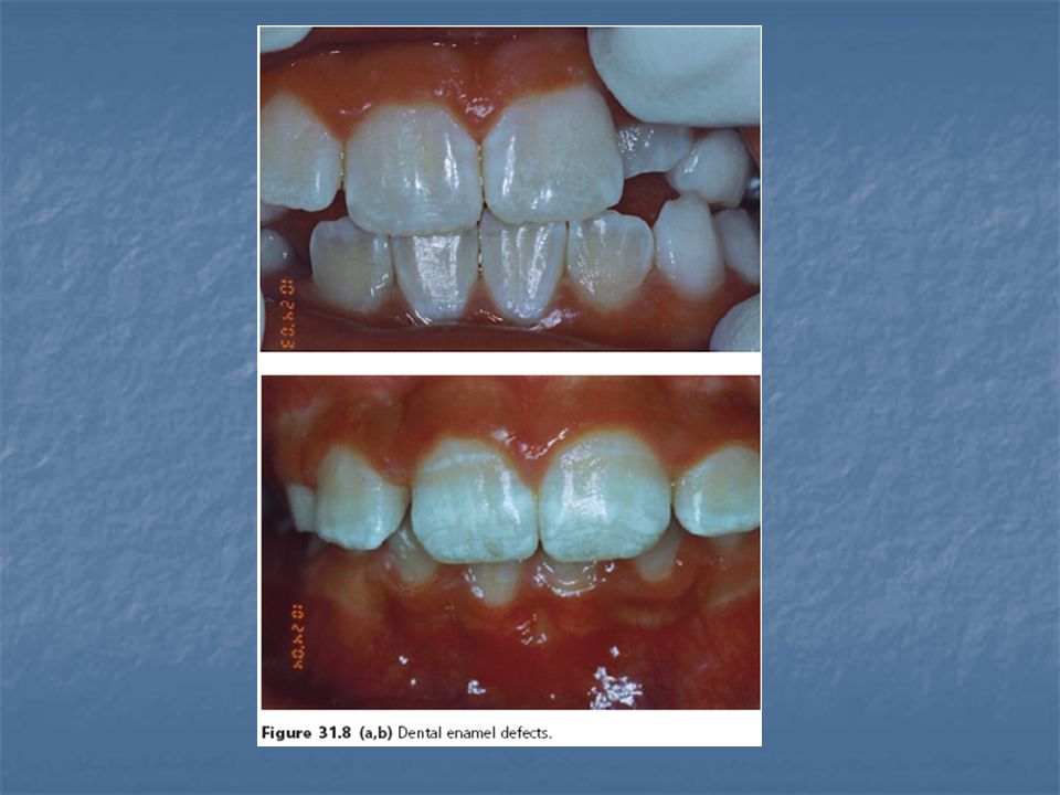
Patients with atypical disease exhibit only minor gastrointestinal complaints. They can display anemia, dental enamel defects, osteoporosis, arthritis, increased transaminases, neurological symptoms, or infertility. However, most of these patients show severe mucosal damage and possess the celiac specific antibody pattern.

9
CLASSIFICATION Asymptomatic (silent) celiac disease :
Patients are often recognized incidentally based upon screenings for antibodies against gliadin or tissue transglutaminase. Although these patients very often display the characteristic architectural remodelling of the intestinal mucosa seen in celiac disease (ie, crypt hyperplasia and villous atrophy), they do not show clinical symptoms. Often minor symptoms (eg, fatigue) are only realized after introduction of a gluten free diet.
, they do not show clinical symptoms. Often minor symptoms (eg, fatigue) are only realized after introduction of a gluten free diet.")
10
CLASSIFICATION Latent celiac disease
There are some patients who have normal jejunal mucosa and minor symptoms or no symptoms at one or more time points while on a normal, gluten-containing diet. Two variants of what has been called latent celiac disease have been identified: Celiac disease was present before, usually in childhood; the patient recovered completely with a gluten-free diet, remaining "silent" even when a normal diet was reintroduced. About 20 percent of such patients continue to have latent disease (asymptomatic with normal villous architecture) into adulthood, while the others re-develop variable degrees of villous atrophy . Latency may be transient and thus regular follow up of such patients is warranted. A normal mucosa was diagnosed at an earlier occasion while ingesting a normal diet, but celiac disease developed later.
 into adulthood, while the others re-develop variable degrees of villous atrophy . Latency may be transient and thus regular follow up of such patients is warranted. A normal mucosa was diagnosed at an earlier occasion while ingesting a normal diet, but celiac disease developed later.")
11
CLINICAL MANIFESTATIONS
Gastrointestinal manifestations — Patients may present with classic signs, including diarrhea with bulky, foul-smelling, floating stools due to steatorrhea and flatulence. These symptoms are paralleled by the consequences of malabsorption, such as growth failure in children, weight loss, severe anemia, neurologic disorders from deficiencies of B vitamins, and osteopenia from deficiency of vitamin D and calcium. However, there is a shift from fewer patients presenting with classic celiac disease to more patients with atypical symptoms or an asymptomatic presentation Adult patients with undiagnosed celiac disease rarely present with profuse diarrhea and severe metabolic disturbances (celiac crisis)
")
12
CLINICAL MANIFESTATIONS
Subclinical disease most patients have mild and unspecific symptoms, such as fatigue, borderline iron deficiency or otherwise unexplained elevations in serum aminotransferases, or no symptoms at all. Those without any specific complaints may be diagnosed during screening programs or during endoscopy performed for other reasons . Establishing the diagnosis of subclinical celiac disease is of potential importance for four reasons: the danger of malignancy the presence of unsuspected nutritional deficiencies the association with low-birth weight infants in affected mothers the occurrence of autoimmune disorders.

13
Nongastrointestinal manifestations
Neuropsychiatric disease Neurologic symptoms most often include headache, dysthymia, and peripheral neuropathy Peripheral neuropathies, characterized by burning, tingling, and numbness in hands and feet, found in 50% of patients and may precede the diagnosis of celiac disease A gluten free diet has been shown to have a favorable effect on headache and dysthymia, but not on peripheral neuropathies The correlation of celiac disease with depression is still unclear. A similar discrepancy in the association of celiac disease with epilepsy was noted. Other cause of neuropathy:In some patients, neurologic deficits can result from vitamin deficiencies, such as deficiencies in vitamins B1 (thiamine), B2 (riboflavin), B3 (niacin), B6 (pyridoxine), B12 (cobalamine), and E. However, vitamin deficiency syndromes are uncommon in the absence of severe and extensive small bowel involvement. In addition, neuropathies may be associated with lymphoma
, B2 (riboflavin), B3 (niacin), B6 (pyridoxine), B12 (cobalamine), and E. However, vitamin deficiency syndromes are uncommon in the absence of severe and extensive small bowel involvement. In addition, neuropathies may be associated with lymphoma.")
14
Nongastrointestinal manifestations
Arthritis — A higher prevalence of osteoarthritis has been described in celiac disease, but whether there is a causal relationship is unclear. Iron deficiency — Celiac disease may be a surprisingly frequent cause of iron deficiency anemia. The incidence was 20 percent in the subgroup of nonresponders to supplemental iron. Some reports have suggested that celiac disease can be associated with occult gastrointestinal bleeding. However, the positive results with colorimetric tests may have been due to excess loss of intestinal cells and/or malabsorption of peroxidase-containing foods rather than loss of red blood cells . Furthermore, one study found that occult bleeding was no more common in patients with celiac disease compared with a control population . Thus, occult gastrointestinal bleeding may not be a major contributor to iron deficiency.

15
Nongastrointestinal manifestations
Metabolic bone disease — Metabolic bone disease is common in celiac disease and can occur in patients without gastrointestinal symptoms In adults, loss of bone density in the peripheral skeleton may persist despite apparent normalization at axial skeletal sites after patients are on a gluten free diet . In contrast, in a study of 30 children and adolescents maintained on a long-term gluten-free diet (average 10.7 years), bone mineral density and serum markers of bone metabolism completely normalized. Data suggest that the risk of fractures is only slightly increased in patients with celiac disease.
, bone mineral density and serum markers of bone metabolism completely normalized. Data suggest that the risk of fractures is only slightly increased in patients with celiac disease.")
16
Nongastrointestinal manifestations
Hyposplenism — Several case reports have described hyposplenism in association with celiac disease the pathogenesis of which is unknown. Prophylactic pneumococcal vaccination has been suggested. Kidney disease — Glomerular IgA deposition is common, occurring in as many as one-third of patients. The great majority of affected patients have no clinical manifestations of renal disease, perhaps because there is no associated activation of complement Idiopathic pulmonary hemosiderosis — Coexistence of celiac disease and idiopathic pulmonary hemosiderosis, also known as Lane-Hamilton syndrome, has been reported in a number of cases, and introduction of a gluten-free diet has been associated with remission of pulmonary symptoms in several patients.

17
RISK OF MALIGNANCY AND MORTALITY
A number of observational studies have noted a small absolute increase in overall mortality in patients with celiac disease compared with the general population . Estimates of the magnitude of risk have differed in various reports, many of which were small, based upon referral populations, and had several methodologic limitations. In addition, the increase in some studies was limited to non-Hodgkin's lymphoma.

18
ASSOCIATED CONDITIONS
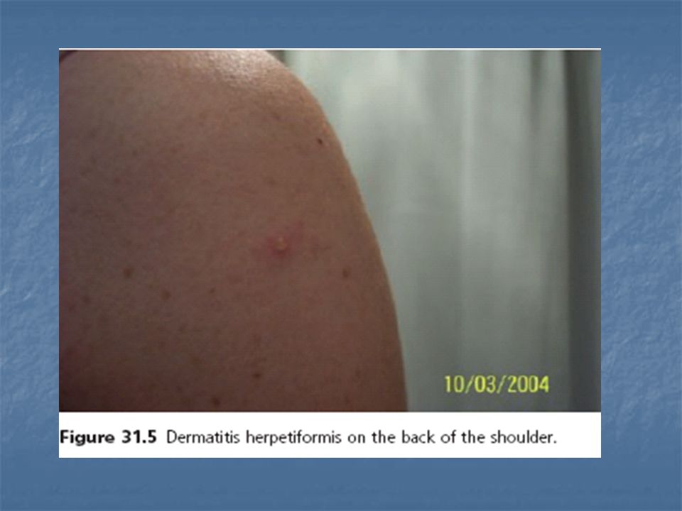
Dermatitis herpetiformis Dermatitis herpetiformis is a condition characterized by pruritic papulovesicles over the external surface of the extremities and on the trunk The diagnosis is confirmed histologically by the demonstration of granular IgA deposits along the nonaffected subepidermal basement membrane. Similar to celiac disease, antibodies against tissue transglutaminase (anti-tTG) are elevated in patients with the disease. The autoantibodies are directed mainly against epidermal transglutaminase, which shows high sequence homology to tTG Dermatitis herpetiformis is common among patients with celiac disease Although the celiac disease in patients with dermatitis herpetiformis is often asymptomatic, the skin lesions in most patients respond to gluten withdrawal
 are elevated in patients with the disease. The autoantibodies are directed mainly against epidermal transglutaminase, which shows high sequence homology to tTG. Dermatitis herpetiformis is common among patients with celiac disease. Although the celiac disease in patients with dermatitis herpetiformis is often asymptomatic, the skin lesions in most patients respond to gluten withdrawal.")
19
Dermatitis herpetiformis
Herpetiform clusters of vesicles on an erythematous, edamatous base with crusts and postinflammatory pigmentation on the upper back and shoulder.

22
ASSOCIATED CONDITIONS
Diabetes mellitus Celiac disease is closely associated with type 1 diabetes mellitus. Between 2.6 and 7.8 percent of adults with type 1 diabetes had IgA autoantibodies to endomysium or to tissue transglutaminase; most such patients were proven to have celiac disease with small bowel biopsy . Many such patients had no overt clinical manifestations of celiac disease. Type 1 diabetes and celiac disease share multiple genetic loci such as HLA-DR3, HLA-DQ2 (HLA-DQ8) and several genetic variations . This suggests that type 1 diabetes and celiac disease have common features in their pathogenesis such as tissue damage from autoimmunity or intolerance to dietary antigens.
 and several genetic variations . This suggests that type 1 diabetes and celiac disease have common features in their pathogenesis such as tissue damage from autoimmunity or intolerance to dietary antigens.")
23
ASSOCIATED CONDITIONS
Whether a gluten-free diet improves diabetes in diabetic patients with celiac disease is unclear Furthermore, the very early supplementation of newborns diet with gluten (<3 months) showed an increased risk for islet autoantibodies, which precede type 1 diabetes mellitus prospective clinical studies are required to clarify the relationship between celiac disease, type 1 diabetes, and other secondary autoimmunities.
 showed an increased risk for islet autoantibodies, which precede type 1 diabetes mellitus. prospective clinical studies are required to clarify the relationship between celiac disease, type 1 diabetes, and other secondary autoimmunities.")
24
ASSOCIATED CONDITIONS
Selective IgA deficiency An association between selective IgA deficiency and celiac disease appears to be well-established as screening programs have detected celiac disease in up to 8 percent of patients On the other hand, selective IgA deficiency occurs in 1 to 2 percent of patients with celiac disease Screening for celiac disease in patients with IgA deficiency is best done using an IgG test for tissue transglutaminase. Down syndrome — There appears to be a strong association between Down syndrome and celiac disease. The prevalence of biopsy proven celiac disease has been reported to be as high as 16 percent, representing a 20-fold increase compared with the general population

25
ASSOCIATED CONDITIONS
Liver disease celiac disease may be associated with nonspecific mild chronic elevation in serum aminotransferase levels (AST ranging from 29 to 80, and ALT ranging from 60 to 130 with the ALT usually slightly greater than AST. A meta-analysis found that in patients with cryptogenic hypertransaminasemia, celiac serologies were positive in six percent and duodenal biopsies suggested celiac disease in four percent In addition, the meta-analysis found abnormal serum transaminases in 27 percent of patients with newly diagnosed celiac disease. When a gluten free diet was followed, serum transaminases normalized in 63 to 90 percent of patients within a year.

26
ASSOCIATED CONDITIONS
Celiac disease has also been associated with advanced liver disease One report, for example, focused on four patients with severe liver disease (due to congenital liver fibrosis, massive steatosis, and progressive hepatitis of unclear origin) and untreated celiac disease. Hepatic dysfunction reversed in all patients following a gluten-free diet Although the number of patients studied is small, these data suggest that celiac disease may contribute to or be the cause of serious liver disease, which may improve following a gluten-free diet.
 and untreated celiac disease. Hepatic dysfunction reversed in all patients following a gluten-free diet. Although the number of patients studied is small, these data suggest that celiac disease may contribute to or be the cause of serious liver disease, which may improve following a gluten-free diet.")
27
ASSOCIATED CONDITIONS
The association between celiac disease and primary biliary cirrhosis (PBC) has also been described in other reports . Two studies suggested a prevalence of 6 to 11 percent in patients with PBC , although these may be overestimates. No cases of celiac disease were detected in 65 patients with PBC in a study from Italy . Recognition of celiac disease in patients with PBC may be important since both diseases impact negatively upon bone mineralization and are risk factors for osteoporosis
 has also been described in other reports . Two studies suggested a prevalence of 6 to 11 percent in patients with PBC , although these may be overestimates. No cases of celiac disease were detected in 65 patients with PBC in a study from Italy . Recognition of celiac disease in patients with PBC may be important since both diseases impact negatively upon bone mineralization and are risk factors for osteoporosis.")
28
ASSOCIATED CONDITIONS
Thyroid disease — There is an increased incidence of autoimmune thyroid disease among patients with celiac disease. Hypothyroidism is more frequent than hyperthyroidism. Gastroesophageal reflux disease — An association of celiac disease with gastroesophageal reflux disease (GERD) has been reported. Eosinophilic esophagitis — The incidence of eosinophilic esophagitis is increased in both children and adults with celiac disease (age-adjusted and sex-adjusted SIR 16.0, 95% CI ) .A diagnosis of eosinophilic esophagitis should be considered in patients with celiac disease and dysphagia or persistent reflux.
 has been reported. Eosinophilic esophagitis — The incidence of eosinophilic esophagitis is increased in both children and adults with celiac disease (age-adjusted and sex-adjusted SIR 16.0, 95% CI ) .A diagnosis of eosinophilic esophagitis should be considered in patients with celiac disease and dysphagia or persistent reflux.")
29
ASSOCIATED CONDITIONS
Inflammatory bowel disease Several case series have demonstrated an association between celiac disease and inflammatory bowel disease, more frequently with ulcerative colitis than Crohn’s disease In one case-control study, the risk of IBD in patients with celiac disease was elevated 10-fold while the risk of celiac disease in patients with IBD was comparable to controls One study demonstrated that first-degree relatives of patients with celiac disease may be at a fivefold increased risk of developing ulcerative colitis as compared to the general population

30
ASSOCIATED CONDITIONS
Menstrual and reproductive issues — Women with untreated celiac disease may have an increased frequency of reproductive abnormalities: later menarche, earlier menopause, secondary amenorrhea, recurrent miscarriage, and infertility Male infertility, characterized by abnormalities in sperm motility and morphology as well as a biochemical picture of androgen resistance (high serum testosterone and high LH concentrations), has been reported in celiac disease
, has been reported in celiac disease.")
31
ASSOCIATED CONDITIONS
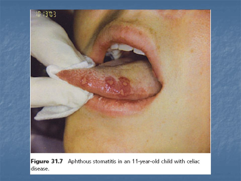
Myocarditis and cardiomyopathy Two reports from Italy suggest that celiac disease, which is often clinically unsuspected, accounts for as many as 5 percent of patients with autoimmune myocarditis or idiopathic dilated Atrophic glossitis — Oral lesions (erythema or atrophy) and a soreness or burning sensation of the tongue have been described in association with celiac disease and respond to a gluten free diet . Oral symptoms are frequent in patients with classical celiac disease, thus the involvement of the oral cavity is a helpful tool in diagnosis of celiac disease. Pancreatitis — A large database study described an increased risk of pancreatitis (both acute and chronic) in patients with celiac disease diagnosed in adulthood . Further studies are needed to clarify the strength of the association and potential mechanisms that underlie it. Ischemic heart disease — Some studies suggest that patients with celiac disease are at increased risk for ischemic heart disease .
 and a soreness or burning sensation of the tongue have been described in association with celiac disease and respond to a gluten free diet . Oral symptoms are frequent in patients with classical celiac disease, thus the involvement of the oral cavity is a helpful tool in diagnosis of celiac disease. Pancreatitis — A large database study described an increased risk of pancreatitis (both acute and chronic) in patients with celiac disease diagnosed in adulthood . Further studies are needed to clarify the strength of the association and potential mechanisms that underlie it. Ischemic heart disease — Some studies suggest that patients with celiac disease are at increased risk for ischemic heart disease .")
33
Diagnosis of celiac disease

34
Categories of celiac disease
The classical form, characterized by fully developed villous atrophy and features of intestinal malabsorption. The atypical form, characterized by fully developed villous atrophy in the setting of milder clinical features such as iron deficiency, osteoporosis, short stature, and/or infertility. Despite the historical title of "atypical", this form is the most common. The silent form in which villous atrophy is found after testing asymptomatic patients (eg, because of a family history of celiac disease or during an upper endoscopy performed for another reason). A potential form in those who have never had a biopsy consistent with celiac disease, but show serologic and/or immunologic abnormalities characteristic for the disorder. This is most often detected in patients with a family history of celiac disease. A latent form in patients who had a previous diagnosis of celiac disease that responded to gluten withdrawal but retained normal villous architecture after gluten reintroduction. The latent form also refers to patients with elevated IgA tTG serology but normal intestinal mucosa who may subsequently develop celiac disease.
. A potential form in those who have never had a biopsy consistent with celiac disease, but show serologic and/or immunologic abnormalities characteristic for the disorder. This is most often detected in patients with a family history of celiac disease. A latent form in patients who had a previous diagnosis of celiac disease that responded to gluten withdrawal but retained normal villous architecture after gluten reintroduction. The latent form also refers to patients with elevated IgA tTG serology but normal intestinal mucosa who may subsequently develop celiac disease.")
35
WHO SHOULD BE TESTED Those with gastrointestinal symptoms
chronic or recurrent diarrhea Malabsorption weight loss abdominal distension or bloating patients with symptoms suggestive for IBS severe lactose intolerance.

36
WHO SHOULD BE TESTED Individuals without other explanations for signs and symptoms Iron deficiency anemia folate or vitamin B12 deficiency persistent elevation in serum aminotransferases, short stature delayed puberty recurrent fetal loss low birthweight infants reduced fertility persistent aphthous stomatitis dental enamel hypoplasia idiopathic peripheral neuropathy nonhereditary cerebellar ataxia recurrent migraine headaches.

37
WHO SHOULD BE TESTED Symptomatic individuals at high risk for celiac disease patients with type 1 diabetes mellitus other autoimmune disorders first- and second-degree relatives of individuals with celiac disease patients with Turner, Down, or Williams syndromes.

38
WHO SHOULD NOT BE TESTED
screening of the general population is not recommended. Screening of patients with osteoporosis is also not recommended in the consensus statement since the prevalence of celiac disease is not significantly increased among the general population of patients with osteoporosis.

41
Serologic evaluation As a general rule, testing should begin with serologic evaluation. As will be discussed below, the most sensitive and specific tests are IgA anti tissue transglutaminase and IgA endomysial antibody, which have equivalent diagnostic accuracy. By contrast, antigliadin antibody tests are no longer used routinely because of their lower sensitivity and specificity. However, a second generation AGA test (Deamidated Gliadin Peptide [DGP]) yielded far higher diagnostic accuracy (sensitivity 94 percent, specificity 99 percent) .the combination of anti-tTG and DGP serology may become a powerful non-invasive pairing for serologic diagnosis of celiac disease. Serologic testing may not be as accurate in children less than age five and is less accurate before age two.
 yielded far higher diagnostic accuracy (sensitivity 94 percent, specificity 99 percent) .the combination of anti-tTG and DGP serology may become a powerful non-invasive pairing for serologic diagnosis of celiac disease. Serologic testing may not be as accurate in children less than age five and is less accurate before age two.")
42
Testing on a gluten-rich diet
All diagnostic tests should be performed while the patient is on a gluten-rich diet. Some patients may have already begun a low gluten diet before undergoing formal evaluation and thus may have normal results from antibody testing. Such patients should be advised to consider resuming a gluten-rich diet for 2 to 12 weeks before antibody titers are drawn Antibody levels remain elevated for varying lengths of time (1 to 12 months) after patients with celiac disease begin a gluten-free diet. Thus, antibody testing in patients who have only recently begun a gluten-free diet is reasonable and may yield a positive result, although testing while on a gluten containing diet is preferable to exclude celiac disease.
 after patients with celiac disease begin a gluten-free diet. Thus, antibody testing in patients who have only recently begun a gluten-free diet is reasonable and may yield a positive result, although testing while on a gluten containing diet is preferable to exclude celiac disease.")
43
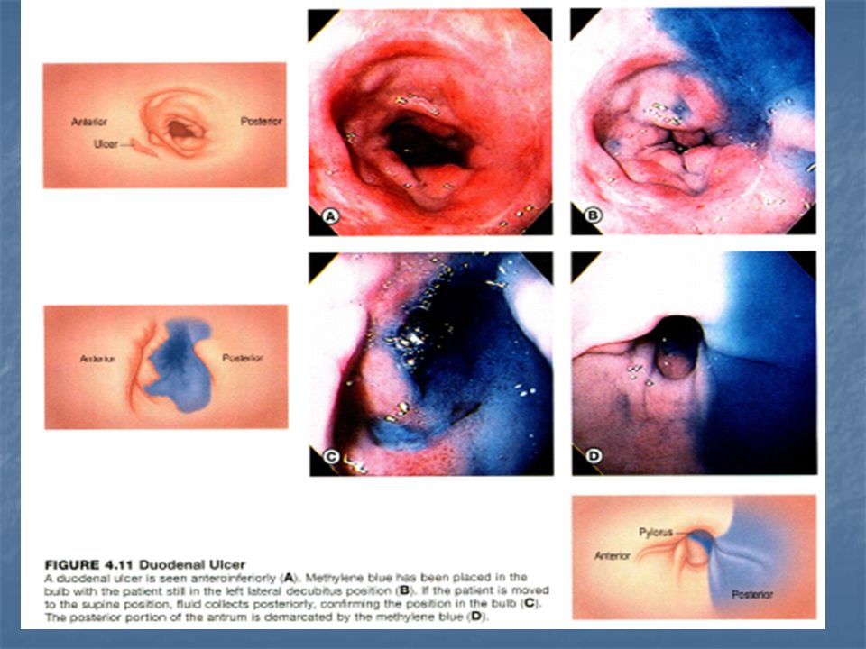
Small bowel biopsy Patients with a positive IgA endomysial or transglutaminase antibody test should undergo a small bowel biopsy. Exceptions are those who have biopsy-proven dermatitis herpetiformis in whom the diagnosis can be established without a small bowel biopsy. Multiple biopsies should be obtained from the duodenal bulb and the second and third portion of the duodenum Duodenal bulb biopsies should be clearly labeled as such to help ensure that the pathologist takes into account the different mucosal architecture of the bulb to avoid false positive reports of villous atrophy.

44
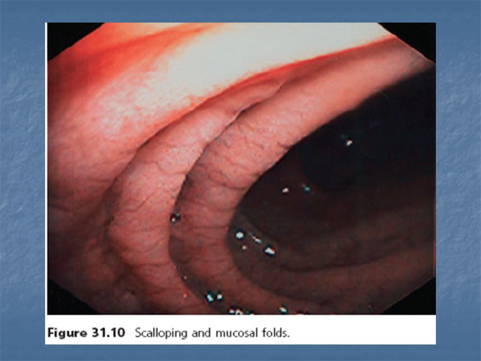
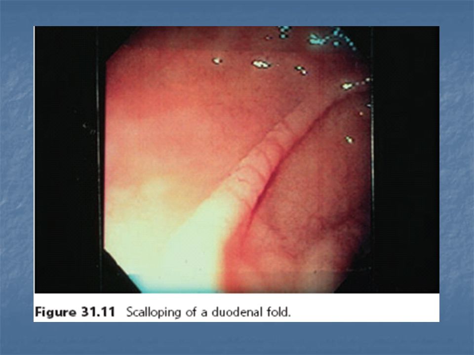
Small bowel biopsy The exact minimal number of duodenal biopsies needed to make a diagnosis of celiac disease is uncertain, although some experts believe that at least four should be obtained The duodenal mucosa may appear atrophic with loss of folds, contain visible fissures, have a nodular appearance or the folds may be scalloped but such findings are not universally present and may be seen with other disorders Staining techniques and high resolution magnification endoscopy can help identify areas of villous atrophy for biopsy.

46
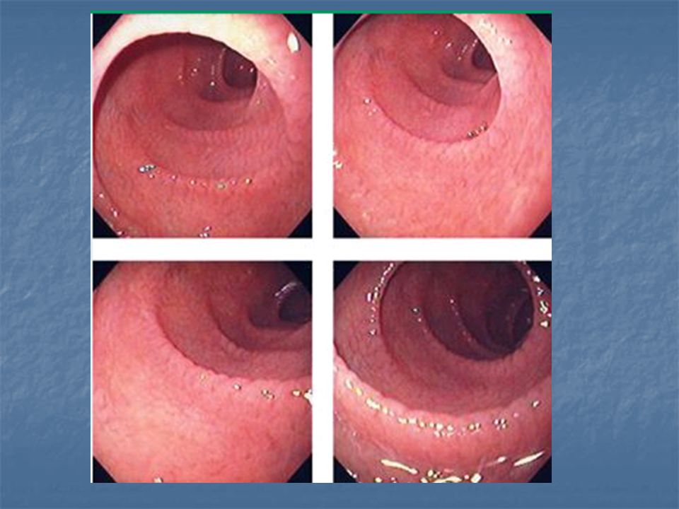
A. The second duodenum is characterized
by circular folds termed valvulae conniventes or Kerckring's valves. The mucosa has a granular appearance. The junction of the second and third duodenum (inferior duodenal angle) is seen in the distance. B. The mucosa may have a frosty white appearance.
 is seen in the. distance. B. The mucosa may have a. frosty white appearance.")
47
C. The duodenal mucosa is characterized by slender villi composed of goblet cells.

52
Marsh-Oberhuber classification
Marsh 0 (preinfiltrative): less than 30 intraepithelial lymphocytes (IELs) per 100 enterocytes, normal villi and crypts. This finding represents to normal histology. Marsh 1 (infiltrative): more than 30 IELs per 100 enterocytes, normal villi and crypts. This finding is nonspecific and can occur in early or mild celiac disease, but it may also be seen in healthy individuals, in patients with small intestinal bacterial overgrowth, in Crohn’s disease, in other autoimmune disorders, in patients infected with Helicobacter pylori, in patients taking nonsteroidal antiinflammatory drugs, and in a variety of other situations. Marsh 2 (infiltrative-hyperplastic): more than 30 IELs per 100 enterocytes, normal villi, crypt hyperplasia
: less than 30 intraepithelial lymphocytes (IELs) per 100 enterocytes, normal villi and crypts. This finding represents to normal histology. Marsh 1 (infiltrative): more than 30 IELs per 100 enterocytes, normal villi and crypts. This finding is nonspecific and can occur in early or mild celiac disease, but it may also be seen in healthy individuals, in patients with small intestinal bacterial overgrowth, in Crohn’s disease, in other autoimmune disorders, in patients infected with Helicobacter pylori, in patients taking nonsteroidal antiinflammatory drugs, and in a variety of other situations. Marsh 2 (infiltrative-hyperplastic): more than 30 IELs per 100 enterocytes, normal villi, crypt hyperplasia.")
53
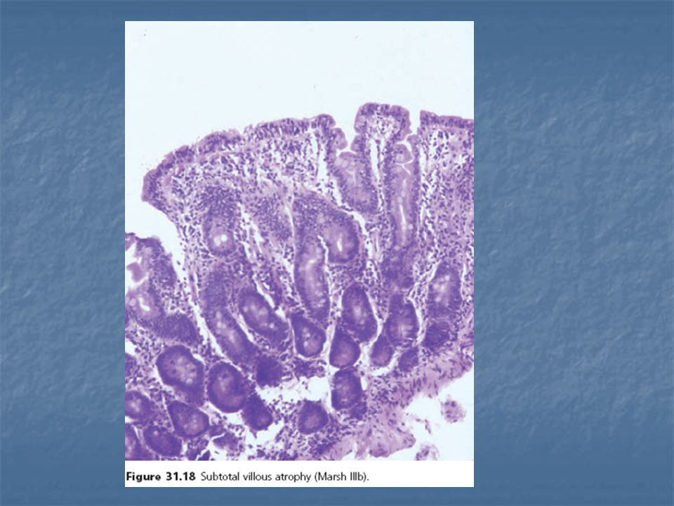
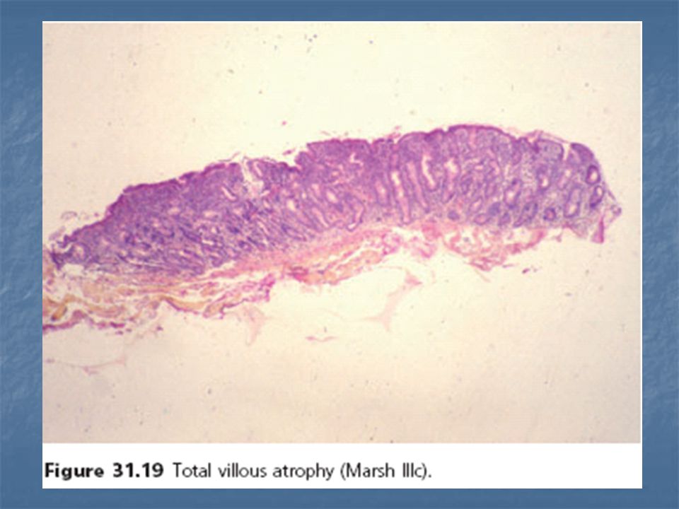
Marsh 3a (flat destructive): more than 30 IELs per 100 enterocytes, mild villous atrophy, crypt hyperplasia Marsh 3b (flat destructive): more than 30 IELs per 100 enterocytes, moderate villous atrophy, crypt hyperplasia Marsh 3c (flat destructive): more than 30 IELs per 100 enterocytes, total villous atrophy, crypt hyperplasia Marsh 4 (atrophic-hypoplastic): more than 30 IELs per 100 enterocytes, total villous atrophy, crypt hypoplasia
: more than 30 IELs per 100 enterocytes, moderate villous atrophy, crypt hyperplasia. Marsh 3c (flat destructive): more than 30 IELs per 100 enterocytes, total villous atrophy, crypt hyperplasia. Marsh 4 (atrophic-hypoplastic): more than 30 IELs per 100 enterocytes, total villous atrophy, crypt hypoplasia.")
58
The diagnosis is presumptively established when there is concordance between the serologic results and the biopsy findings. It is confirmed when symptoms resolve subsequently on a gluten-free diet. Demonstration of histologic normalization is not always required.

59
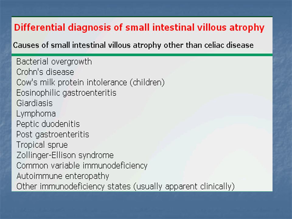
Suggestive clinical features but negative serologic tests
There are three main possibilities in those with suggestive clinical features but negative serologic tests: The individual may have selective IgA deficiency. The individual may already be on a low gluten diet. The patient may not have celiac disease in which case other causes of symptoms or villous atrophy should be considered (table 1 and algorithm 2). here are occasional patients in whom the diagnosis is unclear despite the above. Such patients can undergo testing for HLA haplotypes associated with celiac disease. More than 99 percent of patients with celiac disease have HLA DQ2 and/or DQ8 compared with about 40 percent of the general population. Thus, celiac disease is highly unlikely in patients without these haplotypes
. here are occasional patients in whom the diagnosis is unclear despite the above. Such patients can undergo testing for HLA haplotypes associated with celiac disease. More than 99 percent of patients with celiac disease have HLA DQ2 and/or DQ8 compared with about 40 percent of the general population. Thus, celiac disease is highly unlikely in patients without these haplotypes.")
61
Positive serologic tests but negative small bowel biopsies
False positive tTG results are rare but do occur and are usually low titer (typically less than twice the upper limit of normal). Repeating the test using an assay that uses human tTG as the capture antigen may resolve the discrepancy since older tTG tests using non-human tTG have more frequently been associated with false positive results. The intestinal biopsy should be reviewed by a pathologist familiar with CD to look for subtle abnormalities of CD such as an increase in IELs. If these two steps do not reconcile the results, the patient can be placed on a high gluten diet and, after 6 to 12 weeks, numerous additional biopsies obtained from multiple sites in the mid and distal duodenum since CD enteropathy can be patchy and missed due to sampling error. As noted above, staining techniques and high resolution magnification endoscopy can help identify areas of villous atrophy for biopsy.
. Repeating the test using an assay that uses human tTG as the capture antigen may resolve the discrepancy since older tTG tests using non-human tTG have more frequently been associated with false positive results. The intestinal biopsy should be reviewed by a pathologist familiar with CD to look for subtle abnormalities of CD such as an increase in IELs. If these two steps do not reconcile the results, the patient can be placed on a high gluten diet and, after 6 to 12 weeks, numerous additional biopsies obtained from multiple sites in the mid and distal duodenum since CD enteropathy can be patchy and missed due to sampling error. As noted above, staining techniques and high resolution magnification endoscopy can help identify areas of villous atrophy for biopsy.")
62
Some patients have a positive serologic test and only mild histologic changes supportive of celiac disease. Patients with mild villous atrophy appear to benefit from a gluten-free diet. The response to gluten-free diet in patients with an increase in intraepithelial lymphocytes but normal villi is less clear

63
IgA EMA, IgA tTG, and IgA AGA levels fall with treatment;
as a result, these assays can be used as a noninvasive means of monitoring the response to a gluten-free diet

64
IgA endomysial assay Endomysial antibodies bind to connective tissue surrounding smooth muscle cells Frozen sections of monkey esophagus were initially used for the assay. Currently, many laboratories use sections of human umbilical cord which are more readily available. Serum IgA endomysial antibodies bind to the endomysium, producing a characteristic staining pattern, which is visualized by indirect immunofluorescence . The test result is reported simply as positive or negative since even low titers of serum IgA endomysial antibodies are specific for celiac disease. The target antigen has been identified as a tissue transglutaminase IgA endomysial antibody testing is moderately sensitive and highly specific for untreated celiac disease . Serum levels of IgA endomysial antibody fall on a gluten-free diet and the test often becomes negative in treated patients .

65
Anti-tissue transglutaminase antibodies
The antigen against which antiendomysial antibodies are directed is a tissue transglutaminase (tTG) Anti-tTG antibodies were highly sensitive and specific for the diagnosis of celiac disease in most reports ELISA tests for IgA anti-tTG antibodies are now widely available and are easier to perform and less costly than the immunofluorescence assay used to detect IgA endomysial antibodies. The diagnostic accuracy of IgA anti-tTG immunoassays has been improved further by the use of human tTG in place of the non-human tTG preparations used in earlier immunoassay kits.
 Anti-tTG antibodies were highly sensitive and specific for the diagnosis of celiac disease in most reports. ELISA tests for IgA anti-tTG antibodies are now widely available and are easier to perform and less costly than the immunofluorescence assay used to detect IgA endomysial antibodies. The diagnostic accuracy of IgA anti-tTG immunoassays has been improved further by the use of human tTG in place of the non-human tTG preparations used in earlier immunoassay kits.")
66
Antigliadin antibody assays
Gliadin is a component of the wheat storage protein gluten. Purified gliadin is readily available and is used as the antigen for enzyme-linked immunosorbent assays (ELISA) to detect serum antigliadin antibodies. positive predictive value in a general population is relatively poor Antigliadin antibody test results are reported as a titer or level. A high titer of antigliadin antibody is somewhat more specific for celiac disease than a low titer, but some normal individuals have high serum levels of antigliadin antibody. Antigliadin antibody levels decrease during treatment with a gluten-free diet.
 to detect serum antigliadin antibodies. positive predictive value in a general population is relatively poor. Antigliadin antibody test results are reported as a titer or level. A high titer of antigliadin antibody is somewhat more specific for celiac disease than a low titer, but some normal individuals have high serum levels of antigliadin antibody. Antigliadin antibody levels decrease during treatment with a gluten-free diet.")
67
Assay sensitivity and specificity
A systematic review of the literature estimated that the sensitivity and specificity of IgA endomysial and IgA tissue transglutaminase antibodies were over 95 percent and close to 100 percent, respectively The following data come from our experience: IgA endomysial antibodies – sensitivity 85 to 98 percent; specificity 97 to 100 percent IgA tissue transglutaminase antibodies – sensitivity 90 to 98 percent; specificity 95 to 97 percent IgA antigliadin antibodies – sensitivity 80 to 90 percent; specificity 85 to 95 percent IgG antigliadin antibodies – sensitivity 75 to 85 percent; specificity 75 to 90 percent

68
Assay sensitivity and specificity
It is also important to consider the likelihood that the patient has the disease since, at a given sensitivity and specificity, the pretest probability of disease determines the positive and negative predictive values of the test In addition to laboratory variation, the sensitivity of these tests may depend upon the severity of the disease. In one report, as an example, serum antibodies were determined in 101 patients with biopsy proven celiac disease . The sensitivity of IgA endomysial antibodies varied for 100 percent in patients with total villous atrophy to only 31 percent in those with partial villous atrophy.

69
Role of antibodies in disease pathogenesis
Antigliadin antibodies do not appear to be essential for the pathogenesis of celiac disease . Furthermore, many normal individuals have increased IgA and/or IgG antigliadin levels In contrast, IgA endomysial antibodies are rarely found in the absence of gluten-sensitive enteropathy. However, patients with celiac disease who lack these antibodies do not differ in their clinical presentation from those who are antibody positive. antibodies against tissue transglutaminase are of some pathogenetic importance. Furthermore, tissue transglutaminase enzymatically alters gliadin peptides to increase their propensity to induce helper T-cell activation when presented by DQ2 or DQ8 on antigen presenting cells. Individuals with celiac disease also have increased levels of serum antibodies against other food proteins such as beta-lactoglobulin, casein, and ovalbumin . It is not clear whether this reflects a general aberrant immune responsiveness to food antigens or results from enhanced systemic exposure to these proteins because of increased small intestinal permeability.

70
CLINICAL APPLICATION OF SEROLOGIC TESTS
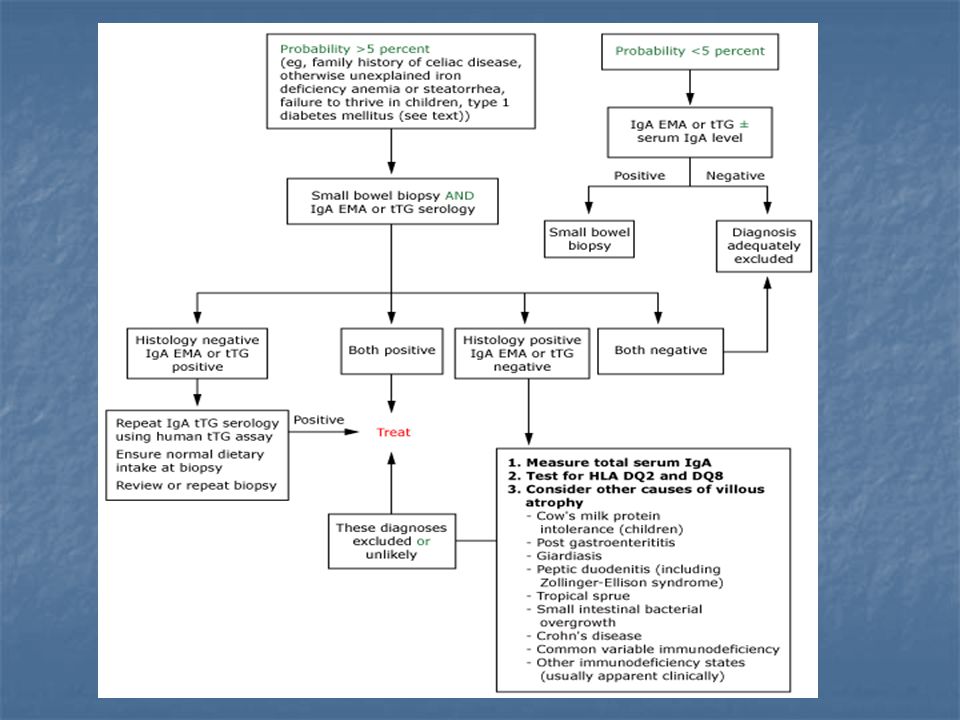
Three different clinical circumstances should be considered when using serologic studies in the diagnosis and management of celiac disease: Evaluation of individuals with a low pretest probability for celiac disease. Such patients may have undergone testing for celiac disease during evaluation of common disorders such as irritable bowel syndrome, reduced fertility or osteoporosis. Evaluation of individuals with a moderate or high pretest probability for celiac disease Monitoring adherence and response to a gluten-free diet

71
Individuals with a low risk for celiac disease
Asymptomatic patients without a family history of celiac disease or laboratory or clinical evidence for malabsorption can be considered to be low risk In the low-risk setting, the IgA endomysial antibody test has the highest diagnostic accuracy but is a little more costly than the IgA tTG ELISA test.As noted above, the serum IgA tissue transglutaminase and IgA endomysial tests have similar sensitivities. A negative result for either test has a high negative predictive value in this situation and may obviate the need for small bowel biopsy. IgG DGP testing can be used in patients with IgA deficiency. The specificities of the IgA endomysial and IgA tissue transglutaminase tests are high. Thus, their positive predictive values are high even in low-risk populations In contrast, the specificities of IgA and IgG antigliadin tests are lower, and positive results have a low positive predictive value in low-risk populations

72
Individuals with a moderate or high risk for celiac disease
When the pretest probability of celiac disease is perceived to be high (ie, greater than 5 percent), diagnosis is based upon serologic tests and histology. Celiac disease is frequently associated with dermatitis herpetiformis, Down syndrome, selective IgA deficiency, and other conditions that have autoimmune features such as type 1 diabetes mellitus, thyroid disease, and autoimmune liver disease. Patients with these conditions or a family history of celiac disease can be considered as being at increased risk. Patients with suggestive clinical features such as severe diarrhea, weight loss, or persistent anemia can also be considered to be at high risk. We recommend performing both IgA endomysial (or TTG) and small bowel biopsy prior to dietary treatment.
, diagnosis is based upon serologic tests and histology. Celiac disease is frequently associated with dermatitis herpetiformis, Down syndrome, selective IgA deficiency, and other conditions that have autoimmune features such as type 1 diabetes mellitus, thyroid disease, and autoimmune liver disease. Patients with these conditions or a family history of celiac disease can be considered as being at increased risk. Patients with suggestive clinical features such as severe diarrhea, weight loss, or persistent anemia can also be considered to be at high risk. We recommend performing both IgA endomysial (or TTG) and small bowel biopsy prior to dietary treatment.")
73
SCREENING IN ASYMPTOMATIC INDIVIDUALS
Screening studies suggest that the incidence of CD in whites of northern European ancestry may be as high as 1:100 to 1:250. The benefit of population screening include: a reduction in risk for enteropathy-associated T-cell lymphoma, a reversal of unrecognized nutritional deficiency states, resolution of mild or ignored intestinal symptoms, avoidance of other autoimmune disorders, an improvement in general well-being, and a possible reduction in mortality. Asymptomatic individuals may not be sufficiently motivated to adhere to a strict gluten-free diet. There may also be adverse psychological effects when asymptomatic individuals receive a diagnosis of a chronic incurable condition that demands substantial lifestyle changes. For these reasons, widespread screening of asymptomatic individuals is not generally advocated at this time, even in populations in which the prevalence of celiac disease is high.

74
OTHER NONINVASIVE TESTS FOR THE DIAGNOSIS OF CELIAC DISEASE
A variety of hematologic and biochemical abnormalities may be found in individuals with untreated celiac disease including iron deficiency, folic acid deficiency, and vitamin D deficiency. These abnormalities reflect nutritional deficiency states secondary to enteropathy-induced malabsorption. Although relevant to patient evaluation and management, none is sufficiently sensitive or specific to serve as useful screening or diagnostic tools An oral xylose and/or lactulose absorption test, fecal fat evaluation, small bowel radiographic study, or capsule endoscopy may also be abnormal in untreated celiac disease, but will not provide a specific diagnosis HLA typing may be useful in patients who are already on a gluten-free diet without having achieved a firm diagnosis. Those without HLA DQ2 or DQ8 are very unlikely to have celiac disease

75
SUMMARY AND RECOMMENDATIONS
An approach to diagnosis of celiac disease is summarized in the following algorithm (algorithm 1). All testing should be performed while patients are on a gluten-containing diet. IgA deficiency is more common in celiac disease (2 to 5 percent) than in the general population (<0.5 percent). The IgA EMA and IgA tTG serology tests will be falsely negative in untreated celiac disease in patients with IgA deficiency. As a result, total serum IgA can be measured in addition to IgA EMA or IgA tTG especially when there is heightened clinical suspicion for celiac disease and IgA markers are negative. If total IgA levels are abnormally low, an IgG-based assay should be used to test for celiac disease. The IgG antigliadin assay has been traditionally used in this circumstance but is not ideal since it yields frequent false positive results. Thus, serum IgG tTG or IgG DGP tests are preferable. Negative results upon testing for HLA DQ2 or DQ8 can also help exclude the diagnosis in this setting.
. All testing should be performed while patients are on a gluten-containing diet. IgA deficiency is more common in celiac disease (2 to 5 percent) than in the general population (<0.5 percent). The IgA EMA and IgA tTG serology tests will be falsely negative in untreated celiac disease in patients with IgA deficiency. As a result, total serum IgA can be measured in addition to IgA EMA or IgA tTG especially when there is heightened clinical suspicion for celiac disease and IgA markers are negative. If total IgA levels are abnormally low, an IgG-based assay should be used to test for celiac disease. The IgG antigliadin assay has been traditionally used in this circumstance but is not ideal since it yields frequent false positive results. Thus, serum IgG tTG or IgG DGP tests are preferable. Negative results upon testing for HLA DQ2 or DQ8 can also help exclude the diagnosis in this setting.")
76
SUMMARY AND RECOMMENDATIONS
A few controversies remain regarding confirmation of the diagnosis. Some authorities recommend that a repeat small intestinal biopsy should be obtained 6 to 24 months after beginning a gluten-free diet to demonstrate histologic improvement. Some also recommended a repeat small bowel biopsy (after a gluten rechallenge) following a gluten-free diet. We do not believe that these approaches are generally required unless the diagnosis remains uncertain based upon the serologic profile, histology and clinical response. Furthermore, these approaches are not recommended in a consensus statement issued by the NIH
 following a gluten-free diet. We do not believe that these approaches are generally required unless the diagnosis remains uncertain based upon the serologic profile, histology and clinical response. Furthermore, these approaches are not recommended in a consensus statement issued by the NIH.")
77
Management of celiac disease in adults

78
Indications — A gluten-free diet is recommended in patients with celiac disease (classic disease, atypical celiac disease, and asymptomatic or silent celiac disease). Patients with latent celiac disease (positive IgA endomysial antibody, but normal small bowel biopsy) are currently not advised to be on a gluten-free diet but should continue to be monitored and rebiopsied if symptoms develop.
 are currently not advised to be on a gluten-free diet but should continue to be monitored and rebiopsied if symptoms develop.")
79
dietary advice Foods containing wheat, rye, and barley should be avoided. Soybean or tapioca flours, rice, corn, buckwheat, and potatoes are safe. Read labels on prepared foods and condiments carefully, paying particular attention to additives such as stabilizers or emulsifiers that may contain gluten. Dairy products may not be well tolerated initially since many patients with celiac disease can have secondary lactose intolerance. As a result, lactose-containing products should initially be avoided in patients whose symptoms appear to be worsened by them.

80
dietary advice Oat consumption should be limited to 50 to 60 g/day in patients with mild disease upon presentation or whose disease is in remission after a stringent gluten-free diet. The latter patients should be followed carefully for clinical or serologic evidence of disease recurrence after reintroducing oats. Patients with severe disease should avoid oats altogether. Although gluten is also found in oats, the toxicity of oats in celiac disease is now in doubt because some studies suggest that pure oat flour can be tolerated without disease recurrence

81
Is strict gluten avoidance necessary?
the ability to tolerate gluten in the diet is highly variable among patients. While some patients are exquisitely sensitive to even small amounts of gluten, other patients can tolerate the reintroduction of small amounts in their diet after achieving remission. Despite this variable response, several arguments favor encouraging strict adherence to a gluten-free diet in most patients with established celiac disease regardless of clinical symptoms

82
MONITORING THE RESPONSE TO A GLUTEN-FREE DIET
Approximately 70 percent of patients have noticeable clinical improvement within two weeks As a general rule, symptoms improve faster than histology, especially when biopsies are obtained in the proximal intestine. The reason is incompletely understood; however, a possible explanation is that the less severely damaged distal small intestine recovers faster than the proximal intestine, which is typically more severely affected due to relatively increased exposure to gluten

83
MONITORING THE RESPONSE TO A GLUTEN-FREE DIET
We suggest that patients be evaluated four to six weeks following the initiation of a gluten-free diet at which time a complete blood count, folate, B12, iron studies, liver chemistries, and serologic testing should be performed. It is important to note that women often experience breast tenderness for three months after starting a gluten-free diet and reassurance should be provided.

84
MONITORING THE RESPONSE TO A GLUTEN-FREE DIET
Serologic testing with increased use of IgA tTG for initial diagnosis this assay is now also increasingly used in monitoring response to gluten-free diet. A normal baseline value is typically reached within 3 to 12 months depending upon the pre-treatment concentrations. Normal IgA tTG levels do not reliably indicate recovery from villous atrophy Conversely, if the levels do not fall as anticipated, the patient is usually continuing to ingest gluten either intentionally or inadvertently

85
Small bowel biopsy The need for a follow-up biopsy in patients with clinical improvement has been debated, especially since serologic testing can be used to monitor recovery and compliance with the diet. Some authorities rely on clinical improvement and changes in serologic markers, reserving re-biopsy for nonresponsive patients in whom there remains diagnostic uncertainty, or those who wish to confirm mucosal healing

86
The author's practice is to repeat a small intestinal biopsy in all patients three to four months after beginning a gluten-free diet to demonstrate histologic improvement. If improvement in small intestinal morphology is not seen, but symptomatic improvement has occurred, the diet should be continued and the small intestinal biopsy should be repeated after six to nine months. Although histological improvement is usually seen, persistent abnormalities have been described, even in patients with symptomatic improvement . The significance is at the moment unclear, but this may be due to low level gluten contamination, a persistent immune response or other unknown mechanisms. Other diagnoses, after a first check for poor compliance, or inadvertent ingestion of gluten should be considered in these patients (see 'Other diagnoses' below).
.")
87
Gluten rechallenge Gluten rechallenge is unnecessary according to a consensus statement issued by the National Institutes of Health and Guidelines issued by the European Society of Paediatric Gastroenterology and Nutrition (ESPGAN) According to the ESPGAN guidelines, gluten rechallenge should only be used in equivocal cases such as when no initial biopsy was performed, biopsies were inadequate or atypical, in communities with high rates of other enteropathies, or in situations when patients plan to abandon a gluten-free diet in an uncontrolled way. Gluten rechallenge should be performed after obtaining a control biopsy on a gluten-free diet and repeat biopsies should be obtained three to six months later with the recognition that relapse can take five to seven years or more. A rare hazard in giving a gluten rechallenge is the development of fulminant diarrhea, with resulting dehydration, acidosis, and other metabolic disturbances (a condition known as "gliadin shock") Such patients should be treated with glucocorticoids. There is a suggestion that use of gluten challenge in young children may be associated with an increased likelihood of development of autoimmune disorders such as IDDM.
 According to the ESPGAN guidelines, gluten rechallenge should only be used in equivocal cases such as when no initial biopsy was performed, biopsies were inadequate or atypical, in communities with high rates of other enteropathies, or in situations when patients plan to abandon a gluten-free diet in an uncontrolled way. Gluten rechallenge should be performed after obtaining a control biopsy on a gluten-free diet and repeat biopsies should be obtained three to six months later with the recognition that relapse can take five to seven years or more. A rare hazard in giving a gluten rechallenge is the development of fulminant diarrhea, with resulting dehydration, acidosis, and other metabolic disturbances (a condition known as gliadin shock ) Such patients should be treated with glucocorticoids. There is a suggestion that use of gluten challenge in young children may be associated with an increased likelihood of development of autoimmune disorders such as IDDM.")
88
NONRESPONDERS Nonresponders are individuals who have persistent symptoms or serologic and/or histologic abnormalities after two years on a gluten-free diet. The majority of patients with celiac disease respond to a gluten-free diet but approximately 5 percent of individuals do not. In patients who are incomplete responders or nonresponders, it is important to consider that not all clinical features of celiac disease respond at the same rate Furthermore, bone loss due to secondary hyperparathyroidism and peripheral neuropathy may only improve partially despite a gluten-free diet [

89
Patients who do not respond to a gluten-free diet fall into five main categories:
Patients with poor compliance or inadvertent gluten ingestion Patients with clinical or histologic features that overlap with celiac disease but are caused by other disorders Patients with concurrent disorders Patients with refractory sprue Patients with ulcerative jejunitis or intestinal lymphoma

90
1)Poor compliance or inadvertent gluten ingestion
The most common reasons for a lack of response are poor compliance or inadvertent gluten ingestion In patients who continue to have symptoms or persistent histologic abnormalities, or in those in whom serum antibody titers have not declined, a meticulous dietary history should be obtained, and dietary counseling pursued with a dietitian specifically trained and experienced in celiac disease.

91
2)Other diagnoses An erroneous diagnosis of celiac sprue may result from false positive serology, specifically IgA antigliadin antibodies. Diseases associated with small bowel villous atrophy should be excluded in patients with persistent symptoms who do not show histologic improvement

92
3)Concurrent disorders
Other concurrent diagnoses should be considered in patients who, despite apparent compliance, continue to have symptoms or do not have histologic improvement Concomitant or secondary lactose intolerance is a possible cause of continued diarrhea and flatulence Patients with celiac disease may have concurrent bowel disturbances such as irritable bowel syndrome, which affects a large proportion of the general population. Small bowel bacterial overgrowth, which may respond to antibiotics, develops in a small percentage of patients with celiac disease Some patients have coexisting pancreatic insufficiency Microscopic colitis is found in 4 percent of patients with celiac disease, which represents a 70-fold increase in risk. These patients had more severe villous atrophy and frequently required glucocorticoids or immunosuppressive drugs to treat the diarrhea

93
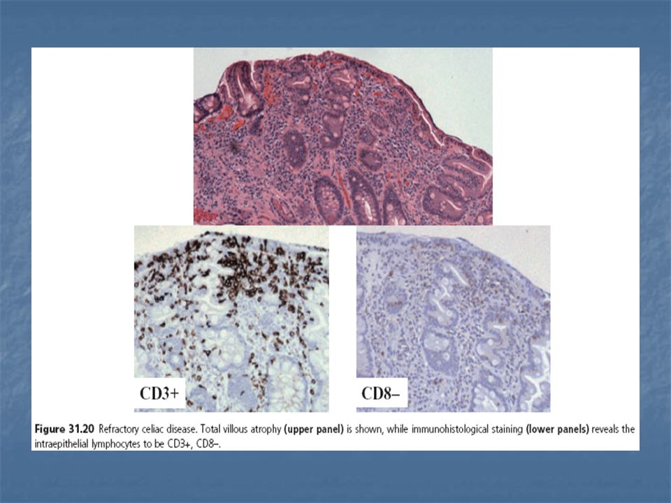
4)Refractory sprue Patients with refractory sprue (also referred to as "unclassified sprue") fall into two clinical categories : Patients who have no initial response to a gluten-free diet Patients who experience initial clinical improvement on a gluten-free diet, but, after a period of remission, develop disease refractory to gluten abstinence Refractory sprue has also been subdivided into two immunologic categories : Type 1 in which there is a normal population of intraepithelial lymphocytes. Type 2 in which there is an aberrant or premalignant population of intraepithelial lymphocytes based upon clonality analysis of T-cell receptors and immunophenotyping. Type 2 can progress to enteropathy-associated T-cell lymphoma, which may present clinically as ulcerative jejunitis

95
Patients with type 1 disease have a less severe presentation and a much better prognosis than patients with type 2 disease Furthermore, type 1 does not appear to evolve into type 2. An illustrative study compared outcomes in 41 patients with type 1 disease to 50 patients with type 2 disease [66]. Five-year survival was higher in the type 1 group (96 versus 58 percent). Most deaths were due to development of T-cell lymphoma (which developed in one-half of patients during follow-up). No patient with type 1 disease developed type 2 disease during an average five years follow-up. Refractory sprue (particularly type 2) can be severe and associated with progressive malabsorption and death. A subset of patients develops subepithelial collagen deposition, a condition referred to as "collagenous sprue"
. Most deaths were due to development of T-cell lymphoma (which developed in one-half of patients during follow-up). No patient with type 1 disease developed type 2 disease during an average five years follow-up. Refractory sprue (particularly type 2) can be severe and associated with progressive malabsorption and death. A subset of patients develops subepithelial collagen deposition, a condition referred to as collagenous sprue")
96
The cause of refractory sprue is unknown.
It is possible that some patients with this condition develop sensitivity to a dietary constituent other than gluten However, identification of the responsible antigens in most patients is difficult and unrewarding. As a result, treatment has focused on immunosuppression, which has traditionally relied upon glucocorticoids. The dose of glucocorticoids required varies among patients, and not all patients respond. In severely ill patients, we usually begin with hydrocortisone (100 mg IV Q6H). Oral dosing (such as 40 to 60 mg of prednisolone daily) can be used in patients who are tolerating an oral diet. After a few weeks, the dose can be reduced by 5 to 10 mg per day in responding patients and subsequently tapered to the lowest dose that keeps the patient in remission.
. Oral dosing (such as 40 to 60 mg of prednisolone daily) can be used in patients who are tolerating an oral diet. After a few weeks, the dose can be reduced by 5 to 10 mg per day in responding patients and subsequently tapered to the lowest dose that keeps the patient in remission.")
97
Other dugs used in case series:
Experience with alternative immunosuppressant therapy in patients who require high doses of glucocorticoids is limited to case reports and clinical experience. However, azathioprine and 6-mercaptopurine appear to be effective steroid-sparing agents Oral budesonide (9 mg, range 6 to 12 mg) has been effective in case series Other dugs used in case series: emental diet in patients with type 1 refractory sprue small intestinal release mesalamine type 2 disease following treatment with alemtuzumab, an anti-CD52 monoclonal antibody Cladribine in patients with type 2 refractory sprue
 has been effective in case series. Other dugs used in case series: emental diet in patients with type 1 refractory sprue. small intestinal release mesalamine. type 2 disease following treatment with alemtuzumab, an anti-CD52 monoclonal antibody. Cladribine in patients with type 2 refractory sprue.")
98
5)Ulcerative jejunitis and intestinal lymphoma
Ulcerative jejunitis and lymphoma should be considered in patients with refractory sprue unresponsive to glucocorticoids. The conditions are thought to share a similar pathogenesis, since both have aberrant T-cell monoclonality The disease most commonly presents in middle-aged patients with underlying celiac disease.

99
Patients with ulcerative jejunitis have multiple chronic, benign-appearing ulcers, most frequently in the jejunum. Clinical manifestations are similar to severe celiac disease; patients may present with new or recurrent symptoms of malabsorption, lassitude, anorexia, weight loss, abdominal pain, diarrhea, and fever despite being on a gluten-free diet Intestinal stricturing can develop with resulting small bowel obstruction. Evaluation should begin with an abdominal computed tomography (CT) enterography or magnetic resonance (MR) enterography scan and upper endoscopy, which, if negative, should be followed by a capsule endoscopy. Ulcerative jejunitis responds poorly to a gluten-free diet and is associated with an unfavorable prognosis. Up to one-third of patients die from complications. The prognosis can be improved if the ulcerated or strictured segment can be resected.
 enterography or magnetic resonance (MR) enterography scan and upper endoscopy, which, if negative, should be followed by a capsule endoscopy. Ulcerative jejunitis responds poorly to a gluten-free diet and is associated with an unfavorable prognosis. Up to one-third of patients die from complications. The prognosis can be improved if the ulcerated or strictured segment can be resected.")
100
Lymphoma should be suspected in patients with celiac disease presenting with the clinical features described above for ulcerative jejunitis. Among patients who initially responded to a gluten-free diet, the diagnostic dilemma is whether the return of symptoms is due to dietary lapses or the development of lymphoma. Clinical manifestations more suggestive of lymphoma, such as fever, hepatomegaly, splenomegaly, duodenal mass(es), or ascites, may help the diagnostic conundrum, but their presence implies more advanced disease. Other presentations of lymphoma include acute perforation, gastrointestinal obstruction, or, less commonly, gastrointestinal hemorrhage. A full-thickness surgical intestinal biopsy may be required to establish the diagnosis in patients in whom clinical suspicion is high, but radiographic and endoscopic testing is inconclusive
, or ascites, may help the diagnostic conundrum, but their presence implies more advanced disease. Other presentations of lymphoma include acute perforation, gastrointestinal obstruction, or, less commonly, gastrointestinal hemorrhage. A full-thickness surgical intestinal biopsy may be required to establish the diagnosis in patients in whom clinical suspicion is high, but radiographic and endoscopic testing is inconclusive.")
101
Five-year survival is approximately 10 percent, with the worst outcomes in patients with previously diagnosed celiac disease Favorable outcomes with multidrug therapy occur only in patients who have minimal gastrointestinal symptoms prior to the diagnosis of lymphoma, and can tolerate therapy Patients should also be maintained on a gluten-free diet.

102
OTHER ASPECTS OF MANAGEMENT
Repletion of nutritional deficiencies Specific dietary deficiencies such as iron, folic acid, calcium, vitamin D and, rarely, thiamine, vitamin B6 and B12 deficiency should be tested for and corrected. Mineral deficiencies including magnesium, zinc, copper and selenium may also occur depending on the disease severity and dietary intake. A gluten-free diet may induce troublesome constipation since it is low in roughage. This usually responds to fiber supplementation with psyllium seed husks.

103
OTHER ASPECTS OF MANAGEMENT
Prevention of bone loss — Bone loss (principally osteopenia and less often osteoporosis) is common in celiac disease, and can occur in patients without gastrointestinal symptoms Much of the bone loss is related to secondary hyperparathyroidism, which is probably due to vitamin D deficiency. Patients with advanced disease may have bone pain, pseudofractures, or deformity, but the majority of patients are asymptomatic or have only raised serum levels of alkaline phosphatase or hypocalcemia It can only be partially reversed with a gluten-free diet; loss of bone density in the peripheral skeleton may persist despite apparent normalization at axial skeletal sites Patients diagnosed with celiac disease should be evaluated for bone loss using a DEXA (dual energy x-ray absorptiometry) scan. Monitoring by repeat DEXA scan after one year is useful in patients with osteopenia since it permits estimation of the rate of change of bone mineral density
 is common in celiac disease, and can occur in patients without gastrointestinal symptoms. Much of the bone loss is related to secondary hyperparathyroidism, which is probably due to vitamin D deficiency. Patients with advanced disease may have bone pain, pseudofractures, or deformity, but the majority of patients are asymptomatic or have only raised serum levels of alkaline phosphatase or hypocalcemia. It can only be partially reversed with a gluten-free diet; loss of bone density in the peripheral skeleton may persist despite apparent normalization at axial skeletal sites. Patients diagnosed with celiac disease should be evaluated for bone loss using a DEXA (dual energy x-ray absorptiometry) scan. Monitoring by repeat DEXA scan after one year is useful in patients with osteopenia since it permits estimation of the rate of change of bone mineral density.")
104
OTHER ASPECTS OF MANAGEMENT
Pneumococcal vaccination — Celiac disease is associated with hyposplenism.Therefore, prophylactic administration of pneumococcal vaccine is recommended. Dermatitis herpetiformis — Celiac disease is associated with a number of skin disorders of which dermatitis herpetiformis is the most common Improvement in dermatitis herpetiformis following withdrawal of gluten may be considerably delayed (6 to 12 months) compared to the response of the intestinal manifestations of the disease . As a result, treatment with sulfones (such as dapsone) in addition to gluten avoidance may be necessary to achieve rapid control .
 compared to the response of the intestinal manifestations of the disease . As a result, treatment with sulfones (such as dapsone) in addition to gluten avoidance may be necessary to achieve rapid control .")
105
SCREENING FAMILY MEMBERS
Relatives of patients with celiac disease are at increased risk for having celiac disease The risk is highest among monozygotic twins (approximately 75 percent) , HLA-identical siblings (approximately 40 percent), and among first-degree relatives of families with at least two affected siblings (17 percent) Among first-degree relatives, the risk has varied from 5 to 11 percent in various reports . Thus, screening of first-degree relatives (particularly siblings) should be considered.
 , HLA-identical siblings (approximately 40 percent), and among first-degree relatives of families with at least two affected siblings (17 percent) Among first-degree relatives, the risk has varied from 5 to 11 percent in various reports . Thus, screening of first-degree relatives (particularly siblings) should be considered.")
116
MALABSORPTION SYNDROME
This occurs when the normal digestion and absorption of food is interrupted. PATHOPHYSIOLOGICAL (MECHANISM): - Is divided into: A) Intraluminal stage Impaired hydrolysis and solubilization of nutrients in the small intestine.
: - Is divided into: A) Intraluminal stage. Impaired hydrolysis and solubilization of nutrients in the small intestine.")
117
1) Impaired fat absorption:
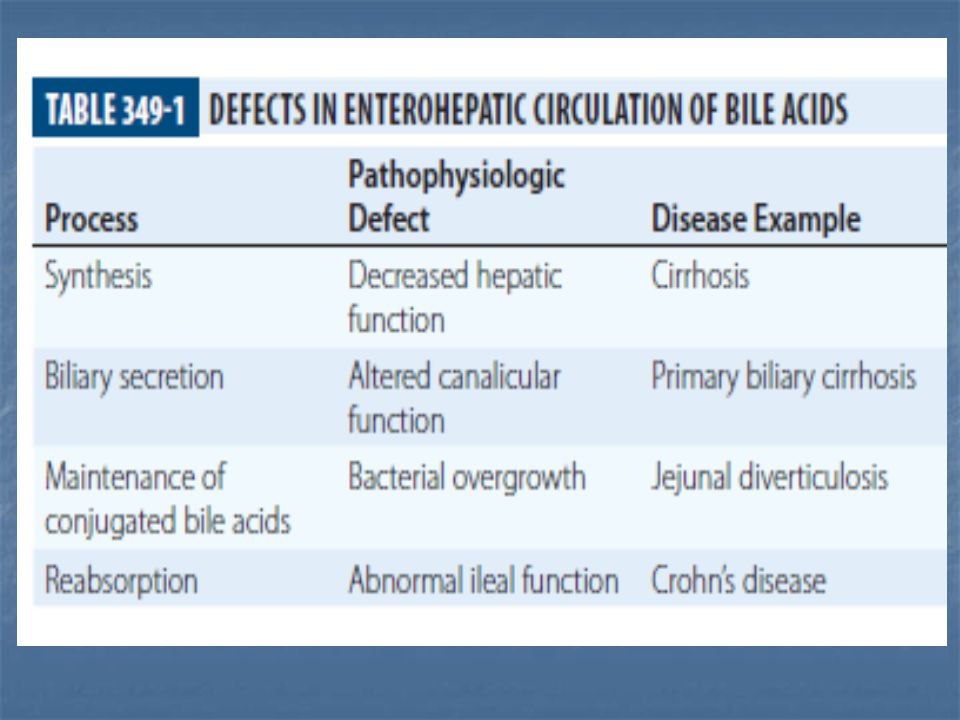
i) Pancreatic lipase is necessary for triglyceride hydrolysis in duodenum. Pancreatic enzyme deficiency leads to fat malabsorption. ii) Inactivation of pancreatic lipase by low gastric luminal pH – fat malabsorption. iii) Interruption of enterohepatic circulation of bile salt – impaired micelle formation – fat malabsorption. Absorption of fat soluble vitamins may be impaired as well.
 Pancreatic lipase is necessary for triglyceride hydrolysis in duodenum. Pancreatic enzyme deficiency leads to fat malabsorption. ii) Inactivation of pancreatic lipase by low gastric luminal. pH – fat malabsorption. iii) Interruption of enterohepatic circulation of bile salt – impaired micelle formation – fat malabsorption. Absorption of fat soluble vitamins may be impaired as well.")
118
2) Impaired carbohydrate absorption:
Most diseases that causes carbohydrate malabsorption do so by affecting intestinal stage. But amylase catalyse hydrolysis of starch to oligosaccharides.

119
3) Impaired protein absorption:
Hydrolysis of polypeptides occurs mainly in small intestine by action of pancreatic enzyme trypsin, chymotrypsin. Deficiency of pancreatic proteases – impaired protein absorption. Diseases like: Chronic pancreatitis Cystic fibrosis Ca. pancreatic resection - Protein malnutrition

120
B) Intestinal stage 1) Abnormalities of small intestinal mucosa. Lactase deficiency e.g. Congenital or acquired Result – malabsorption of lactose. Acquired:- i) Coeliac disease ii) Crohn’s disease iii) Infective enteritis
 Coeliac disease. ii) Crohn’s disease. iii) Infective enteritis.")
121
2) Impaired epithelial cell transport:
Many diseases cause loss of intestinal surface area - malabsorption of many nutrients. e.g i) Coeliac disease ii) Tropical spure iii) Extensive surgical resection iv) Drugs
 Coeliac disease. ii) Tropical spure. iii) Extensive surgical resection. iv) Drugs.")
122
C) Lymphatic transport:
Lymphatic obstruction – fat malabsorption e.g. i) Intestinal lymphangiectasia iii) Tuberculous enteritis iv) Intestinal lymphoma
 Intestinal lymphangiectasia. iii) Tuberculous enteritis. iv) Intestinal lymphoma.")
123
D) Decreased availability of ingested nutrients and
cofactors for absorption. i) Vitamin B12 malabsorption if intrinsic factor is deficient. e.g. gastrectomy, antiparietal cell Ab. ii) Bacterial overgrowth –can bind B12. iii) Patient infected with fist tapeworm – B deficiency.
 Vitamin B12 malabsorption if intrinsic factor is deficient. e.g. gastrectomy, antiparietal cell Ab. ii) Bacterial overgrowth –can bind B12. iii) Patient infected with fist tapeworm – B12 deficiency.")
124
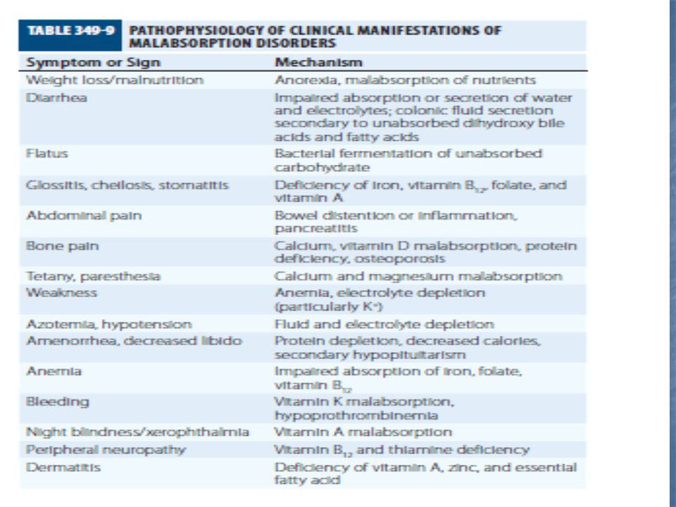
CLINICAL MANIFESTATIONS History:
Diarrhea/steatorrhoea Weight loss Symptoms of anaemia Diarrhoea – bulky, floating, malodorous stool – difficult to flush. Weight loss – may be profound, usually associated with anorexia. Anaemia – B12, iron, folate malabsorption. Patient may complain of dizziness, dyspnoea and fatigue

125
Important part of history: Recent travel - giardiasis
Drug abuse/multiple blood transfusions or ethanol abuse surgical resection - small bowel - gastric Malabsorption + chronic lung disease = cystic fibrosis Fever + weight loss = TB, lymphoma.

126
O/E: Normal. Pallor - muscle wasting Sign of vitamin deficiency
glossitis – B deficiency ecchymoses parasthesia tetany

127
Investigations: General: - CBC - Blood film - Ca. - B12, folate
- Iron study - LFT, PT, PTT

128
Investigations: Specific: Tests of fat absorption:
Quantitative fecal fat Patient should be on daily diet containing grams of fat. Fecal fat estimated on 72 H collection. 6 grams or more of fat/day is abnormal. May be due to: - Pancreatic - Small intestinal - Hepatobiliary disease

129
14C-Triolein Test: Is triglyceride which is hydrolysed by pancreatic lipase. absorption of metabolism ↑ 14CO2 lung

130
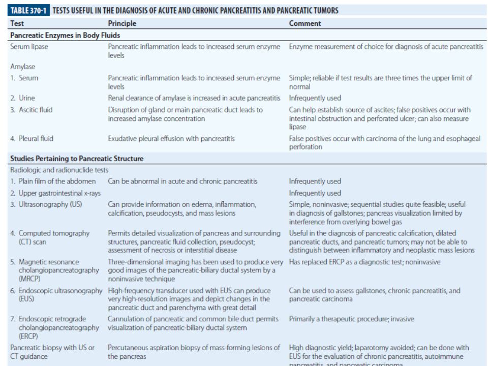
Tests for pancreatic function:
1) Bentiromide test: Chymotrypsin PABA + pepside PABA absorbed and conjugated in liver urine excretion 2) Schilling test
 Bentiromide test: Chymotrypsin. PABA + pepside. PABA absorbed and conjugated in liver. urine excretion. 2) Schilling test.")
131
3) Pancreatic stimulation test
Secretin stimulation – 4) Radiographic techniques: - Plain abdominal X-ray - U/S abdomen - ERCP - CT abdomen
 Radiographic techniques: - Plain abdominal X-ray. - U/S abdomen. - ERCP. - CT abdomen.")
132
Carbohydrate absorption test
1) Hydrogen breath test Hydrogen excretion ↑ in bacterial overgrowth small intestinal malabsorption
 Hydrogen breath test. Hydrogen excretion ↑ in. bacterial overgrowth. small intestinal malabsorption.")
133
Carbohydrate absorption test
2) D-xylose test 5-carbon sugar excreted unchanged in urine 25 grams given Urine collected for 5 hours Normally 25% is excreted In patients with fat malabsorption, this test differentiates pancreatic from small intestinal malabsorpton. D-xylose is normal in pancreatic disease Serum level of D-xylose at 1-2 hours after ingestion can be measured.
 D-xylose test. 5-carbon sugar excreted unchanged in urine. 25 grams given. Urine collected for 5 hours. Normally 25% is excreted. In patients with fat malabsorption, this test. differentiates pancreatic from small intestinal malabsorpton. D-xylose is normal in pancreatic disease. Serum level of D-xylose at 1-2 hours after ingestion can be measured.")
134
Test for bacterial overgrowth:
Intestinal aspiration and culture Breath test C-D xylose breath test

135
Radiography of small intestine:
Barium swallow and follow-through – to see - Blind loop - Stricture - J. diverticular

136
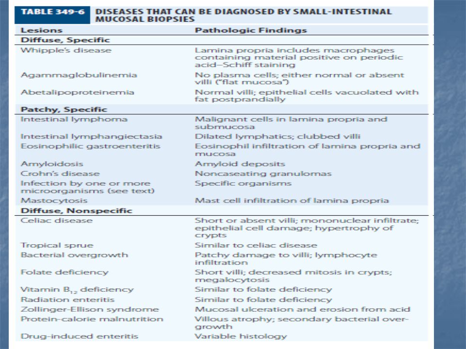
Intestinal mucosal biopsy:
- using crossby capsule - endoscopy Coeliac disease: - Villous atrophy Tropical spure: - short villi and increased lymphocyte

137
Selection of tests in evaluation malabsorption
Quantitaive fecal fat Abnormal Normal D-xylose test Abnormal Normal 14 C-D-xylose test Abd. Radiograph Bentiromide test CT-abd. Abnormal Jej culture Tetracyclin Then repeat breath test Normal Small intestinal Bx

138
Classification of Malabsorption Syndrome
Inadequate digestion: Postgastrectomy steatorrhea. Exocrine Pancreatic insufficiency. Reduced bile salt concentration in intestine: I.) Liver Disease II.) Cholestasis III.) Bacterial over growth IV.) Interruption of enterohepatic circulation of bile salt.
 Liver Disease. II.) Cholestasis. III.) Bacterial over growth. IV.) Interruption of enterohepatic circulation of bile salt.")
139
Inadequate absorptive surface: Resection
Diseased intestine Lymphatic obstruction. e.g Lymphoma D. Primary mucosal defects. Crohn’s disease Coeliac disease Tropical Sprue Disaccharide Deficiency Lymphoma TB

140
Malabsorption due to bacteral over growth of small bowel
Normal small intestine is bacterial sterile due to: Acid Int. peristalsis (major) Immunoglobulin Cause of bacterial growth. e.g. Small intestinal diverticuli Blind loop Strictures DM/ Scleroderma
 Immunoglobulin. Cause of bacterial growth. e.g. Small intestinal diverticuli. Blind loop. Strictures. DM/ Scleroderma.")
141
Pathophysiology Bacterial over growth: Metabolize bile salt resulting in deconjugation of bile salt Bile Salt Impaired intraluminal micelle formation Malabsorption of fat. Intestinal mucosa is damaged by Bacterial invasion Toxin Metabolic products Damage villi may cause total villous atrophy.

142
Clinically: Steatorrhea Anaemia B12 def. Reversed of symptom after antibiotic treatment. Diagnosis: Breath test Cxylose test Culture of aspiration (definitive) Treatment: Antibiotic Tetracyclin Ciproflexacin Metronidazole Amoxil
 Treatment: Antibiotic. Tetracyclin. Ciproflexacin. Metronidazole. Amoxil.")
143
Intestinal Lymphoma Primary 2nd Affect male = 50 Y.
Feature of malabsorption Biopsy resemble coeliac sprue Abdominal pain Fever Incomplete respond to gluten free diet. Absent features of generalized lymphoma.

144
Diffuse small intestinal mucosa disease.
Malabsorption may be due to: Diffuse small intestinal mucosa disease. Obstruction of lymphatic channels Stenosis bacterial overgrowth. Fever Diagnosis: History/Endoscopic Biopsy - CT scan of abdomen Laparotomy Some form secretion - heavy chain Ig A.

145
Perforation Treatment: Chemotherapy Complication: Bleeding
Intestinal obstruction Treatment: Chemotherapy Surgery

Similar presentations