Download presentation
Presentation is loading. Please wait.

1
PowerPoint File available: http://bl831.als.lbl.gov/ ~jamesh/powerpoint/ BNL_2011.ppt

2
Acknowledgements Ken Frankel Rick Donahue Howard Padmore Alastair MacDowell 8.3.1 creator: Tom Alber 8.3.1 PRT head: Jamie Cate Center for Structure of Membrane Proteins (PSI) Membrane Protein Expression Center II Center for HIV Accessory and Regulatory Complexes W. M. Keck Foundation Plexxikon, Inc. M D Anderson CRC University of California Berkeley University of California San Francisco National Science Foundation University of California Campus-Laboratory Collaboration Grant Henry Wheeler The Advanced Light Source is supported by the Director, Office of Science, Office of Basic Energy Sciences, Materials Sciences Division, of the US Department of Energy under contract No. DE-AC02-05CH11231 at Lawrence Berkeley National Laboratory.

3
The optimum wavelength for macromolecular crystallography Higher? or Lower?

4
Charged Particle Equilibrium (CPE) Johns, H. & Cunningham, J. (1974). The physics of radiology. Thomas Springfield, Illinois.
. The physics of radiology. Thomas Springfield, Illinois..")
5
dose Johns, H. & Cunningham, J. (1974). The physics of radiology. Thomas Springfield, Illinois. ~1 cm at 1 MeV f NH Charged Particle Equilibrium (CPE)
.")
6
satisfies CPE collimator crystal X-ray e-e- violates CPE Assume a spherical crystal…

7
is there a “problem” with violating CPE? ICRU report 31 “Average Energy Required to Produce an Ion Pair” (1979) for air: W ~ 30 eV/ion-pair yet, final ions are thermalized (<0.1 eV each) Where does 99% of the energy go? Answer: non-ionizing excitations
 for air: W ~ 30 eV/ion-pair yet, final ions are thermalized (<0.1 eV each) Where does 99% of the energy go. Answer: non-ionizing excitations.")
8
Secondary ionization e-e-

9
e-e- e-e- +

10
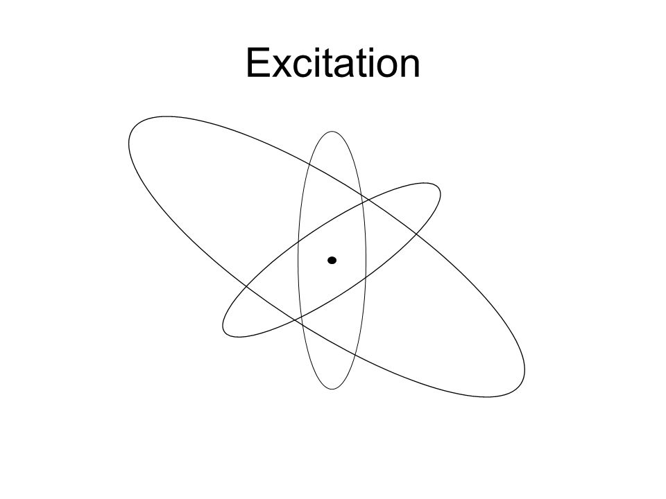
Excitation e-e-

11
e-e-

12
e-e-

14
ionizing interactions e-e- + Violating CPE: two kinds of “dose”? non-ionizing interactions ICRU report 36 “Microdosimetry” (1984)
.")
15
Charged Particle Equilibrium (CPE) Johns, H. & Cunningham, J. (1974). The physics of radiology. Thomas Springfield, Illinois. skin not burned
. The physics of radiology. Thomas Springfield, Illinois. skin not burned.")
16
Ionization track

17
particle transport simulation using MCNP collimator crystal X-ray

18
Where do photons go? beamstop Transmitted (98%) Protein 1A x-rays
 Protein 1A x-rays")
19
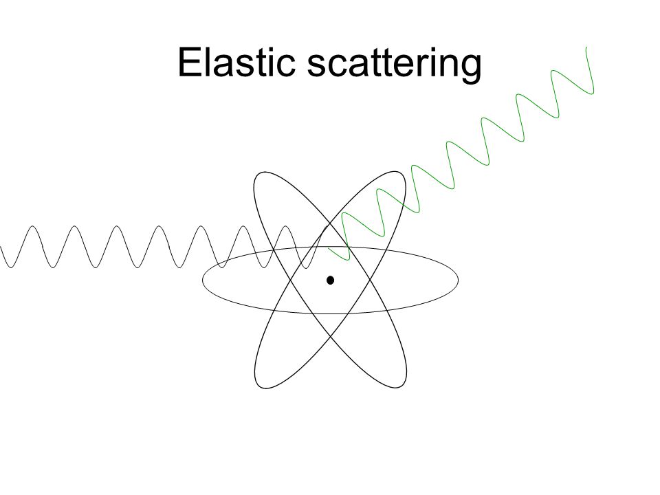
Where do photons go? beamstop elastic scattering (6%) Transmitted (98%) Protein 1A x-rays
 Transmitted (98%) Protein 1A x-rays")
20
Elastic scattering

22
Inelastic scattering e-e- +

23
Where do photons go? beamstop elastic scattering (6%) Transmitted (98%) Protein 1A x-rays
 Transmitted (98%) Protein 1A x-rays")
24
Where do photons go? beamstop elastic scattering (6%) Transmitted (98%) inelastic scattering (7%) Protein 1A x-rays
 Transmitted (98%) inelastic scattering (7%) Protein 1A x-rays.")
25
Where do photons go? beamstop elastic scattering (6%) Transmitted (98%) inelastic scattering (7%) Protein 1A x-rays Re-emitted (99%)Absorbed (~0%)
 Transmitted (98%) inelastic scattering (7%) Protein 1A x-rays Re-emitted (99%)Absorbed (~0%).")
26
Photoelectric absorption

27
e-e- +

28
Where do photons go? beamstop elastic scattering (6%) Transmitted (98%) inelastic scattering (7%) Protein 1A x-rays Re-emitted (99%)Absorbed (~0%)
 Transmitted (98%) inelastic scattering (7%) Protein 1A x-rays Re-emitted (99%)Absorbed (~0%).")
29
Where do photons go? beamstop elastic scattering (6%) Transmitted (98%) inelastic scattering (7%)Photoelectric (87%) Protein 1A x-rays Re-emitted (99%)Absorbed (~0%)
 Transmitted (98%) inelastic scattering (7%)Photoelectric (87%) Protein 1A x-rays Re-emitted (99%)Absorbed (~0%).")
30
Where do photons go? beamstop elastic scattering (6%) Transmitted (98%) inelastic scattering (7%)Photoelectric (87%) Protein 1A x-rays Re-emitted (~0%)Absorbed (99%) Re-emitted (99%)Absorbed (~0%)
 Transmitted (98%) inelastic scattering (7%)Photoelectric (87%) Protein 1A x-rays Re-emitted (~0%)Absorbed (99%) Re-emitted (99%)Absorbed (~0%).")
31
Fluorescence +

32
e-e- +

33
Auger emission +

34
++ e-e-

35
MCNP cuts off at 1 keV 1 MeV100 GJ/molMedical radiation therapy 100 keV10 GJ/molMedical imaging 10 keV1 GJ/molX-ray crystallography 1 keV100 MJ/molS and P K-edges 100 eV10 MJ/mol“water window” 10 eV1 MJ/molC≡C bond 1 eV100 kJ/molC-C bond, visible light 100 meV10 kJ/molhydrogen bond 10 meV1 kJ/molheat (~300 K)
")
36
bonding affects absorption Almkvist, et al. (2010)."K-edge XANES analysis of sulfur compounds: an investigation of the relative intensities using internal calibration", J. Sync. Rad. 17, 683-688. MCNP model
. K-edge XANES analysis of sulfur compounds: an investigation of the relative intensities using internal calibration , J. Sync. Rad. 17, MCNP model.")
37
particle transport simulation using MCNP collimator crystal X-ray

38
dose reduction with 1 Å radiation Crystal diameter (µm) Dose capture fraction f NH ←???→ 1 keV e - pathlength
 Dose capture fraction f NH ← → 1 keV e - pathlength")
39
100 μm crystal vs energy Photon Energy Dose capture fraction 1 keV 10 keV 100 keV 1 MeV f NH

40
dose reduction vs energy Photon Energy Dose capture fraction 1 keV 10 keV 100 keV 1 MeV

41
2 variables Dose capture fraction

42
Critical escape diameter Photon Energy (eV) Crystal diameter (µm)
 Crystal diameter (µm)")
43
Critical escape diameter Photon Energy (eV) Crystal diameter (µm)
 Crystal diameter (µm)")
44
Critical escape diameter Photon Energy (eV) Crystal diameter (µm)
 Crystal diameter (µm)")
45
Critical escape diameter Photon Energy Crystal diameter (µm) 1 keV 10 keV 100 keV 1 MeV 10 MeV
 1 keV 10 keV 100 keV 1 MeV 10 MeV")
46
Darwin’s Formula I(hkl)- photons/spot (fully-recorded) I beam - incident (photons/s/m 2 ) r e - classical electron radius (2.818x10 -15 m) V xtal - volume of crystal (in m 3 ) V cell - volume of unit cell (in m 3 ) λ- x-ray wavelength (in meters!) ω- rotation speed (radians/s) L- Lorentz factor (speed/speed) P- polarization factor (1+cos 2 (2θ) -Pfac∙cos(2Φ)sin 2 (2θ))/2 A- absorption factor exp(-μ xtal ∙l path ) F(hkl)- structure amplitude (electrons) P A | F(hkl) | 2 I(hkl) = I beam r e 2 V xtal V cell λ 3 L ωV cell Darwin, C. G. (1914)."The theory of X-ray reflexion. Part I", Philos. Mag. 27, 315-333.
. The theory of X-ray reflexion. Part I , Philos. Mag. 27,")
47
Darwin’s Formula I(hkl)- photons/spot (fully-recorded) I beam - incident (photons/s/m 2 ) r e - classical electron radius (2.818x10 -15 m) V xtal - volume of crystal (in m 3 ) V cell - volume of unit cell (in m 3 ) λ- x-ray wavelength (in meters!) ω- rotation speed (radians/s) L- Lorentz factor (speed/speed) P- polarization factor (1+cos 2 (2θ) -Pfac∙cos(2Φ)sin 2 (2θ))/2 A- absorption factor exp(-μ xtal ∙l path ) F(hkl)- structure amplitude (electrons) P A | F(hkl) | 2 I(hkl) = I beam r e 2 V xtal V cell λ3 Lλ3 L ωV cell Darwin, C. G. (1914)."The theory of X-ray reflexion. Part I", Philos. Mag. 27, 315-333.
. The theory of X-ray reflexion. Part I , Philos. Mag. 27,")
48
Darwin’s Formula I(hkl)- photons/spot (fully-recorded) I beam - incident (photons/s/m 2 ) r e - classical electron radius (2.818x10 -15 m) V xtal - volume of crystal (in m 3 ) V cell - volume of unit cell (in m 3 ) λ- x-ray wavelength (in meters!) ω- rotation speed (radians/s) L- Lorentz factor (speed/speed) P- polarization factor (1+cos 2 (2θ) -Pfac∙cos(2Φ)sin 2 (2θ))/2 A- absorption factor exp(-μ xtal ∙l path ) F(hkl)- structure amplitude (electrons) P A | F(hkl) | 2 I(hkl) = I beam r e 2 V xtal V cell λ 3 L ωV cell Darwin, C. G. (1914)."The theory of X-ray reflexion. Part I", Philos. Mag. 27, 315-333.
. The theory of X-ray reflexion. Part I , Philos. Mag. 27,")
49
Dose Formula dose- absorbed energy (Gy) I beam - incident (photons/s/μm 2 ) t exp - exposure time (s) λ- x-ray wavelength (in Å) dose ≈ I beam ·t exp λ2λ2 2000
 I beam - incident (photons/s/μm 2 ) t exp - exposure time (s) λ- x-ray wavelength (in Å) dose ≈ I beam ·t exp λ2λ2 2000")
50
Dose Formula D max - maximum dose (Gy) I beam - incident (photons/s/μm 2 ) t dataset - accumulated exposure time (s) λ- x-ray wavelength (in Å) D max ≈ I beam ·t dataset λ2λ2 2000
 I beam - incident (photons/s/μm 2 ) t dataset - accumulated exposure time (s) λ- x-ray wavelength (in Å) D max ≈ I beam ·t dataset λ2λ2 2000")
51
Dose Formula D max - maximum dose (Gy) I beam - incident (photons/s/μm 2 ) t dataset - accumulated exposure time (s) R- radius of crystal T en - transmission of a sphere ~ exp(-μ en ·2R) - density of crystal E ph - photon energy q e - electron charge D max = I beam ·t dataset 3q e E ph 4R (1-T en )
 I beam - incident (photons/s/μm 2 ) t dataset - accumulated exposure time (s) R- radius of crystal T en - transmission of a sphere ~ exp(-μ en ·2R) - density of crystal E ph - photon energy q e - electron charge D max = I beam ·t dataset 3q e E ph 4R (1-T en )")
52
Darwin’s Formula I(hkl)- photons/spot (fully-recorded) I beam - incident (photons/s/m 2 ) r e - classical electron radius (2.818x10 -15 m) V xtal - volume of crystal (in m 3 ) V cell - volume of unit cell (in m 3 ) λ- x-ray wavelength (in meters!) ω- rotation speed (radians/s) L- Lorentz factor (speed/speed) P- polarization factor (1+cos 2 (2θ) -Pfac∙cos(2Φ)sin 2 (2θ))/2 A- absorption factor exp(-μ xtal ∙l path ) F(hkl)- structure amplitude (electrons) P A | F(hkl) | 2 I(hkl) = I beam r e 2 V xtal V cell λ 3 L ωV cell Darwin, C. G. (1914)."The theory of X-ray reflexion. Part I", Philos. Mag. 27, 315-333.
. The theory of X-ray reflexion. Part I , Philos. Mag. 27,")
53
Darwin’s Formula D max - maximum dose (kGy) t dataset - accumulated exposure (s) r e - classical electron radius (2.818x10 -15 m) V xtal - volume of crystal (in m 3 ) V cell - volume of unit cell (in m 3 ) λ- x-ray wavelength (in meters!) ω- rotation speed (radians/s) L- Lorentz factor (speed/speed) P- polarization factor (1+cos 2 (2θ) -Pfac∙cos(2Φ)sin 2 (2θ))/2 A- absorption factor exp(-μ xtal ∙l path ) F(hkl)- structure amplitude (electrons) P A | F(hkl) | 2 I(hkl) = r e 2 V xtal V cell 2 λ L ωV cell D max t dataset Darwin, C. G. (1914)."The theory of X-ray reflexion. Part I", Philos. Mag. 27, 315-333.
. The theory of X-ray reflexion. Part I , Philos. Mag. 27,")
54
Darwin’s Formula D max - maximum dose (kGy) r e - classical electron radius (2.818x10 -15 m) V xtal - volume of crystal (in m 3 ) V cell - volume of unit cell (in m 3 ) λ- x-ray wavelength (in meters!) 2π- rotation range (radians) L- Lorentz factor (speed/speed) P- polarization factor (1+cos 2 (2θ) -Pfac∙cos(2Φ)sin 2 (2θ))/2 A- absorption factor exp(-μ xtal ∙l path ) F(hkl)- structure amplitude (electrons) P A | F(hkl) | 2 I(hkl) = r e 2 V xtal V cell 2 λ L 2πV cell D max Darwin, C. G. (1914)."The theory of X-ray reflexion. Part I", Philos. Mag. 27, 315-333.
. The theory of X-ray reflexion. Part I , Philos. Mag. 27,")
55
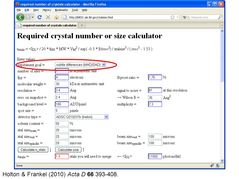
Holton & Frankel (2010) Acta D 66 393-408.
 Acta D")
57
Where: I DL - average damage-limited intensity (photons/hkl) at a given resolution 10 5 - converting R from μm to m, r e from m to Å, ρ from g/cm 3 to kg/m 3 and MGy to Gy r e - classical electron radius (2.818 x 10 -15 m/electron) h- Planck’s constant (6.626 x 10 -34 J∙s) c- speed of light (299792458 m/s) f decayed - fractional progress toward completely faded spots at end of data set ρ- density of crystal (~1.2 g/cm 3 ) R- radius of the spherical crystal (μm) λ- X-ray wavelength (Å) f NH - the Nave & Hill (2005) dose capture fraction (1 for large crystals) n ASU - number of proteins in the asymmetric unit M r - molecular weight of the protein (Daltons or g/mol) V M - Matthews’s coefficient (~2.4 Å 3 /Dalton) H- Howells’s criterion (10 MGy/Å) θ- Bragg angle a 2 - number-averaged squared structure factor per protein atom (electron 2 ) M a - number-averaged atomic weight of a protein atom (~7.1 Daltons) B- average (Wilson) temperature factor (Å 2 ) μ- attenuation coefficient of sphere material (m -1 ) μ en - mass energy-absorption coefficient of sphere material (m -1 ) Self-calibrated damage limit Holton & Frankel (2010) Acta D 66 393-408.
 at a given resolution converting R from μm to m, r e from m to Å, ρ from g/cm 3 to kg/m 3 and MGy to Gy r e - classical electron radius (2.818 x m/electron) h- Planck’s constant (6.626 x J∙s) c- speed of light ( m/s) f decayed - fractional progress toward completely faded spots at end of data set ρ- density of crystal (~1.2 g/cm 3 ) R- radius of the spherical crystal (μm) λ- X-ray wavelength (Å) f NH - the Nave & Hill (2005) dose capture fraction (1 for large crystals) n ASU - number of proteins in the asymmetric unit M r - molecular weight of the protein (Daltons or g/mol) V M - Matthews’s coefficient (~2.4 Å 3 /Dalton) H- Howells’s criterion (10 MGy/Å) θ- Bragg angle a 2 - number-averaged squared structure factor per protein atom (electron 2 ) M a - number-averaged atomic weight of a protein atom (~7.1 Daltons) B- average (Wilson) temperature factor (Å 2 ) μ- attenuation coefficient of sphere material (m -1 ) μ en - mass energy-absorption coefficient of sphere material (m -1 ) Self-calibrated damage limit Holton & Frankel (2010) Acta D")
58
wavelength dependence crystal diameter (μm) Å Minimum size for complete data set f NH = 1
 Å Minimum size for complete data set f NH = 1")
59
Where: I DL - average damage-limited intensity (photons/hkl) at a given resolution 10 5 - converting R from μm to m, r e from m to Å, ρ from g/cm 3 to kg/m 3 and MGy to Gy r e - classical electron radius (2.818 x 10 -15 m/electron) h- Planck’s constant (6.626 x 10 -34 J∙s) c- speed of light (299792458 m/s) f decayed - fractional progress toward completely faded spots at end of data set ρ- density of crystal (~1.2 g/cm 3 ) R- radius of the spherical crystal (μm) λ- X-ray wavelength (Å) f NH - the Nave & Hill (2005) dose capture fraction (1 for large crystals) n ASU - number of proteins in the asymmetric unit M r - molecular weight of the protein (Daltons or g/mol) V M - Matthews’s coefficient (~2.4 Å 3 /Dalton) H- Howells’s criterion (10 MGy/Å) θ- Bragg angle a 2 - number-averaged squared structure factor per protein atom (electron 2 ) M a - number-averaged atomic weight of a protein atom (~7.1 Daltons) B- average (Wilson) temperature factor (Å 2 ) μ- attenuation coefficient of sphere material (m -1 ) μ en - mass energy-absorption coefficient of sphere material (m -1 ) Self-calibrated damage limit Holton & Frankel (2010) Acta D 66 393-408.
 at a given resolution converting R from μm to m, r e from m to Å, ρ from g/cm 3 to kg/m 3 and MGy to Gy r e - classical electron radius (2.818 x m/electron) h- Planck’s constant (6.626 x J∙s) c- speed of light ( m/s) f decayed - fractional progress toward completely faded spots at end of data set ρ- density of crystal (~1.2 g/cm 3 ) R- radius of the spherical crystal (μm) λ- X-ray wavelength (Å) f NH - the Nave & Hill (2005) dose capture fraction (1 for large crystals) n ASU - number of proteins in the asymmetric unit M r - molecular weight of the protein (Daltons or g/mol) V M - Matthews’s coefficient (~2.4 Å 3 /Dalton) H- Howells’s criterion (10 MGy/Å) θ- Bragg angle a 2 - number-averaged squared structure factor per protein atom (electron 2 ) M a - number-averaged atomic weight of a protein atom (~7.1 Daltons) B- average (Wilson) temperature factor (Å 2 ) μ- attenuation coefficient of sphere material (m -1 ) μ en - mass energy-absorption coefficient of sphere material (m -1 ) Self-calibrated damage limit Holton & Frankel (2010) Acta D")
60
wavelength dependence crystal diameter (μm) Å Minimum size for complete data set
 Å Minimum size for complete data set")
61
wavelength dependence crystal diameter (μm) Å Minimum size for complete data set
 Å Minimum size for complete data set")
62
wavelength dependence crystal diameter (μm) Å Å Minimum size for complete data set
 Å Å Minimum size for complete data set")
63
molecular weight crystal diameter (μm) 100 Da 1 kDa 10 kDa 100 kDa 1 MDa 10 MDa Minimum size for complete data set 1 Å X-rays 2 Å spots B = 24 50% solvent
 100 Da 1 kDa 10 kDa 100 kDa 1 MDa 10 MDa Minimum size for complete data set 1 Å X-rays 2 Å spots B = 24 50% solvent")
64
Prediction: Exploiting Nave-Hill effect will require multi-crystal datasets

65
Other reasons for high energy room temperature

67
Zero-parallax microscope pinhole mirror microscope

68
High energy compresses pattern 2 Å data

69
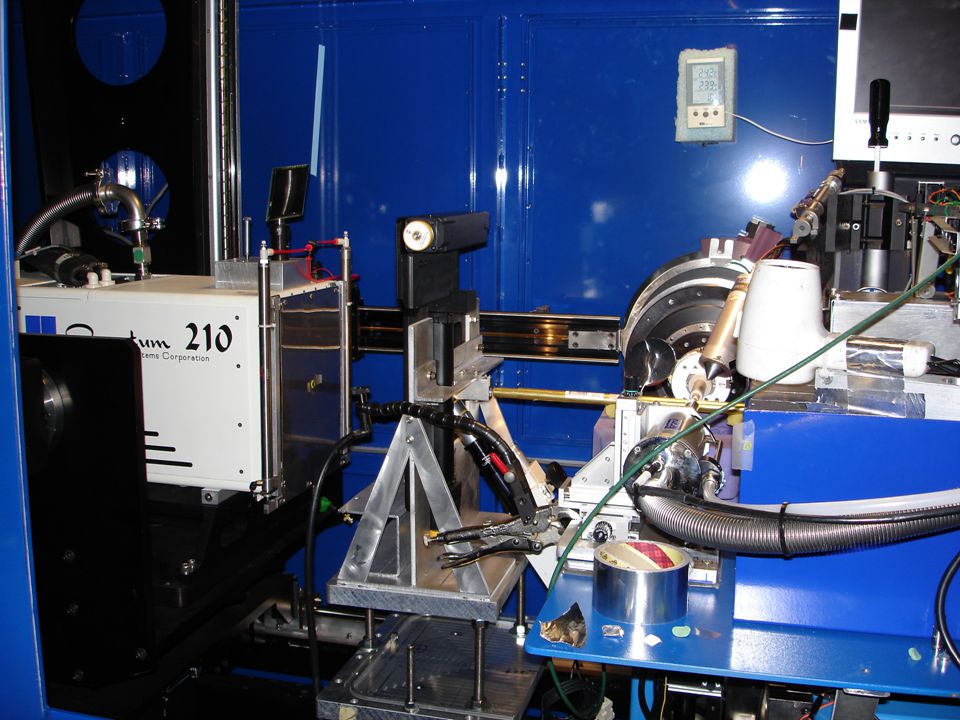
Example Room-temperature Data lysozyme 50 μm beam 37 Gy/s (0.775 Å) 30s exposures at 20C 90° of data, 97% complete I/ = 1.5 at 1.9 Å R merge 18% (overall) 5% (low) ΔB same as 20 min at 100K B factor -2 -4 -6 scale vs batch 210210
 30s exposures at 20C 90° of data, 97% complete I/ = 1.5 at 1.9 Å R merge 18% (overall) 5% (low) ΔB same as 20 min at 100K B factor scale vs batch")
70
high energy myth: less background elastic background/spot intensity ratio is wavelength-independent!

71
photon energy (eV) normalized figure of merit obliquity effect
 normalized figure of merit obliquity effect")
72
high energy myth: less background elastic background/spot intensity ratio is wavelength-independent! However: inelastic and fluorescence are less, as is absorption

73
Other reasons for high energy room temperature high pressure?

74
Room temperature damage rates dose rate (kGy/s) dose at half intensity (MGy)
 dose at half intensity (MGy)")
75
Room temperature damage rates dose rate (kGy/s) dose at half intensity (MGy)
 dose at half intensity (MGy)")
76
Room temperature damage has a size dependence?

77
Radiation Damage “scale factor” F 2 alone cannot explain change in overall scale!

78
Darwin’s Formula I(hkl)- photons/spot (fully-recorded) I beam - incident (photons/s/m 2 ) r e - classical electron radius (2.818x10 -15 m) V xtal - volume of crystal (in m 3 ) V cell - volume of unit cell (in m 3 ) λ- x-ray wavelength (in meters!) ω- rotation speed (radians/s) L- Lorentz factor (speed/speed) P- polarization factor (1+cos 2 (2θ) -Pfac∙cos(2Φ)sin 2 (2θ))/2 A- absorption factor exp(-μ xtal ∙l path ) F(hkl)- structure amplitude (electrons) C. G. Darwin (1914) P A | F(hkl) | 2 I(hkl) = I beam r e 2 V xtal V cell λ 3 L ωV cell
 P A | F(hkl) | 2 I(hkl) = I beam r e 2 V xtal V cell λ 3 L ωV cell.")
79
“scale factor” implies that damaging motions are larger than unit cell Room temperature damage has a size dependence? high pressure will hold it together? suggests micro-fracture mechanism of spot fading

80
Radiation Damage prediction large displacements cannot be faster than the speed of sound

81
Timescales of radiation damage Garret et. al. (2005) Chem. Rev. 105, 355-389
 Chem. Rev. 105,")
82
Two types of reactions Garret et. al. (2005) Chem. Rev. 105, 355-389 nonhomogeneous homogeneous
 Chem. Rev. 105, nonhomogeneous homogeneous")
83
Timescales of radiation damage Garret et. al. (2005) Chem. Rev. 105, 355-389 LCLS ALS bunch
 Chem. Rev. 105, LCLS ALS bunch")
84
Room temperature damage rates dose rate (kGy/s) dose at half intensity (MGy)
 dose at half intensity (MGy)")
85
Room temperature damage rates dose rate (kGy/s) dose at half intensity (MGy)
 dose at half intensity (MGy)")
86
Nozzle ground to provide large X-ray diffraction angle Droplets freeze at 10 6 o /sec. in vacuum to vitreous ice if cryoprotectant added. Hand-grinding to micron gives large take-off angle for X-rays ! D. DePonte X-rays 5 microns Flow rate 12 microliters per minute. An environmental SEM image of operating protein-beam injector for LCLS.

87
What about very low energy?

88
sample shadow on detector Cu

89
sample shadow on detector

90
X-ray of cryo stream

91
low-energy X-rays Cu λ=2d sinθ 2.5 Å data with 5 Å X-rays International Tables for Crystallography, Vol. C, 2nd ed., chapter 6.3

92
Detector d = 2.5 Å sample injector Mirrors λ = 5 Å d = 2.5 Å h,k,l -h,-k,-l Colliding Beam Anomalous Measurement

93
0 20 40 60 80 100 Anomalous differences are resilient to non-isomorphism R iso (%) 1.0 0.8 0.6 0.4 0.2 0 Correlation Coefficient of ΔF ano 100 x 100 lysozyme PDBs
 Correlation Coefficient of ΔF ano 100 x 100 lysozyme PDBs")
94
The optimum wavelength for macromolecular crystallography Maximize data/damage→15-50 keV Turn the crank → 12.68 keV Native element phasing→ 2-2.5 keV the future: multi-crystal data sets

95
PowerPoint File available: http://bl831.als.lbl.gov/ ~jamesh/powerpoint/ BNL_2011.ppt

Similar presentations

 and e/m (e-beam expt.). Photons as discrete Particles.>")
 Pure metal target (Cu) Electrons remover inner-shell electrons from target. Other electrons “fall”>")


 Characterization of Nanomaterials.>")


1.6 1.7 4.0 0.020SDCORR1.0 2.2 0.065 36.8PADFPH31.46.>")
