Download presentation
Presentation is loading. Please wait.

1
-Stem Cell Therapy - Introduction and Practical Guide to Use in Small Animal Practice
This is an introductory slide set intended to be used by Vet-Stem credentialed vets in presenting stem cell therapy to other vets.

2
What Do Stem Cells Do? 10 million cells die in your body every minute of every day. Your own stem cells replace them so you can continue living. This is what stem cells do for a living. Simple explanation for what adult stem cells are doing every day in every body.

3
What are Stem Cells? Stem Cells are Primitive cells present in almost every tissue: Self Renewing Able to become different tissue types Trophic Factories – Growth Factors These are the three major distinctions of what define stem cells.

4
Embryonic vs Adult Stem Cells
Stem Cells from Embryos Intended to form whole animal - not for repair Can form tumors – UNPREDICTABLE Maybe someday Adult Stem Cells – many sources Intended for self repair Do not form tumors when injected Now Gruen L and Grabel L, Stem Cell 2006;24; Adult stem cells are the bodies natural healing cells for repair. They are found in fat, bone marrow, skin and most other tissues of the body.

5
Enabling Innovation Dr. Bill Futrell, et al – Univ of Pittsburgh 1998 “Discovered true stem cells in fat that could create new tissues like bone and cartilage” Stem Cells from fat Extracted can become bone Fix fractures in the lab The original discovery of stem cells from fat occurred at the Univ of Pittsburgh in the plastic surgery department by Dr. Bill Futrell and his group. They found that cells derived from extracted fat tissue could become bone, cartilage, fat and nerves. Patents were filed and this field was born. The question was whether we could harness this to fix a clinical problem like a non-union fracture.

6
Why Adipose as Stem Cell Source?
High healing cell count – No culturing required 1000X stem cell concentration as bone marrow Family of healing cells - heterogeneous Rapid, Easy to access Over 1700 peer reviewed papers published

7
What Cells Do We Use?1 Contrary to the older press and literature, approximately 1/3 of the nucleated cells derived from a sample of fat are stem cells. There are also over 28% of the cells that are regenerative progenitor type cells with the ability to provide growth factors and assist in healing. This is over 1000X more concentrated than bone marrow and is the reason you use the cells fresh, without need for cell culture. However, if you run out of fresh or frozen cells, you can grow additional doses from a small retention sample kept on all patients.

8
Mechanisms of Repair Trophic support - growth factors and cytokines
Anti-inflammatory Differentiation into tissue Homing to injury site Immune System Modulation These are the five main mechanisms of action of adult stem cells. We usually think of differentiation into tissue as the first and most important mechanism, and it is important, but maybe not the most dominant.

9
The Injury Response Cascade
Time Fibrosis Inflammation “Regeneration” Acute Injury Scar Complete Magnitude QUICK FIX In a simple schematic, a typical injury causes a regenerative response, for example in a heart attack. The tissue tries to regenerative for a short period of time and then resorts to scar tissue formation to “patch” the injury. Courtesy, A Caplan, Case Western Reserve

10
Stem Cells in Injury Response
“Regeneration” Acute Injury Scar Reduced Time Magnitude Inflammation Fibrosis In stem cell therapy, we are attempting to boost the number of stem cells and increase the length of time that the body attempts to regenerate a more normal/natural tissue in its repair. Stem Cells Courtesy, A Caplan, Case Western Reserve

11
Roles / Functions “Stem cells are injury-specific, perfectly choreographed pharmaceutical factories” Dr. Arnold Caplan Case Western Reserve Influence by injury micro-environment Stem cells respond to specific injury micro-environment.

12
Stem Cells and Joint Therapy Mechanisms
Chondrogenesis1 - new cartilage formation Lubricin2 - lubricate joint, improve range of motion IRAP Secretion3 - blocks IL-1 inflammation mediator Decrease Inflammation4 - reduce pain and swelling So how do we believe these cells are working in the joints. First, arthroscopy data shows that new cartilage can and is formed. But many times the therapy seems to work much faster than one would expect from this mechanism. Lee et all showed in 2008 that stem cells can produce Lubricin, a key joint lubricating molecule. Ortiz et al in 2007 published that stem cells can also produce IRAP, a potent molecule that blocks IL-1 action, a major contributor to joint inflammation and cartilage degradation. Finally, Tholpady and many others have shown that stem cells can produce a range of anti-inflammatory cytokines and down regulate inflammation. 1 Wei et al, Cytotherapy, 2007 2 Lee et al, BBRC, 2008 3 Ortiz et al, PNAS 2007 4 Tholpady, Plast Surg, 2006

13
Evidence-based Cell Therapy
DATA TYPE AVAILABLE Canine Osteoarthritis Canine Tendon and Ligament In-vitro (lab bench) X Lab Animal Case Studies Retrospective Studies Non-Random, Prospective Randomized, Controlled (equine) Human Clinical Studies STATUS Supported Today we talk about evidence-based practice of medicine. So what types of evidence do we have to support this type of adipose stem cell therapy. For canine arthritis we have in-vitro, lab animal, case studies, retrospective studies, non-randomized but prospective studies, randomized placebo controlled blinded studies, and human clinical studies. For soft tissue tendon and ligament injury, we have the same data, but the controlled, randomized studies we have are equine only.
 X. Lab Animal. Case Studies. Retrospective Studies. Non-Random, Prospective. Randomized, Controlled. (equine) Human Clinical Studies. STATUS. Supported. Today we talk about evidence-based practice of medicine. So what types of evidence do we have to support this type of adipose stem cell therapy. For canine arthritis we have in-vitro, lab animal, case studies, retrospective studies, non-randomized but prospective studies, randomized placebo controlled blinded studies, and human clinical studies. For soft tissue tendon and ligament injury, we have the same data, but the controlled, randomized studies we have are equine only")
14
Rabbit Osteochondral Defect Repair Model
Nathan et al, “Cell-based therapy in the repair of osteochondral defects: A novel use for adipose tissue” Tissue Engineering, 2003.

15
Elbow Dysplasia - DJD Annie – 9 YO
Chronic elbow dysplasia – non-NSAID controlled pain Stem Cell Therapy at Day 0 and 14. Now able to jump in and out of car; took 2 hour run in the woods before her 90 day study exam!

16
Bilateral Stifle / Hips
4 YO Rat Terrier Bilateral stifle and hip degeneration since 6 months old NSAIDS for 12 months and still painful =============== Intra-articular RX with Stem cells in all four joints

17
Bilateral Stifle / Hips
4 YO Rat Terrier Post Treatment: 2 weeks – Dramatic pain reduction Now NSAID free for >24 months

18
Human Therapy “Autologous stem cells (adipose) and fibrin glue used to treat widespread traumatic calvarial defects: case report” A young girl with major trauma to skull cap that could not be repaired with bone graft was treated with her own adipose stem cells and re-grew the skull bones. Journal of Cranio-Maxillofacial Surgery (2004) 32, 370–373 One of the first reports confirming this creation of tissue in a human came in A young girl had lost most of her normal skull cap due to a major trauma and traditional bone grafting had failed. With a single application of fresh, adipose-derived stem and regenerative cell mix, they were able to grow back the majority of her calvarium.
 32, 370–373. One of the first reports confirming this creation of tissue in a human came in A young girl had lost most of her normal skull cap due to a major trauma and traditional bone grafting had failed. With a single application of fresh, adipose-derived stem and regenerative cell mix, they were able to grow back the majority of her calvarium")
19
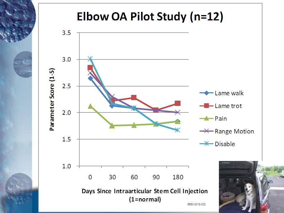
180 Day Prospective, Non-randomized Study Uni or Bilateral Elbow OA
Intra-articular stem cells 1x Vet and Owner assessments at pre, 30, 60, 90, 180 14 dog study The first study to review is a prospective study, but without controls, using baseline values as comparison. 14 dogs were treated in 5 referral clinics in the US. A single intraarticular injection was given and dogs were followed for 180 days.

20
All measured outcomes were improved from baseline.
21
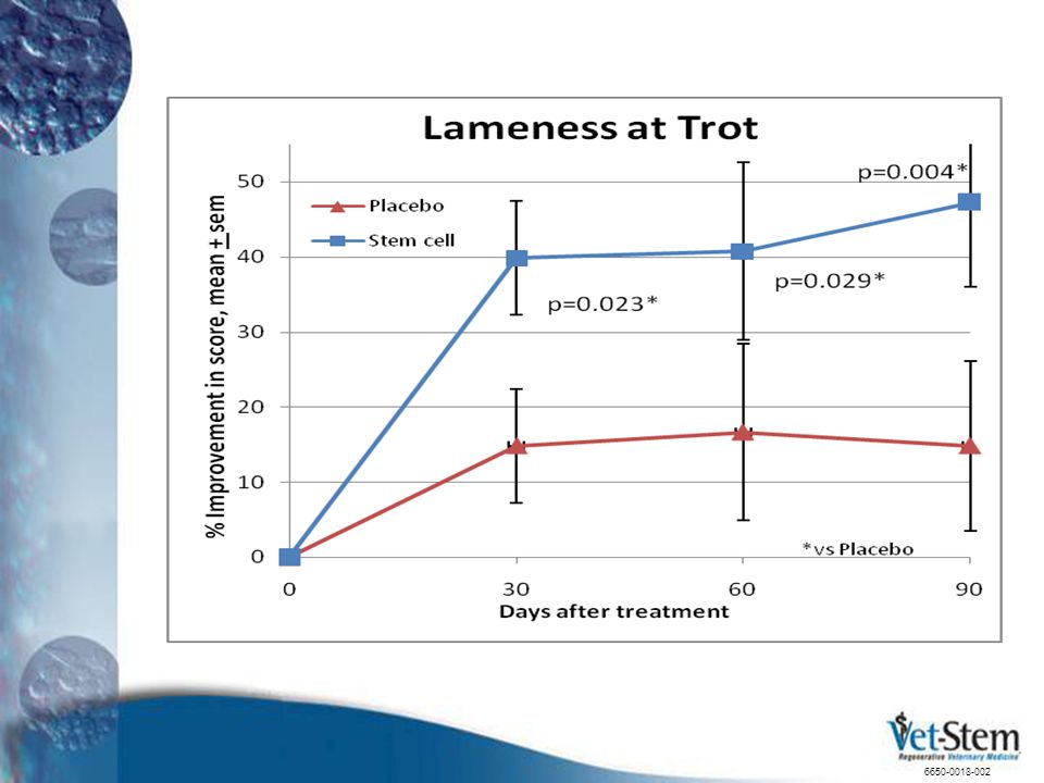
180 Day Study – Blinded/Placebo Bilateral Hip OA – 18 dogs
Intra-articular stem cells – 1X Vet/Owner assessments at pre, 30, 60, 90 The second OA study to review was published in Veterinary Therapeutics in 2007 and is a double-blinded, randomized, placebo-controlled study of OA in the hip. 18 dogs were treated for bilateral hip OA with a single intraarticular injection per joint. The study was blinded for 90 days.

22
This graph shows the improvement over the 90 day period in treated versus saline-injected control dogs. You can see there was a placebo effect, highlighting why placebo-controlled studies are important. The stem cell treated group out-performed the saline group at all time points.
23
Chronic Knee – Post-surgical OA
Pilot Study (n=9) 180 Day Pilot Study, 9 dogs Chronic Post-Surgical OA (>3 Mo) Intraarticular stem cells – 1X Vet and Owner assessments at pre, 30, 90, 180 Not Blinded The last OA study in dogs to review was done in a single clinic by a single surgeon. All dogs had previous surgery in the stifle, had not responded favorable, and now had OA. They had all been re-scoped to confirm that no additional surgery would help. A single injection was made into each joint and improvements from baseline over 180 days were made.
 180 Day Pilot Study, 9 dogs. Chronic Post-Surgical OA (>3 Mo) Intraarticular stem cells – 1X. Vet and Owner assessments at pre, 30, 90, 180. Not Blinded. The last OA study in dogs to review was done in a single clinic by a single surgeon. All dogs had previous surgery in the stifle, had not responded favorable, and now had OA. They had all been re-scoped to confirm that no additional surgery would help. A single injection was made into each joint and improvements from baseline over 180 days were made")
24
Again, all parameters measured improved
Again, all parameters measured improved. “V” means the veterinarian measured the lameness, range of motion and pain and “O” indicates owner measured.

25
Tendon / Ligament Clinical Data
Equine – Blinded, placebo-controlled study1 Equine – Retrospective2 – tendonitis Equine – Retrospective3 - ligament therapy Canine – Clinical case studies4 Canine – Retrospective analysis5 Canine – Prospective, non-randomized6 – shoulder instability In the area of tendon/ligament repair, there are equine and canine data that demonstrate repair of the joint-related soft tissue by adipose stem and regenerative cells. 1 Nixon et al, AJVR, 2007 Meredith et al, ACVIM, 2006 Harman, VOS, 2007 Bausman, IFATS, 2008 5 Harman et al, IFATS, 2007 6 Canapp, AVMA, 2009

26
8 Horses – 4 treated / 4 controls
Collagenase induced injury – Rx 10 days later Fully blinded histology Controls were treated with same volume of saline This is the first reported blinded, placebo controlled study of adipose stem cells in tendon repair in the horse. It was conducted at Cornell University by Dr. Linda Dahlgren and staff in Dr. Alan Nixon’s lab. 8 horses were treated in two groups. Controls were blinded and saline injected. The injury was created by injection of collagenase into the tendons of lab horses. The injury was not treated with stem cells until 10 days after the injection.

27
Outcome: Less scar - More normal tendon
Stem Cell Treated Saline Control Outcome: Less scar - More normal tendon Statistically improved tendon healing

28
Why Use Vet-Stem? Began treating horses in 2003
Global patents/licenses (over 55 issued patents) Over 3,000 horses and 2,000 dogs treated Global leader in stem cell therapy Peer-reviewed, published controlled studies High quality laboratory Human stem cell alliances
 Over 3,000 horses and 2,000 dogs treated. Global leader in stem cell therapy. Peer-reviewed, published controlled studies. High quality laboratory. Human stem cell alliances")
29
Safety Profile* Horses: 7/3,359 (0.2%) Only local swelling/pain
No systemic effects Dog: /1,695 (0.1%) Only local swelling/pain *probable adverse events, through 6/1/09 Always we should ask about the risk/benefit profile for any therapy. Since the cells are from the same animal, rejection and disease transmission are minimized. In horses, 7/3,359 treated horses have had a local reaction that could be probably attributed to the cell therapy injections. No systemic reactions have been seen. In the dog, 2/1,695 dogs treated showed a local joint reaction after treatment that could be probably attributed to the cell injections. Overall, a very nice safety profile.
 Only local swelling/pain. *probable adverse events, through 6/1/09. Always we should ask about the risk/benefit profile for any therapy. Since the cells are from the same animal, rejection and disease transmission are minimized. In horses, 7/3,359 treated horses have had a local reaction that could be probably attributed to the cell therapy injections. No systemic reactions have been seen. In the dog, 2/1,695 dogs treated showed a local joint reaction after treatment that could be probably attributed to the cell injections. Overall, a very nice safety profile")
30
Quality Standards Written standard operating procedures
Independent Quality Assurance Unit Routine audits of all procedures Sample release criteria Formal training systems Environmental monitoring All Handling Done In Sterile Hoods Vet-Stem has strict quality standards and we follow the FDA Good Tissue Practices Act as it applies to autologous cell therapy.

31
Resources from Vet-Stem
Technical training – course & consultation Case selection - consultation Cell banking Lifetime cell supply – Cell Culture Service Marketing Kit Owner support Clinic DVD – waiting room Brochures, posters, counter-top displays Power Point presentations Co-op Advertising Newsletters

32
Keys to Success Credentialing Course (www.vet-stem.com)
Complete Diagnostic Work-up Risk Assessment of Concurrent Diseases Proper Collection – close dead space Proper use of Kit Proper Injection Technique Follow up and Rehab Program

33
Case Selection for OA Ideal Stem Cell Case
Clearly defined disease – lameness Non-surgical candidate – surgery first, if needed Limited intra-articular osteophytes One or two joints No other major systemic diseases No major spinal disease NSAID – non-responsive or lack of tolerance Owner has rational expectations For osteoarthritis, these are the best criteria for selection of the ideal case.

34
Cruciate Ligament Diseases and the Uses of Stem Cells
Surgery – always assess need and make appropriate surgical repairs At surgery – adjunct to reduce inflammation and scar tissue After surgery – treat the synovitis and encourage cartilage repair Chronic ACL inflammation and synovitis Cruciate repair in the dog should be approached with caution. If the case needs surgery, stem cells are not a replacement. If the injury is less than a 25% repair, then stem cells may be indicated to stimulate repair of the torn cruciate. If the joint injury is older and already has OA, then stem cells can be used to treat the pain, inflammation and damage. Stem cells are a great adjunct therapy to surgical repair as well. 34

35
Current Supported Indications Canine/Feline
Osteoarthritis Polyarthritis Tendonitis Ligament injury

36
Service Overview The Vet-Stem Service is simple. The credentialed veterinarian collects a small sample of fat (60-90 grams), sends it by Fed Ex to the Vet-Stem lab in San Diego. The cell technicians will receive the fat, extract the cells, do quality checks and counts, and send cells back THE SAME DAY with the appropriate number of doses loaded into syringes in the appropriate volume for each patient. The Vet will receive the return FedEx shipment approximately 48 hours after collection, and inject the cells into the injured joint, tendon, or IV. Any excess cells are banked at Vet-Stem and stored in liquid nitrogen ready for use in the future.
, sends it by Fed Ex to the Vet-Stem lab in San Diego. The cell technicians will receive the fat, extract the cells, do quality checks and counts, and send cells back THE SAME DAY with the appropriate number of doses loaded into syringes in the appropriate volume for each patient. The Vet will receive the return FedEx shipment approximately 48 hours after collection, and inject the cells into the injured joint, tendon, or IV. Any excess cells are banked at Vet-Stem and stored in liquid nitrogen ready for use in the future")
37
Vet-Stem Adipose Processing Lab
1. Immediate use Summarizing, the lab sends back doses ready to inject for immediate use and banks any extra cells for future use. The number of cells for any individual animal depends on the amount of fat submitted and on the number of cells/gram of fat for that individual animal. Animals with systemic disease such as Cushing's tend to have a lower number of cells/gram of fat. 3. Lifetime supply by culture 2. Frozen for future

38
Real World Dog OA Cases This graph shows the improvement in the first 90 days after treatment of a large group of commercial dogs. The data was retrieved by survey from the attending vets. You can see that the dogs went, on average, from a moderate degree of pain, lameness and range of motion to between normal and mild.

39
Change in NSAID Usage in Dogs Treated with Stem Cells for Osteoarthritis
90 Days Post Stem Cell Treatment 246 Days Post Stem Cell Treatment survey range: days, N= 170 owner voluntary responses 10/08- 05/09 survey range: days, N= 139

40
Conclusion: At 90 and 246 days after treatment
Greater than 33% of dogs discontinued the use of NSAIDs completely Greater than 28% of dogs decreased their dependency on NSAIDs

41
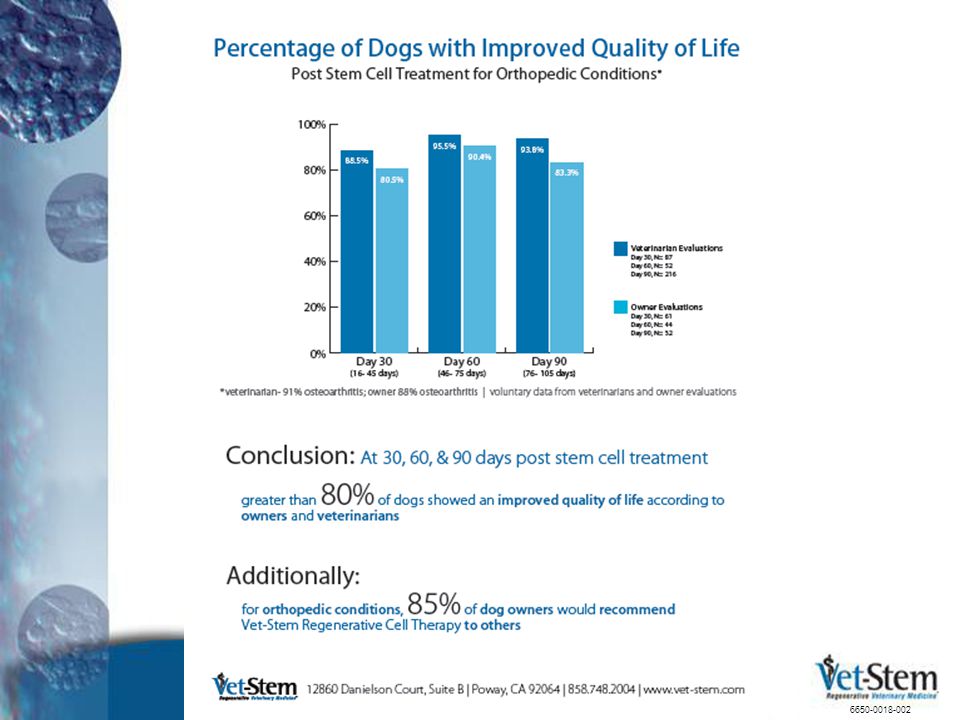
This graphic demonstrates that over 80% of dogs showed an improvement in quality of life according to survey results from owners AND vets. Additionally, over 85% of owners would recommend Vet-Stem therapy to others.
42
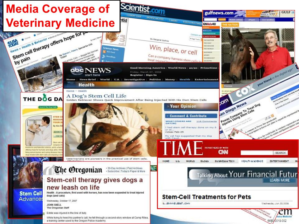
Media Coverage of Veterinary Medicine
43
Additional Slides The additional slides that follow are intended to be supplementary information and can be used to emphasize a particular area of the talk or to modify the theme or direction of the talk for the intended audience.

44
Mechanisms of Repair 1. Trophic support – growth factors and cytokines
Anti-scarring - MMPs (reduce scar tissue formation) Angiogenic - VEGF (induces new blood vessels) Anti-apoptotic (block cell death after injury) Stimulation of resident tissue stem cells Caplan and Dennis, J Cell Biochem, 2006 Anti-scarring – these cells produce MMP, matrix metaleoproteinases, that are natural scar tissue remodelers. The cells produce VEGF, vascular endothelial growth factor, that induces growth of new blood vessels. Anti-apoptosis – this is programmed cell death from injury like ischemia – stem cells via growth factors and cell-to-cell transfer of mitochondria can “rescue” dying cells. They also stimulate stem cells resident in the injured tissue to begin dividing and assisting in the healing/regeneration.
 Angiogenic - VEGF (induces new blood vessels) Anti-apoptotic (block cell death after injury) Stimulation of resident tissue stem cells. Caplan and Dennis, J Cell Biochem, Anti-scarring – these cells produce MMP, matrix metaleoproteinases, that are natural scar tissue remodelers. The cells produce VEGF, vascular endothelial growth factor, that induces growth of new blood vessels. Anti-apoptosis – this is programmed cell death from injury like ischemia – stem cells via growth factors and cell-to-cell transfer of mitochondria can rescue dying cells. They also stimulate stem cells resident in the injured tissue to begin dividing and assisting in the healing/regeneration")
45
Mechanisms of Repair 2. Anti-inflammatory
Decrease pro-inflammatory mediators Increase anti-inflammatory mediators Tholpady et al, Clin Plastic Surg, 2006 Stem cells truly down regulate and buffer an over-active inflammatory reaction. Decrease of TNF alpha and interferon gamma are examples of turning down the proinflammatory cytokines.

46
Mechanisms of Repair 3. Differentiation into tissue Nerve Bone
Cartilage Liver Fat-derived Stem Cells Cardiac Fat Angiogensis/ Anti-apoptosis Gene Therapy Muscle Clearly, in the lab, we can create all these tissues from adipose-derived or bone marrow derived stem cells This has been proven many times in many labs around the world. Tholpady et al, Clin Plastic Surg, 2006 (Photo courtesy Cytori Therapeutics)
")
47
Mechanisms of Repair 4. Homing to injury site Damaged cartilage MSCs
This slide demonstrates that MSCs, or mesenchymal stem cells, are able to home to a damaged site in cartilage, even down into the fibrilated cartilage. Photo Courtesy Cognate Therapeutics

48
Mechanisms of Repair 5. Immune System Modulation
Stem cells can dramatically modulate immune system function. In this example reported in 2009, non-expanded adipose stem cells were used to treat Multiple Sclerosis patients with just an intravenous treatment which dramatically reduced the symptoms, pain, and dysfunction caused by MS autoimmune attack on nerve sheath myelin. Stem cells down-regulate the immune system attack on its own nerve sheath myelin.

49
Composite Score: Lameness at walk Lameness at trot
Five parameters were measured by the veterinarian and this graph shows the composite scores and the significant differences between treated and placebo-controls Composite Score: Lameness at walk Lameness at trot Pain on manipulation Range of motion Functional disability

50
Vet-Stem Adipose Collection Kit
This is the simple collection kit with an ice block (freeze 24 hours before use), sterile collection tubes, and your paperwork for lab submission. Call to schedule the case so the lab is ready for receiving your sample. Sterile Collection tubes Cryo-block Submission form Owner Consent form
, sterile collection tubes, and your paperwork for lab submission. Call to schedule the case so the lab is ready for receiving your sample. Sterile Collection tubes. Cryo-block. Submission form. Owner Consent form")
51
Adipose Tissue Harvest Falciform Fat
Most preferred collection location due to low risk of seroma and adequate fat source Fat can be collected from falciform ligament. This reduces the chance of a dead space seroma formation and generally yields the highest volume of fat, even in a think dog.

52
Adipose Tissue Harvest Thoracic Approach
As an alternative, the lateral thorax, behind the shoulder, high on the chest wall, is a good site.

Similar presentations


, also termed autologous platelet gel, plasma rich in growth factors (PRGF) increased.>")


