Download presentation
Presentation is loading. Please wait.

1
A IRBORNE I NFECTION

2
A IRBORNE INFECTIONS : Contracted by inhalation of microorganisms or spores suspended in air on water droplets or dust particles

3
R ESPIRATORY TRACT INFECTIONS Infections involving the respiratory tracts Classified as an upper respiratory tract or a lower respiratory tract infections Lower respiratory infections, such as pneumonia, tend to be far more serious conditions than upper respiratory infections, such as the common cold

4
URTI Infections in the: Nose Sinuses Pharynx Larynx Middle ear

5
URTI T YPICAL INFECTIONS Tonsillitis Pharyngitis Laryngitis Sinusitis (can be cause by fungi) Otitis media (can be cause by fungi) Influenza Common cold
 Otitis media (can be cause by fungi) Influenza Common cold")
6
S YMPTOMS OF URTI S Cough Sore throat Runny nose Nasal congestion Headache Low grade fever Sneezing

7
F UNGAL INFECTIONS OF THE UPPER RESPIRATORY TRACTS

8
Fungal Ear infections Fungal nasal sinusitis Fungal infections of the oral cavity Fungal keratitis

9
F UNGAL E AR I NFECTIONS “O TOMYCOSIS ” Otitis externa & Otitis media

10
O TITIS EXTERNA Fungal infection of the external ear canal World-wide, but more common in tropical and sub-tropical regions

11
E TIOLOGY Caused mainly by: Aspergillus fumigatus Aspergillus niger Candida albicans Candida tropicalis

12
O THER CAUSES MAY INCLUDE Malassezia species Pseudallescheria boydii Absidia species Acremonium species Penicillium species Rhizopus species Scopulariopsis brevicaulis

13
C LINICAL MANIFESTATION Inflammation Itching Scaling Discomfort Masses of debris containing hyphae Pain

14
Otitis Externa

15
L ABORATORY DIAGNOSIS Direct examination of epithelial debris Hyphae and in some instances the fruiting structures of the etiologic agent Culture: Sabouraud dextrose agar incubated at 30°C (without cycloheximide)
")
16
M ANAGEMENT Removal of debris and cleaning Topical azole cream Gauze packs soaked in amphotercib B + natamycin or imidazole

17
F UNGAL P ARANASAL S INUSITIS

18
F UBGAL PARANASAL SINUSITIS Sinusitis caused by different fungi Especially in patients with a history of allergic rhinitis or immunosuppression

19
C AUSATIVE AGENTS Dematiaceous fungi (phaeohyphomycosis): Bipolaris species Curvularia species Alternaria species Non Dematiaceous fungi (haylohyphomycosis): Aspergillus species Zygomycetes
: Bipolaris species Curvularia species Alternaria species Non Dematiaceous fungi (haylohyphomycosis): Aspergillus species Zygomycetes")
20
Curvularia geniculata (Atlas of Clinical Fungi, De Hoog et al. 2000)
")
21
Curvularia lunata

22
Bipolaris

23
Alternaria

24
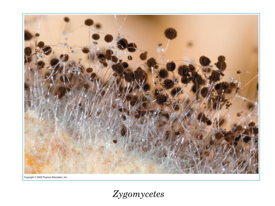
Zygomycetes

26
Zygomycetes in tissues

27
M ANAGEMENT OF P ARANASAL SINUSITIS Surgery Antifungal (Amphotericin B or Azoles)
")
28
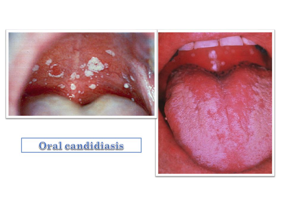
O RAL THRUSH Oral candidiasis or candidosis

29
O RAL CANDIDIASIS OR CANDIDOSIS (O RAL THRUSH ) Over growth of C. albicans in the oral cavity Whitish removable layer cover reddish, eroded, easily bleeding mucosa May extend to the esophagus Mainly seen in: Prolonged use of broad spectrum antibiotics Impaired T-cell immunity

31
T REATMENT For healthy adults and children Eating unsweetened yogurt Taking acidophilus capsules or liquid For adults with weakened immune systems Azoles Amphotericin B

32
K ERATOMYCOSIS mycotic keratitis

33
K ERATOMYCOSIS Corneal infection caused by either filamentous fungi or yeast The most important risk factors: Trauma (generally with plant material) Chronic ocular surface diseases Contact lens usage Surgery Eye-drops abuse Immunodeficiencies Condition related to warm climates
 Chronic ocular surface diseases Contact lens usage Surgery Eye-drops abuse Immunodeficiencies Condition related to warm climates")
34
Keratitis

35
Fungi typeMouldsYeasts Predisposing factorsOutdoor or vegetable-related trauma Contact lens usage Eye surgery Chronic ocular surface diseases Chronic mucocutaneous candidiasis Immunosuppression, including AIDS Corneal anesthetic abuse Most common etiologic agents Fusarium spp Aspergillus spp Acremonium Other Candida albicans Candida parapsilosis Candida tropicalis E PIDEMIOLOGICAL AND CLINICAL DIFFERENCES BETWEEN THE TWO FORMS OF THE INFECTION

36
L ABORATORY DIAGNOSIS Microscopic examination Hyphae in corneal scrapings Fungi are usually deep within the corneal structure, not on the surface. Extensive debridement may be necessary to obtain satisfactory clinical material (swabs are unsatisfactory)
.")
37
Septate hyphae The fungus was seen in several repeated corneal samplings

38
M ANAGEMENT Drug of choice is Natamycin Amphotericin B a second alternative Systemic therapy with azoles Surgery may be necessary

39
L OWER R ESPIRATORY T RACTS I NFECTIONS

41
Generally more serious than upper respiratory infections The leading cause of death among all infectious diseases The two most common LRIs: Bronchitis and pneumonia

42
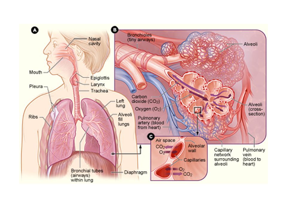
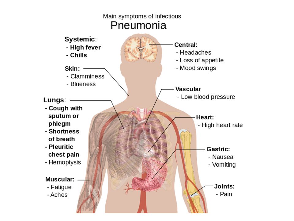
P NEUMONIA Pneumonia is an inflammatory condition of the lung Especially affecting the microscopic air sacs (alveoli) Associated with fever, chest symptoms, and a lack of air space (consolidation) on a chest X-ray
 Associated with fever, chest symptoms, and a lack of air space (consolidation) on a chest X-ray")
43
C AUSES Microbial infections: Bacteria, Viruses Fungi Parasites Other causes

44
T YPICAL SYMPTOMS Cough Chest pain Fever Difficulty breathing

45
D IAGNOSIS X-rays Sputum examination

47
C LASSIFICATION Community-acquired Aspiration Hospital-acquired Ventilator-associated pneumonia Lobar pneumonia Bronchial pneumonia By the causative organism

48
C AUSATIVE AGENTS Viruses and bacteria (most common) Fungi and parasites (less common) Mixed infections with both viruses and bacteria: Up to 45% of infections in children 15% of infections in adults Causative agent is not isolated in approximately half of cases
 Fungi and parasites (less common) Mixed infections with both viruses and bacteria: Up to 45% of infections in children 15% of infections in adults Causative agent is not isolated in approximately half of cases")
49
F UNGAL PNEUMONIA

50
Uncommon Occur in individuals with weakened immune systems due to: AIDS Immunosuppressive drugs Other medical problems

51
F UNGAL SPECIES Opportunistic: Aspergillus species Candida species Pneumocystis jiroveci Primary: Histoplasma capsulatum Blastomyces dermatitidis Coccidioides immitis

52
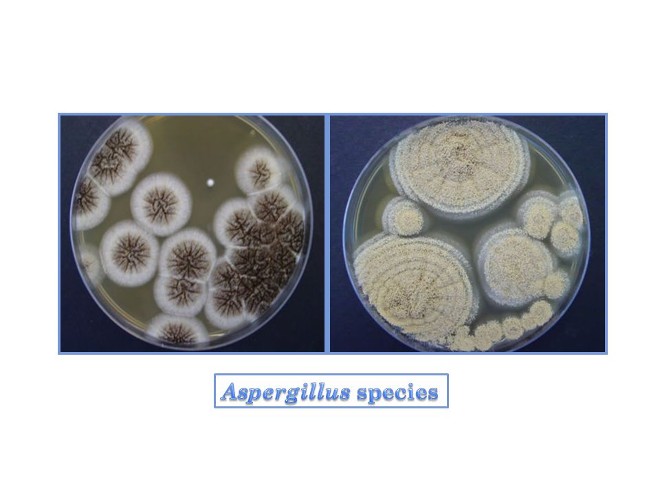
A SPERGILLUS SPECIES Pulmonary Aspergillosis: Allergic, aspergilloma and invasive aspergillosis The common etiological agents are: Aspergillus fumigatus Aspergillus flavus Aspergillus niger Aspergillus nidulans Aspergillus terreus

53
Aspergillosis of the lung Methenamine silver stained tissue section showing dichotomously branched, septate hyphae (left) and a conidial head of A. fumigatus (right)
.")
55
O THER OPPORTUNISTIC FUNGAL INFECTIONS : C ANDIDA SPECIES C. albicans (50-60 % of all yeast infections) C. glabrata C. tropicalis C. parapsilosis
 C. glabrata C. tropicalis C. parapsilosis.")
56
Candida albicans in the lung of an immunocompromised patient, PAS stain

57
Pneumocystis jiroveci

58
P NEUMOCYSTIS JIROVECII Yeast-like fungus of the genus Pneumocystis Pneumocystis pneumonia Prior to its discovery as a human-specific pathogen, P. Jirovecii was known as P. carinii

59
P ATHOGENICITY AND CLINICAL SIGNIFICANCE Pneumocystis is one of the major causes of opportunistic mycoses in immunocompromised Clinical forms: Asymptomatic infections Infantile (interstitial plasma cell) pneumonia Pneumonia in immuno-compromised host Extra-pulmonary infections
 pneumonia Pneumonia in immuno-compromised host Extra-pulmonary infections")
60
D IAGNOSIS OF P. JIROVECI PNEUMONIA Depend of morphologic identification Culture is difficult Trophic (trophozoite) Intracystic spores
 Intracystic spores.")
61
Pneumocystis jiroveci morphology The cysts of P. jiroveci are spherical in shape and measure approximately 4-7 µm Gomori's Methenamine Silver Stain X 1000

62
Cysts of Pneumocystis jiroveci in lung tissue GMS stain The walls of the cysts are stained black and often appear crescent shaped or like crushed ping-pong balls

63
Pneumocystis jiroveci and artifacts Yeast cells Pneumocystis jiroveci

64
Pneumocystis in induced sputum; wright stain stains trophozoites Pneumocystis in bronchoalveolar lavage; toluidine blue highlights cyst forms

65
E ND

Similar presentations

>")


, and the parenchyma.>")


