Download presentation
Presentation is loading. Please wait.

1
Peripheral arterial disease Ahmad Osailan

2
Pathophysiology Form of atherosclerosis Progressive disease May occur suddenly if an embolism occurs or when a blood clot rapidly develops in a blood vessel restricted by an atherosclerotic plaque, and the blood flow is quickly cut off.

3
Peripheral arterial disease Cerebrovascular disease (ischaemic stroke, transient ischaemic attack) Coronary artery disease (stable/unstable angina, myocardial infarction) PAD (intermittent claudication, critical leg ischaemia, amputation, gangrene, necrosis) Atherothrombosis = thrombus formation on top of existing atherosclerosis Occurs in multiple arterial beds
 Coronary artery disease (stable/unstable angina, myocardial infarction) PAD (intermittent claudication, critical leg ischaemia, amputation, gangrene, necrosis) Atherothrombosis = thrombus formation on top of existing atherosclerosis Occurs in multiple arterial beds")
4
Introduction Peripheral arterial disease arises when there is significant narrowing of arteries distal to the arch of the aorta. Incidence (Including asymptomatic- assessed by non-invasive tests) PAD affects 20% of people >70years age(Cochrane 2007). 13.9-16.9% in men 11.4-20.5% in women over 55 years of age Usually about 60% of affected will be asymptomatic. Mechanisms Atherosclerosis Vasospasm Inflammation/ vasculitis Thrombosis/ Embolism
 PAD affects 20% of people >70years age(Cochrane 2007) % in men % in women over 55 years of age Usually about 60% of affected will be asymptomatic. Mechanisms Atherosclerosis Vasospasm Inflammation/ vasculitis Thrombosis/ Embolism.")
5
Case Presentation ID/CC: 83 yo Caucasian female with HTN, s/p aortic valve replacement in generally excellent health who complains of one year of right thigh pain with “ambulation around the grocery store but not around the house.”

6
Symptoms of PAD Leg or hip pain during walking (intermittent claudication). The pain stops when you rest. Numbness, tingling or weakness in the legs. Burning or aching pain in feet or toes when resting. Sore on leg or foot that won’t heal. Cold legs or feet. Color change in skin of legs or feet. Loss of hair on legs.

7
Claudication DEFIt is the ischaemic pain due to decompensation of the blood supply typically occurring with physical activity. Determining how much physical activity is needed before the onset of pain is crucial. It is most common with the distal superficial femoral artery (located just above the knee joint), which corresponds to claudication in the calf muscle area (the muscle group just distal to the arterial disease). If proximal vessels are involved, pain might be felt in thighs and buttocks too. There should be no weakness or numbness. Pain resolves quickly on rest(<5mins). Spinal pain( nerve root irritation, spinal stenosis) usually gives symptoms localised to a muscle group, accompanied by weakness or heaviness on walking and often by leaning forward.
, which corresponds to claudication in the calf muscle area (the muscle group just distal to the arterial disease). If proximal vessels are involved, pain might be felt in thighs and buttocks too. There should be no weakness or numbness. Pain resolves quickly on rest(<5mins). Spinal pain( nerve root irritation, spinal stenosis) usually gives symptoms localised to a muscle group, accompanied by weakness or heaviness on walking and often by leaning forward..")
8
Risk Factors Smoking biggest risk factor. DM is a close second. Rest are hypertension, hyperlipidaemia, family history, sedentary lifestyle

9
Noninvasive diagnosis of PAD Ankle Brachial Index (ABI) ABPI = Ankle systolic pressure/ Brachial systolic pressure. In the absence of significant stenosis or occlusion in these vessels the two values are usually within 10 mmHg of each other even in the presence of more proximal disease. The maximum cuff pressure at which the pulse can just be heard with the probe is recorded. BP measures in both arms and the higher of both used. Interpretation of values Symptom free - 1 or more Intermittent claudication - 0.95 - 0.5 Rest pain - 0.5 - 0.3 Gangrene and ulceration - <0.2

10
How to do an ABPI? http://www.youtube.com/watch?v=bTVYl9UR dSI&feature=related

12
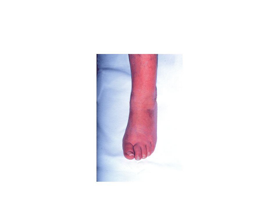
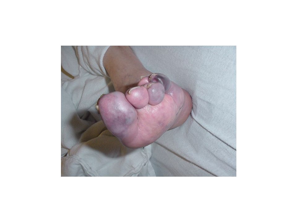
Critical limb ischaemia

14
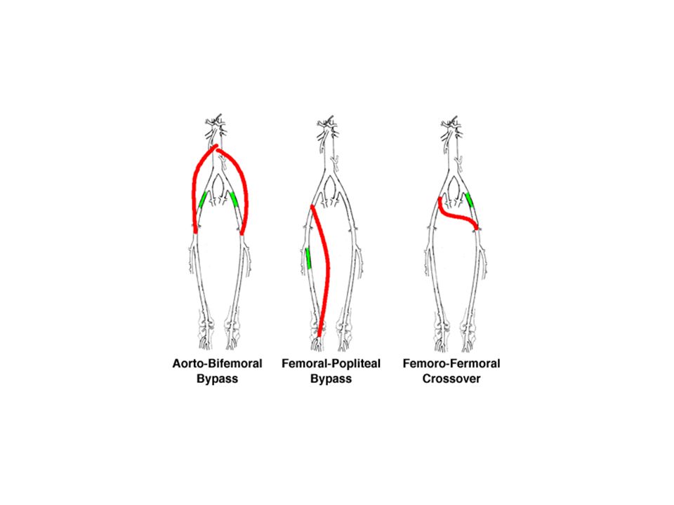
Treatments Risk factor reduction Exercise Medications Percutaneous translumenal angioplasty (PTA) Arterial bypass surgery
 Arterial bypass surgery")
15
Exercise Numerous studies demonstrating clear benefits A meta-analysis in JAMA (1995) showed an increase of 179% (from 125 to 350 meters) to onset of claudication pain and an increase of 122% (from 325-723 meters) to maximal claudication pain Equal to an additional 4 blocks by treadmill P<.001
 showed an increase of 179% (from 125 to 350 meters) to onset of claudication pain and an increase of 122% (from meters) to maximal claudication pain Equal to an additional 4 blocks by treadmill P<.001")
16
Exercise Prescription Training Intensity Initial Set by result of peak treadmill. Starting exercise work load brings on claudication pain. Subsequent Speed or grade increased if patient walks > 10 minutes. Grade increased first if speed > 2 mph. Speed increased first if < 2 mph.

17
Exercise Prescription Duration Initial 35 minutes (intermittent walking) Subsequent Add 5 minutes every session until 50 minutes (intermittent walking) is possible
 Subsequent Add 5 minutes every session until 50 minutes (intermittent walking) is possible")
18
Exercise Prescription Frequency 3-5 times per week. Specificity of Activity Treadmill walking is the recommended exercise.

19
Surgical Treatments for PVD Thrombectomy Bypass Grafts

21
Peripheral venous diseases

22
Peripheral venous disease Peripheral venous disease is a term describing damage, defects or blockage in the veins that carry blood from the hands and feet to the heart. Peripheral venous disease can occur almost anywhere in the body but is mostly seen in the arms and legs. The most common cause of peripheral venous disease is a blood clot that blocks a vein. A clot forms when vein walls become weak and blood flow slows. When the clot is in a vein deep within the body, it is called deep vein thrombosis. When the clot is in a vein closer to the skin, it is called superficial thrombophlebitis. blood clotdeep vein thrombosissuperficial thrombophlebitis

23
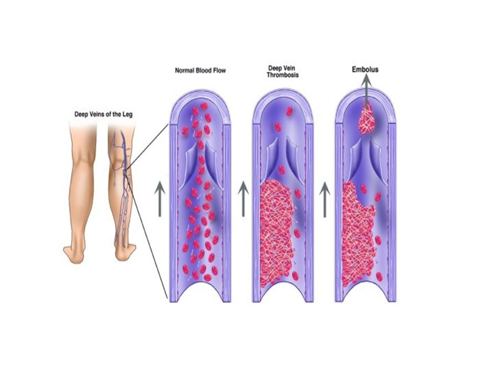
Deep Venous Thrombosis A blood clot in a deep vein. May form on the valves within the vein, and may subsequently increase in size to totally occlude the vein. Sometimes parts of the clot may break off and travel in the bloodstream to the lungs and cause serious health problems (pulmonary embolism). DVT is perhaps the most dangerous problem.
. DVT is perhaps the most dangerous problem..")
25
Varicose Veins Caused because either the blood flow is too slow making the vein pile up with blood or the valve in the vein is not working well so the blood falls down due to gravity and piles up in the veins of the legs. Sclerotherapy: Irritant chemical is injected into the veins, causing them to scar and seal off. This “detours” the blood to nearby healthier veins. Stripping: Procedure used to remove larger varicose veins. Parts of the vein can be removed or tied off, or the entire vein can be removed.

27
Phlebitis Inflammation of the leg veins. Two types: Inflammation of the veins on the surface of the leg (more common). Inflammation of the deep veins of the leg. Phlebitis is caused by an infection or injury. Can cause a blood clot to form and this clot can then embolize and result in pulmonary embolism. This is the worst thing that can happen if you have phlebitis.
. Inflammation of the deep veins of the leg. Phlebitis is caused by an infection or injury. Can cause a blood clot to form and this clot can then embolize and result in pulmonary embolism. This is the worst thing that can happen if you have phlebitis..")
28
Physical therapy treatment For varicos veins: Elevating the affected leg above your heart to help pooled blood drain properly. Avoiding long periods of standing or sitting. If you must sit for a long period, stretch and flex your legs every 5 minutes or so to keep blood flowing. Wearing elastic compression stockings that squeeze the veins and keep the blood flowing in your legs, which makes it more difficult for blood clots to form.

29
Physical therapy treatment For DVT: There is no specific Exercises for DVT BUT there are precautions to exercise patients with DVT, they are: 1.Never elevate the affected extremities to assisted gravity position for blood flow. 2.Do not exercise patient in supine position, always perform exercises while sitting 3. never do any massage technique on the affected extremity 4.Encourage walking more than anything else.

Similar presentations


, fibrous material and.>")


>")

 A National Public Awareness Campaign from the P.A.D. Coalition and the National Heart,>")


