Download presentation
Presentation is loading. Please wait.

1
Lower Extremity Venous Disease: Peripheral Venous Insufficiency
Aman K Kakkar MD FACC Heart and Vascular Care, Inc

2
30 Million people suffer from venous reflux disease, the underlying cause for most varicose veins

3
Prevalence and Etiology of Venous Insufficiency
Venous reflux disease is 2x more prevalent than coronary heart disease (CHD) and 5x more prevalent than peripheral arterial disease (PAD)1 Annual U.S. Incidence U.S. Prevalence Notes: Market research indicates that over 2 million work days are lost annually in the US and $1.4 billion is spent each year on this common medical condition (Sources: American Heart Association, SIR,Brand et al. “The Epidemiology of Varicose Veins: The Framingham Study”) Millions
 and 5x more prevalent than peripheral arterial disease (PAD)1. Annual U.S. Incidence. U.S. Prevalence. Notes: Market research indicates that over 2 million work days are lost annually in the US and $1.4 billion is spent each year on this common medical condition (Sources: American Heart Association, SIR,Brand et al. The Epidemiology of Varicose Veins: The Framingham Study ) Millions.")
4
Prevalence of Venous Insufficiency
Of the estimated 30 million people with symptomatic superficial venous reflux1 : Only 1.9 million seek treatment annually2 Only 500K Patients receive treatment Over 28 million go untreated Prevalence by Age and Gender3,4 Age Female Male 8% 1% 41% 24% 72% 43% Notes: Statistics show that of the 25 million people in the U.S. who suffer from symptomatic reflux, only about 5% seek treatment annually; 2/3 of patients who do seek treatment have saphenous reflux When left untreated, venous reflux can lead to significant clinical issues, like pain, swelling, varicose veins, skin changes, and ulcers With advancing age, especially with females, the prevalence of venous disease grows The typical female patient is in her 40’s and has had multiple pregnancies It is estimated that in America, 72% of women and 42% of men will experience varicose veins by the time they reach their 60s; prevalence is highly correlated to age and gender (Barron HC, Ross BA. Varicose Veins: A guide to prevention and treatment. NY, NY: Facts on File, Inc. [An Infobase Holdings Company]; 1995;vii.)
")
5
Venous System Venous blood flows from the capillaries to the heart
Deep femoral v. Femoral v. Popliteal v. Small saphenous v. Great saphenous v. Perforating v. Venous blood flows from the capillaries to the heart Flow occurs against gravity Muscular compression of the veins Negative intrathoracic pressure Calf muscle pump Low flow, low pressure system Notes: This diagram shows the interrelation between superficial and deep venous systems and the perforators that connect the two systems Deep venous system Superficial venous system Saphenous veins Lateral venous complex Perforating veins Image source: Fundamentals of Phlebology: Venous Disease for Clinicians. Illustration by Linda S. Nye. American College of Phlebology 2004.

6
Pathophysiology of Venous Insufficiency
Notes: Healthy leg veins contain valves that open and close to assist the return of blood back to the heart Venous insufficiency or venous reflux disease develops when the valves that keep blood flowing out of the legs and back to the heart become damaged or diseased Venous insufficiency is the result of over-dilation of the venous vessels in the legs. This dilation eventually prevents the valve cusps from closing properly, resulting in reflux. The pooling of blood results in ineffective flow back to the heart. In some cases the reflux is caused not only by the over-dilation of the vessel wall, but also by damaged or absent valves. In this case, the valves have been so badly damaged, or degenerated, that they are almost nonexistent and no longer function To assess if venous reflux is present, a duplex ultrasound scan is performed

8
Risk Factors and Symptoms of Venous Insufficiency
Risk factors of venous insufficiency: Gender Age Heredity Pregnancy Standing occupation Obesity Prior injury or surgery Sedentary lifestyle Symptoms of venous insufficiency: Leg pain, aching, or cramping Burning or itching of the skin Leg or ankle swelling “Heavy” feeling in legs Skin discoloration or texture changes Open wounds or sores Restless legs Varicose Veins Notes: Gender: Approximately four times as many women as men are affected by varicose veins, suggesting that female hormones may be a risk factor Age: Generally, most elderly individuals show some degree of varicose vein occurrence Heredity: Weak vein walls and valves, as well as shortage of vein valves, seem to be inherited characteristics, and may play a role in determining who develops varicose veins and at what age Pregnancy: is associated with an increase in blood volume. Also, added pressure on the veins in the legs by the weight of the growing uterus and the relaxation effects of the hormones estrogen and progesterone on the vein walls contribute to the development of varicose veins during pregnancy Standing occupation: causes a great amount of pressure to develop in the leg veins Obesity: the added weight causes a great amount of pressure on the veins in the legs Prior trauma or surgery to the leg could cause interruption of the normal blood flow channels Sedentary lifestyle/prolonged sitting: The calf muscles are inactive and therefore can’t help push venous blood back up to the heart. This causes blood to pool in the veins, thus resulting in increased pressure on the vein walls

9
Manifestations of Venous Insufficiency
Superficial venous reflux is progressive and if left untreated, may worsen over time. Below are manifestations of the disease.5 Varicose Veins Swollen Legs Skin Changes Skin Ulcers Notes: Although often underestimated as a cosmetic problem, venous insufficiency can produce significant clinical problems for the patient An estimated 25 million people in the United States have varicose veins, 2 to 6 million have more advanced forms of chronic venous insufficiency (swelling, skin changes), and nearly 500,000 have painful venous ulcers. (White JV, Ryjewski C. Chronic venous insufficiency. Perspect Vasc Surg Endovasc Ther 2005;17:319-27) Overall, as the severity of the disease progresses, quality of life decreases 20+ million 2 to 6 million 500,000 Photos courtesy of Rajabrata Sarkar, MD, PhD.
, and nearly 500,000 have painful venous ulcers. (White JV, Ryjewski C. Chronic venous insufficiency. Perspect Vasc Surg Endovasc Ther 2005;17:319-27) Overall, as the severity of the disease progresses, quality of life decreases. 20+ million. 2 to 6 million. 500,000. Photos courtesy of Rajabrata Sarkar, MD, PhD.")
10
PATHOPHYSIOLOGY

11
CEAP Classifications Clinical Classifications of Venous Insufficiency (CEAP) Class 0 - No visible or palpable signs of venous disease Class 1 - Telangiectasias or reticular veins Class 2 - Varicose veins Class 3 - Edema Class 4 - Skin changes (4a) Skin changes including pigmentation or venous eczema (4b) Skin changes with lipodermatosclerosis Class 5 - Healed venous ulceration Class 6 - Active venous ulceration Notes: CEAP Classifications are used to determine the severity of venous disease. C= Clinical signs (grade 0-6) E= Etiologic Classification (congenital, primary, or secondary) A= Anatomic Distribution (superficial, deep, perforator; alone or in combination) P= Pathophysiologic Dysfunction (reflux of obstruction; alone or in combination) CEAP Class 0 - No visible or palpable signs of venous disease Patient is asymptomatic. CEAP Class 1 – Telangiectasias or reticular veins Patient presents with “spider veins” which are very small diameter vessels that appear as starburst-like lines. CEAP Class 2 - Varicose Veins Varicose veins are elongated, dilated, tortuous, pouched and thickened veins with incompetent valves. In addition to varicose veins, a high percentage of patients also have incompetence of one or more of the key “gatekeeper” valves, e.g., the terminal valves of the saphenofermoral junction or saphenopopliteal junction, and/or perforating veins. CEAP Class 3 - Edema This next progressive state of venous insufficiency occurs as the result of venous hypertension forcing fluid into the lymphatic and interstitial spaces resulting in extreme reflux and poor antegrade flow. This causes swelling of the limb. Pain and discomfort are typical of this classification, particularly in the lower leg (calf & ankle) where proximity of nerves exacerbates the situation. In addition to superficial involvement, these stages may include some portion of the deep system (including perforators). CEAP Class 4a and 4b - Skin Changes Including Pigmentation and Venous Ezcema or with Lipodermatosclerosis CEAP 5 and 6 – Healed and Active Venous Ulcer This is the most severe form of venous insufficiency and typically involve both the deep (including perforators) and superficial systems. Extreme reflux and venous hypertension result in changes in the microcirculation of the skin eventually leading to severe ulceration. It is believed that communication via the perforator veins between the deep and superficial systems is a primary component of these classes.
 Skin changes including pigmentation or venous eczema. (4b) Skin changes with lipodermatosclerosis. Class 5 - Healed venous ulceration. Class 6 - Active venous ulceration. Notes: CEAP Classifications are used to determine the severity of venous disease. C= Clinical signs (grade 0-6) E= Etiologic Classification (congenital, primary, or secondary) A= Anatomic Distribution (superficial, deep, perforator; alone or in combination) P= Pathophysiologic Dysfunction (reflux of obstruction; alone or in combination) CEAP Class 0 - No visible or palpable signs of venous disease Patient is asymptomatic. CEAP Class 1 – Telangiectasias or reticular veins. Patient presents with spider veins which are very small diameter vessels that appear as starburst-like lines. CEAP Class 2 - Varicose Veins. Varicose veins are elongated, dilated, tortuous, pouched and thickened veins with incompetent valves. In addition to varicose veins, a high percentage of patients also have incompetence of one or more of the key gatekeeper valves, e.g., the terminal valves of the saphenofermoral junction or saphenopopliteal junction, and/or perforating veins. CEAP Class 3 - Edema. This next progressive state of venous insufficiency occurs as the result of venous hypertension forcing fluid into the lymphatic and interstitial spaces resulting in extreme reflux and poor antegrade flow. This causes swelling of the limb. Pain and discomfort are typical of this classification, particularly in the lower leg (calf & ankle) where proximity of nerves exacerbates the situation. In addition to superficial involvement, these stages may include some portion of the deep system (including perforators). CEAP Class 4a and 4b - Skin Changes Including Pigmentation and Venous Ezcema or with Lipodermatosclerosis. CEAP 5 and 6 – Healed and Active Venous Ulcer. This is the most severe form of venous insufficiency and typically involve both the deep (including perforators) and superficial systems. Extreme reflux and venous hypertension result in changes in the microcirculation of the skin eventually leading to severe ulceration. It is believed that communication via the perforator veins between the deep and superficial systems is a primary component of these classes.")
12
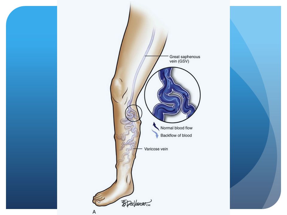
Great Saphenous Vein : The Culprit

13
American Venous Forum (AVF) Guidelines: Diagnosis
 Guidelines: Diagnosis")
14
Great Saphenous Vein (GSV) : Reflux
 : Reflux")
15
Small Saphenous Vein (SSV): Reflux
: Reflux")
16
TREATMENT ALGORITHM

17
AVF Guidelines: Treatment

18
AVF Guidelines: Treatment

19
Vein Closure: Indications, Risks
The Catheter ablation is intended for endovascular coagulation of blood vessels in patients with superficial venous reflux Contraindications: Patients with thrombus in the vein segment to be treated Potential Risks & Complications: Potential complications include, but are not limited to, the following: vessel perforation, thrombosis, pulmonary embolism, phlebitis, hematoma, infection, adjacent nerve injury, skin burns, deep vein thrombosis

20
Vein Closure: Options Stripping Foam Sclerotherapy
Radiofrequency Ablation Laser Ablation

21
RECOVERY Trial7: Pain A Prospective, Multi-Center, Randomized Study
Overall Maximum Pain Score (0 none to 10 max) p < 4 Notes: This slide looks at the average Maximum Pain Score from all participants in the trial. The patients were asked during their follow-up visits to rate their maximum pain level on a scale from zero to ten. This graph shows the average maximum score of all patients and all visits. 2 ClosureFAST Laser
 p < Notes: This slide looks at the average Maximum Pain Score from all participants in the trial. The patients were asked during their follow-up visits to rate their maximum pain level on a scale from zero to ten. This graph shows the average maximum score of all patients and all visits. 2. ClosureFAST Laser.")
22
RECOVERY Trial7: Ecchymosis A Prospective, Multi-Center, Randomized Study
Moderate to Severe Ecchymosis (Bruising) After Treatment Moderate to severe ecchymosis is defined as bruising over greater than 25% of the treated surface area p < 51.3% Notes: Ecchymosis was evaluated at each follow-up visit by visual evaluation by the practicing physician. They categorized their observations into 6 categories: no ecchymosis >0 but </=25% of treated surface area ecchymosed >25% but </= 50% >50% but </= 75% >75% but </=100% >100% (if the ecchymosis went beyond the treated area) This graph shows the “Moderate to Severe Ecchymosis” which is defined as >25% of the area ecchymosed. There were cases of ecchymosis in the lower 2 categories for CLF and laser, but this could be due to the infiltration of tumescent anesthesia. In order to compensate for tumescent, which is used in both CLF and laser procedures, we looked at those individuals who had >25% ecchymosis after the procedure. 2.2% ClosureFAST Laser
 After Treatment. Moderate to severe ecchymosis is defined as bruising over greater than 25% of the treated surface area. p < % Notes: Ecchymosis was evaluated at each follow-up visit by visual evaluation by the practicing physician. They categorized their observations into 6 categories: no ecchymosis. >0 but </=25% of treated surface area ecchymosed. >25% but </= 50% >50% but </= 75% >75% but </=100% >100% (if the ecchymosis went beyond the treated area) This graph shows the Moderate to Severe Ecchymosis which is defined as >25% of the area ecchymosed. There were cases of ecchymosis in the lower 2 categories for CLF and laser, but this could be due to the infiltration of tumescent anesthesia. In order to compensate for tumescent, which is used in both CLF and laser procedures, we looked at those individuals who had >25% ecchymosis after the procedure. 2.2% ClosureFAST Laser.")
23
RadioFrequency Ablation
The Radiofrequency Closure System is a minimally invasive treatment alternative for patients with symptomatic superficial venous reflux and varicose veins Using a catheter-based approach, catheter delivers radiofrequency (RF) energy to the vein wall RF energy creates conductive heating that contracts the vein wall collagen, thereby occluding the vein Notes: Collagen Contraction The application of heat to human tendon tissue causes collagen tissue to be shortened. (Vangsness, CT Jr. Et al:”Collagen Shortening: An Experimental Approach with Heat,” Clinical Orthopaedics and Related Research, No. 337, , 1997) VNUS animal research has demonstrated that the application of controlled heating to the vein wall causes the collagen fibrils to contract (shorten) and thicken, and the vein to significantly shrink in diameter. Effects of controlled heating of the vein wall Thermal energy is quickly transferred from the ClosureFAST catheter’s heating element to the vein wall through conduction. Heating of the vein wall tissue causes endothelial destruction and collagen contraction that result in vein occlusion. Thermal energy causes collagen to undergo the following changes: Heat sensitive bonds break at 60ºC Crystalline extended structure begins to uncoil, causing the collagen fibrils to shorten and thicken. As the molecule contracts, its diameter increases, causing a reduction in vein lumen diameter Histological Effects of RF heating Controlled thermal injury to the vessel wall causes the following changes in the histology of the vessel resulting in vein contraction. When the vein wall is exposed to sufficient thermal energy it causes: Endothelial denudation, Collagen denaturation, Smooth muscle necrosis, Vein wall shrinkage and thickening, and Vessel lumen reduction. Following these immediate effects, the treated vessel undergoes an inflammatory response, fibroblast infiltration, new collagen deposition, and eventual fibrosis.
 energy to the vein wall. RF energy creates conductive heating that contracts the vein wall collagen, thereby occluding the vein. Notes: Collagen Contraction. The application of heat to human tendon tissue causes collagen tissue to be shortened. (Vangsness, CT Jr. Et al: Collagen Shortening: An Experimental Approach with Heat, Clinical Orthopaedics and Related Research, No. 337, , 1997) VNUS animal research has demonstrated that the application of controlled heating to the vein wall causes the collagen fibrils to contract (shorten) and thicken, and the vein to significantly shrink in diameter. Effects of controlled heating of the vein wall. Thermal energy is quickly transferred from the ClosureFAST catheter’s heating element to the vein wall through conduction. Heating of the vein wall tissue causes endothelial destruction and collagen contraction that result in vein occlusion. Thermal energy causes collagen to undergo the following changes: Heat sensitive bonds break at 60ºC. Crystalline extended structure begins to uncoil, causing the collagen fibrils to shorten and thicken. As the molecule contracts, its diameter increases, causing a reduction in vein lumen diameter. Histological Effects of RF heating. Controlled thermal injury to the vessel wall causes the following changes in the histology of the vessel resulting in vein contraction. When the vein wall is exposed to sufficient thermal energy it causes: Endothelial denudation, Collagen denaturation, Smooth muscle necrosis, Vein wall shrinkage and thickening, and Vessel lumen reduction. Following these immediate effects, the treated vessel undergoes an inflammatory response, fibroblast infiltration, new collagen deposition, and eventual fibrosis.")
24
Procedure using the Radiofrequency Ablation Catheter
Notes: The procedure is performed under general and/or local anesthesia Using ultrasound, the Closure catheter is positioned into the diseased vein through a small opening in the skin The slender catheter is powered by radiofrequency (RF) energy which delivers heat to the vein wall As thermal energy is delivered, the vein wall shrinks and the vein is sealed closed Once the diseased vein is closed, blood is re-routed to other healthy veins Consider showing the VNUS ClosureFAST animation video to further detail the treatment
 energy which delivers heat to the vein wall. As thermal energy is delivered, the vein wall shrinks and the vein is sealed closed. Once the diseased vein is closed, blood is re-routed to other healthy veins. Consider showing the VNUS ClosureFAST animation video to further detail the treatment.")
25
Procedure

26
Summary Easily Diagnosed but vastly untreated
Although many aspects of treatment have remained the same for over a century Recent advances have provided new and better treatment options for patients with chronic venous disease.

Similar presentations