Download presentation
Presentation is loading. Please wait.

1
Peritoneum and Mesentery
Azin Shayganfar .MD

2
Definitions and Anatomy

3
Peritoneum serosal membrane: subserosal tissue :
a single layer of flat mesothelial cells supported by submesothelial connective tissue. subserosal tissue : 1-fat cells, 2- lymphatics, blood vessels 3-inflammatory cells like lymphocytes and plasma cells.

4
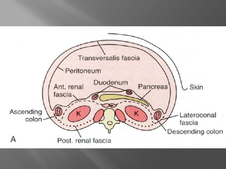
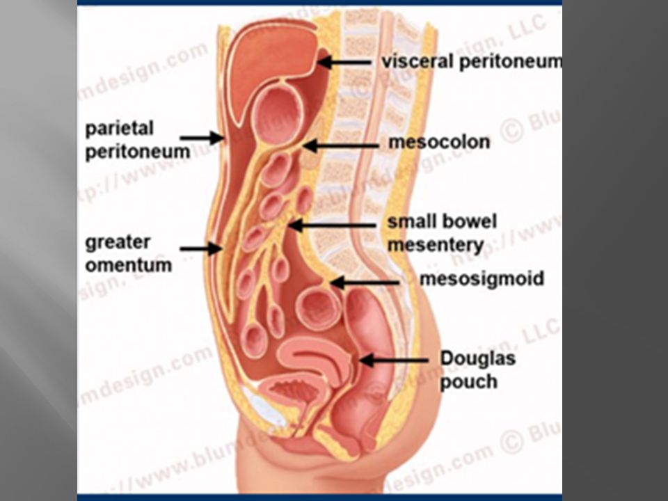
The visceral peritoneum lines all the organs that are intraperitoneal.
The parietal peritoneum lines the anterior, lateral and posterior walls of the peritoneal cavity. The peritoneal cavity is a potential space between the parietal peritoneum, and the visceral peritoneum.

5
The deepest portion: the pouch of Douglas in women and the retrovesical space in men both in the upright and supine position. The cavity is closed except for the fallopian tubes and contains 50 to 75 mL of clear fluid Peritoneal ligaments, mesentery, and omentum divide the peritoneum into two compartments: the main region, called the greater sac, and , omental bursa, or lesser sac.

9
90% of peritoneal fluid is cleared at the subphrenic space by the submesothelial lymphatics.

10
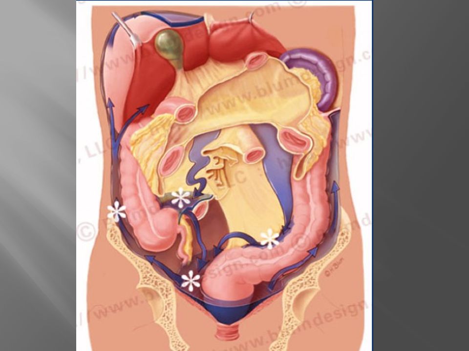
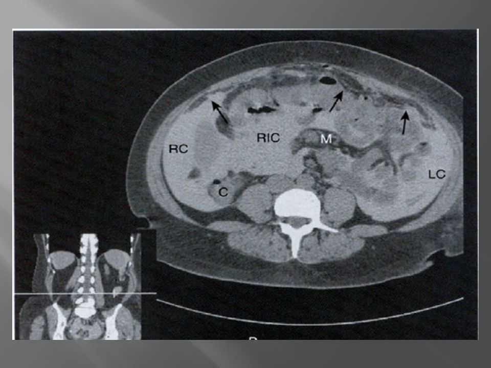
watershed regions There are in the peritoneal cavity that are areas of fluid stasis: Ile ocolic region Root of the sigmoid mesentery Pouch of Douglas When you are staging a patient for GI malignancy you have to look for disease in these areas of stasis.

12
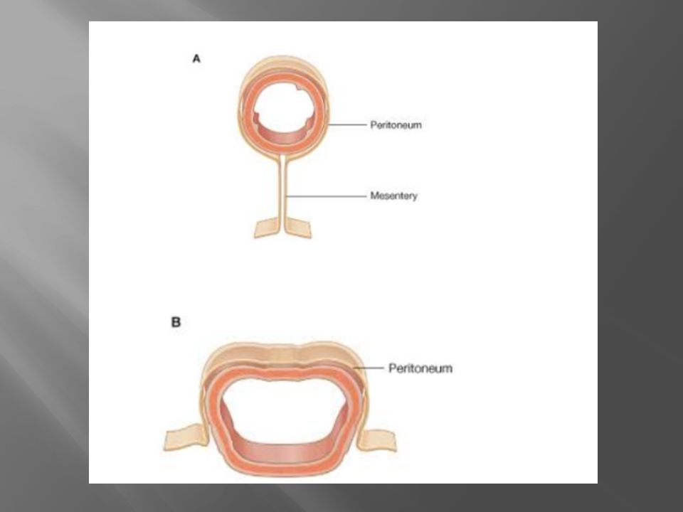
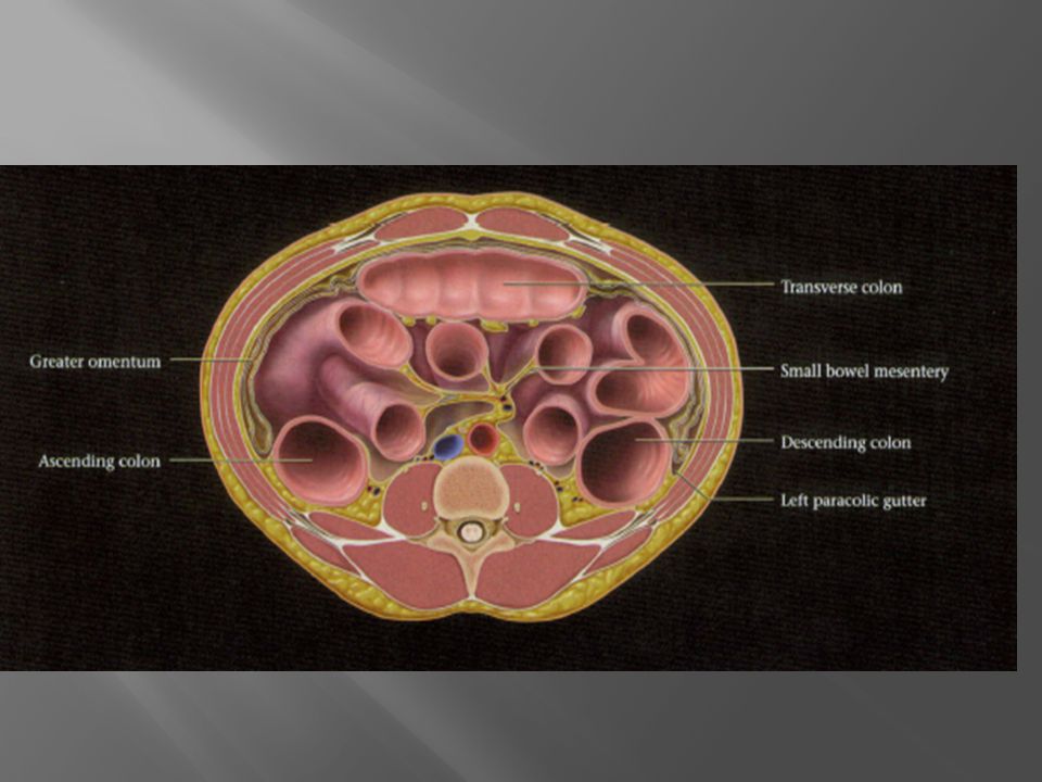
The mesentery is a double fold of the peritoneum

14
True mesenteries Specialized mesenteries
connect to the posterior peritoneal wall. 1-The small bowel mesentery 2-The transverse meso colon 3-The sigmoid mesentery Specialized mesenteries do not connect to the posterior peritoneal wall. 1-The greater omentum: connects the stomach to the colon 2-The lesser omentum: connects the stomach to the liver 3-The meso appendix: connects the apendix to the ileum

17
Omentum divided into the greater and lesser omentum

18
greater omentum originates along the margin of the greater curvature of the stomach and can cover a broad expanse of the anterior abdominal wall. normally is usually imperceptible on routine scans exhibits fat density infectious processes or neoplasms can increases in density and produce a mass effect on the small bowel loops.

22
The lesser omentum is subdivided into:
Gastro hepatic ligament : connects the left lobe of the liver to the lesser curvature of the stomach. Hepato duodenal ligament : free edge of the omentum, which contains the portal vein, hepatic artery and common bile duct .

24
1-The lesser omentum 2-Transverse mesocolon 3-Small bowel mesentery 4-Sigmoid mesentery

25
Pathology OF Peritoneum and Mesentery

26
Imaging Modalities US: may depict peritoneal collections or ascites and is used to guide drainage of ascites and large superficial fluid collections CT : is the most common imaging modality used to detect diseases of the peritoneum To fully delineate peritoneal anatomy and the extent of disease, we prefer to perform isotropic imaging with coronal and sagittal reformations.

27
CT did not show: 1- microscopic lesions, masses <1CM 2- omental metastases, such as pancreatic and gastric carcinomas, that are immediately adjacent to primary masses. A positive CT scan is a useful guide for the surgeon, but a negative study does not obviate the need for a second-look surgical procedure.

28
biopsy of the diaphragmatic areas is the best method for the early detection of peritoneal seeding .

29
Magnetic resonance (MRI)
Disadvantages of MR imaging include: 1- motion artifacts caused by respiration and peristalsis 2- chemical shift artifacts at the bowel-mesentery interface. 3-the spatial resolution of MR imaging is lower than that of CT, a characteristic that may make it difficult to assess small peritoneal lesions. 4-Patients who are ill may not tolerate prolonged MR imaging examinations

30
Differential Diagnosis of peritoneal mass
The first step : separate them into cystic and solid. The secound : we have to realize that any loculated fluid collection due to abscess or as a result of pancreatitis,bowel perforation can simulate a cystic mass. Especially fluid collections in the lesser sac can simulate a cystic mass. Lastly we have to know which cystic masses are common and look for specific features of these masses.

32
Metastatic Disease common with neoplasms originating in
the ovary, sto mach, pancreas, and colon Sites of Implants : 1- The falciform ligament 2- gastro hepatic ligament 3- ileo colic region 4- posterior and dependent sites of the peritoneum 5- broad ligaments

33
diagnostic signs : rounded, ill defined, cakelike, or stellate.
If the tumor is of muci nous origin, such as the ovary or colon, it may show soft tissue or fluid density. mucinous or other treated tumors can produce small calcifications throughout the peritoneum.

34
Low-density mass adjacent to the falciform ligament and on the surface of the liver below the diaphragm due to metastatic ovarian cancer

35
Mass in the hilum of the spleen due to metastatic ovarian cancer

36
Calcined peritoneal metastases on the undersurface of diaphragm

37
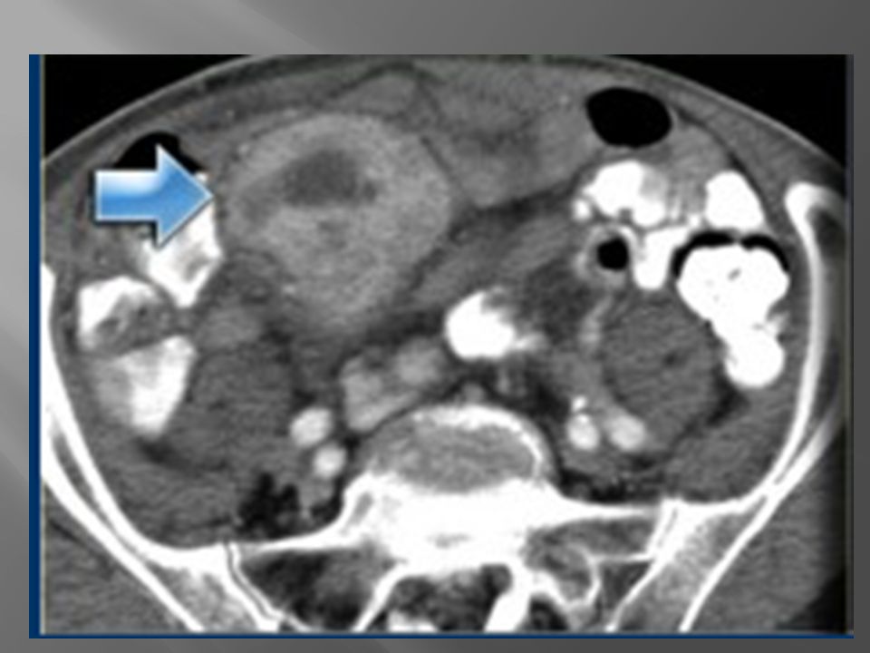
Patient with carcinoma of the pancreas has a metastatic implant adjacent to the ileocecal valve

38
Two metastatic implants on the right and left sides of the sigmoid mesentery

39
extensive tumor implantation produces a thick soft tissue density displacing the colon from the anterior peritoneum.

41
Lymphoma NHL is the most common cause of lymphadenopathy
Usually there are other sites with lymphoma. The CT attenuation at diagnosis is very homogeneous in most cases with minimal to no enhancement. Heterogeneous attenuation is seen only in cases with aggressive histology or in during the treatment

43
Carcinoid is a slow-growing neuroendocrine tumour most commonly found in the small bowel. Less than 10% of patients with carcinoid will develop the carcinoid syndrome Carcinoid metastasizes to the mesentery, which at times is easier to appreciate than the primary tumor in the small bowel . Carcinoid metastasizes to the mesentery is associated bowel wall thickening due to a desmoplastic reaction

44
typical carcinoid with central calcification (blue arrow) Notice the bowel retraction and wall thickening
 Notice the bowel retraction and wall thickening")
45
Gastrointestinal Stromal Tumor GIST
Primary small bowel tumors can extend into the mesentery and the typical example of that is the GIST. a large mesenteric component and such a small attachment to the bowel. On CT they are of mixed density due to necrosis and hemorrhage and they tend to be well vascularized.

47
Mesenteric Fibromatosis or desmoid tumor
locally aggressive but benign proliferative tumor that does not metastasize. the small bowel mesentery is the most common site of intra-abdominal fibro matosis Most cases are sporadic (10% to 15% occur in FAP) About 83% of patients with mesenteric fibromatosis and FAP have a history of abdominal surgery, most commonly total cole ctomy. Only about 10% of patients with sporadic form have had previous abdominal surgery
 About 83% of patients with mesenteric fibromatosis and FAP have a history of abdominal surgery, most commonly total cole ctomy. Only about 10% of patients with sporadic form have had previous abdominal surgery.")
48
CT findings: a focal mesenteric mass which may have :
- highly collagenous stroma(soft tissue density) a myxoid stroma (more hypodense) On MRI low to intermediateT1 signal and intermediateT2 signal, with variable contrast enhancement after injection
 a myxoid stroma (more hypodense) On MRI. low to intermediateT1 signal and. intermediateT2 signal, with variable contrast enhancement after injection.")
49
Mesenteric Fibromatosis

52
Malignant mesothelioma
Suggestive features are a sheet-like peritoneal thickening and absence of lymphadenopathy. Just like pleural mesothelioma, it is associated with asbestos exposure.

53
Notice the sheet-like thickening of the peritoneum
Notice the sheet-like thickening of the peritoneum. The diagnosis was suggested because of the pleural calcifications

54
Primary Peritoneal Serous Carcinoma
It occurs exclusively in women. This tumor is histologically identical to malignant ovarian surface epithelial tumors. As a radiologist you should consider this diagnosis if you think of metastatic ovarian cancer but the ovaries are normal

55
There is ascites and omental involvement, so your first thought is ovarian cancer, but the ovaries were normal.

57
Mucinous Carcinomatosis
is the most common cystic tumor to affect the peritoneal cavity. we see tumor nodules along the peritoneal lining, omental tumor deposits, and bowel obstruction. Usually arise from mucinous carcinomas of the ovary or of the GI tract (stomach, colon), pancreas. The prognosis is poor.
, pancreas. The prognosis is poor.")
58
when low-grade mucinous adenocarcinoma of the appendix spreads to the peritoneal cavity, the consequence is typically pseudomyxoma peritonei, which is a distinct tumor with a better prognosis

59
Mucinous Carcinomatosis

60
Pseudomyxoma peritonei
result of a mucinous adeno carcinoma of the appendix, which presents as a mucocele It is a clinical syndrome, characterized by recurrent and recalcitrant voluminous mucinous ascites due to surface growth on the peritoneum without significant invasion of underlying tissues. A typical feature of pseudomyxoma peritonei is scalloped indentation of the surface of the liver and spleen. Unlike peritoneal metastases, there are no tumor nodules. There may be some calcifications.

61
Pseudomyxoma peritonei

62
Pseudomyxoma peritonei
Often is confused with mucinous carcinomatosis. Unlike carcinomatosis : does not have true omental tumor deposits presenting as omental cake or peritoneal tumor deposits

63
Mesenteric cyst - Lymphangioma
Mesenteric cyst is a descriptive term for any cystic lesion within the mesentery. Usually it is a lymphangioma Other mesenteric cysts : enteric duplication cyst enteric cyst non pancreatic pseudo cyst meso thelial cyst are very uncommon

64
Lymphangioma is a benign lesion of vascular origin with enhancing septa Most lymphangiomas are located in the neck, but 5% of lymphangiomas are abdominal. Unlike in cystic peritoneal metastases, ascites is not a feature of lymphangioma. When you see a septated cystic lesion without ascites the most likely diagnosis is a lymphangioma Lymphangioma is often closely associated with the small bowel.

66
Notice that CT does not always appreciate the septations although the specimen clearly shows multiple septations. USor MR depict these septations better than CT

67
Enteric Duplication Cyst
is a cyst with a wall that has all three layers of the bowel wall Although we commonly think of duplication cysts when we see a cystic mass adjacent to the bowel, we have to realize, that these are rare lesions. They may occur anywhere in the mesentery, so either adjacent to or away from the bowel.

68
an enteric duplication cyst It is located in the transverse mesocolon
an enteric duplication cyst It is located in the transverse mesocolon. This patient was suspected of having a cystic pancreatic tumor. The specimen demonstrates all the bowel wall layers.

69
Nonpancreatic Pseudocyst
is a residual of an old hematoma or infection. Most of these patients have a history of prior abdominal trauma. Often there is a thickened wall and there can be some debris within the lesion.

70
The patient had a car accident eight months before.
This isprobably an old mesenteric hematoma Notice the thickened wall on the CT and the debris on the US

71
nonpancreatic pseudocyst Notice the thick wall Probably this is an old hematoma or abscess. You can suggest this diagnosis when you have a positive history and you see this thickened wall or debris

72
Peritoneal Inclusion Cyst
Also called Benign cystic mesothelioma. This is an uncommon benign primary peritoneal tumor It occurs in premenopausal women with prior gynaecological surgery or infection that results in peritoneal scarring. The hormonally active ovaries secrete fluid that becomes loculated in the pelvis.

73
3- Peritoneal surfaces of uterus,bladder
The imaging features of a peritoneal inclusion cyst are non-specific except that it has to be located in the pelvis: 1- Multicystic pelvic mass 2- Enhancing septa 3- Peritoneal surfaces of uterus,bladder 4-May extend into upper abdomen

74
Sometimes the ovary is seen 'trapped' with the septate fluid collection

75
When become very large, they may extend into the upper abdomen Notice that the left ovary is encircled by the cyst There are also some enhancing septa

76
There is a multi-cystic mass extending from the pelvis along the right paracolic gutter to the upper abdomen. similar to images of a pseudomyxoma peritonei But you will not see scalloping of the surface of the liver.

77
THE END

Similar presentations


:>")