Download presentation
Presentation is loading. Please wait.

1
The Chest X-ray Still common In patient Portable imaging ‘Morning Portables’ Pre-op Post-op Out patient Still effective but possibly overused

2
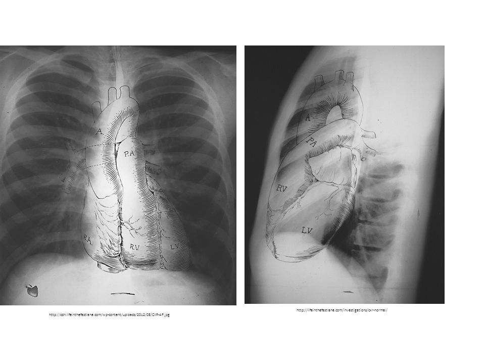
http://cdn.lifeinthefastlane.com/wp-content/uploads/2012/08/CXR-AP.jpg http://lifeinthefastlane.com/investigations/cxr-normal/
3
Right Atrium Right Ventricle Left Ventricle Left Atrium The Chambers

4
The Pulmonary and Aortic Vessels Main Pulmonary Artery RDPA Main Pulmonary Artery LDPA Aortic Arch Descending Aorta

5
This Lecture Pulmonary Hypertension Ventricular Septal Defect Mitral Stenosis vs Mitral Regurgitation Aortic Stenosis

6
Pulmonary HTN Increase in pulmonary vascular resistance Mean pulmonary artery pressure (PAP) >25mmHg at rest At least 30 mmHg during exercise Mean pulmonary wedge pressure < 15mmHg
 >25mmHg at rest At least 30 mmHg during exercise Mean pulmonary wedge pressure < 15mmHg")
7
Causes Group 1 Idiopathic Pulmonary Arterial Hypertension Group 2 Left Heart Disease Pulmonary Venous Hypertension Group 3 Hypoxemia COPD Group 4 Group 5

8
Clinical Indications Dyspnea on exertion Dizziness Syncope Angina

9
Radiographic Indicators Prominent main pulmonary artery Convex shadow along the left cardiac border (PA) Right ventricle enlargement Increased cardiac border in contact with anterior chest wall (Lat) Right descending pulmonary enlargement Transverse diameter is greater than 16mm Pruning of peripheral pulmonary vessels Abrupt tapering of distal pulmonary vessels with loss of side branches Pleural effusion/Pulmonary Edema
 Right ventricle enlargement Increased cardiac border in contact with anterior chest wall (Lat) Right descending pulmonary enlargement Transverse diameter is greater than 16mm Pruning of peripheral pulmonary vessels Abrupt tapering of distal pulmonary vessels with loss of side branches Pleural effusion/Pulmonary Edema")
10
Case courtesy of Dr Frank Gaillard, Radiopaedia.org Prominent Main Pulmonary Artery and Trunk Enlarged RDPA Pleural Effusion

11
http://posterng.netkey.at/esr/viewing/index.php?module=viewing_poster&task=viewsection&pi=100743& ti=311759&searchkey= Enlarged Right Ventricle Enlarged Pulmonary Trunk

12
Miniati et al. Accuracy of chest radiography in predicting pulmonary hypertension: A case-control study. M. Miniati et al. / Thrombosis Research 133 (2014) 345–351
 345–351.")
13
http://www.vcuthoracicimaging.com/Historyanswer.aspx?qid=40&fid=1

14
Ventricular Septal Defect Holes in the ventricular septum Conoventricular Perimembranous Inlet Muscular Most common congenital cardiac malformation Up to 50% in congenitally malformed hearts

15
Causes Can exist in isolation Or existing with other malformations: Tetralogy of Fallot Double outlet right ventricle Transposition Univentricular hearts

16
Clinical Indications Dependent on size May be asymptomatic May be cyanotic Retardation of growth Pulmonary HTN Pulmonary edema Wheezing Tachypnea

17
Radiographic Indicators Cardiomegaly Can be difficult to diagnose due to the thymus Increased pulmonary vascular markings

18
Normal http://www.scielo.br/scielo.php?pid=S0100-39842006000600012&script=sci_arttext&tlng=en Ventricular Septal Defect http://radiopaedia.org/articles/ventricular-septal-defect-1

19
Normal Cardiomegaly RADIOLOGIC CLINICS OF NORTH AMERICA VOLUME 37 * NUMBER 6 * NOVEMBER 1999

20
Ventricular Septal Defect http://radiopaedia.org/cases/ventricular- septal-defect Case courtesy of Dr Frank Gaillard, Radiopaedia.org

21
Spicer et al. Orphanet Journal of Rare Diseases (2014) 9:144
 9:144")
22
Tetralogy of Fallot http://radiopaedia.org/cases/tetralogy-of-fallot-1">Tetralogy of Fallot Ventricular Septal Defect Right Ventricular outflow track obstruction Overriding Aorta Right Ventricular Hypertrophy Let’s Think What Happens

23
Mitral Stenosis Leaflet thickening resulting in narrowing of the mitral valve. Causes Rheumatic Fever Common in developing countries Degenerative Common in developed countries

24
Healthy Mitral Valve Mitral Valve Stenosis Fish Mouth Appearance

25
Mitral Regurgitation Systolic retrograde flow from the left ventricle into the left atrium. Causes Non-Ischemic Degenerative Endocarditis Rheumatic Ischemic

26
http://www.vetmed.ucdavis.edu/vmth/small_animal/cardio_kittleson/case s/case9/figures.htm Healthy Mitral Valve http://nethealthbook.com/cardiovascular-disease/heart- disease/mitral-valve-disease/ Mitral Valve Prolapse

27
Clinical Indicators Dyspnea Exercise Intolerance Long Murmur Pulmonary HTN

28
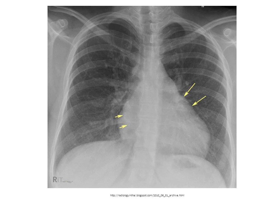
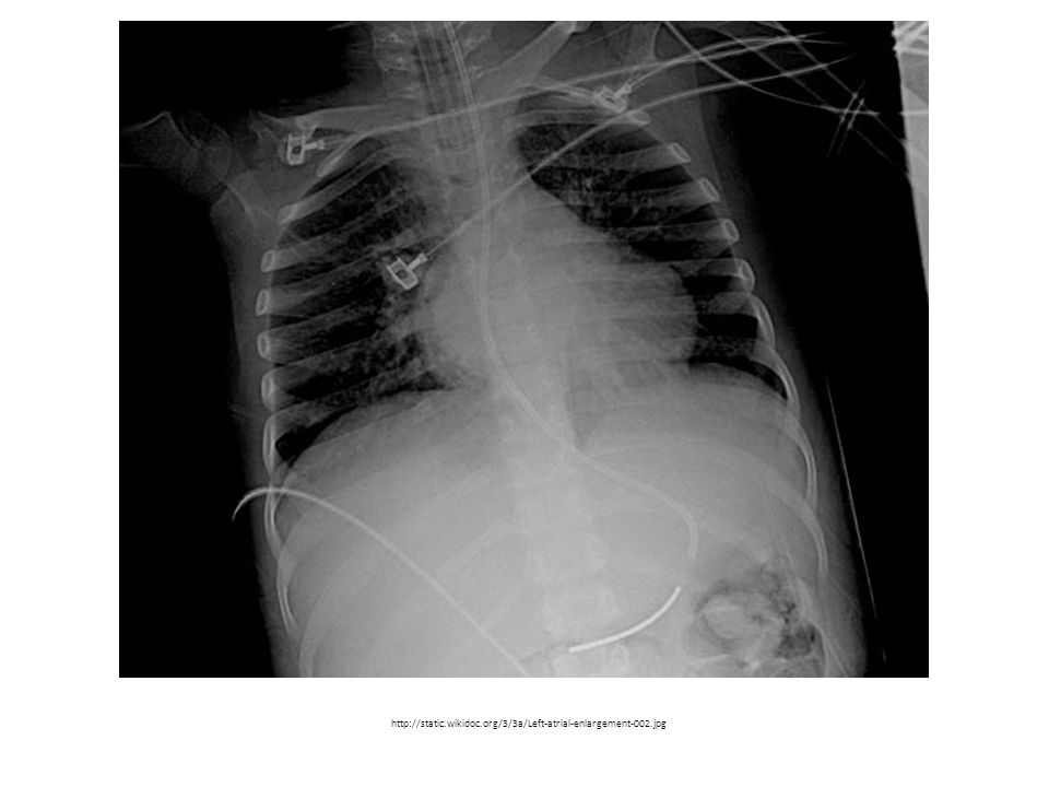
Radiographic Indicators Left atrial enlargement Double Density Splayed Carina Decreased aortic knob

29
LA LV RV RA LA LV RVRA Double Density and Splayed Carina

30
http://radiopaedia.org/articles/mitral-valve-regurgitation

31
http://www.learningradiology.com/lectures/cardiaclectures/valvularlesions2012/Valvular%20Lesions%20of%20the%20Heart/Valvular%20Lesions %20of%20the%20Heart.html

32
http://radiologyinthai.blogspot.com/2010_06_01_archive.html
33
http://static.wikidoc.org/3/3a/Left-atrial-enlargement-002.jpg
34
Aortic Stenosis Narrowing of the aortic valve. Causes: Age (Calcific AS, Senile) Similar to atherosclerosis Rheumatic Valve Congenital
 Similar to atherosclerosis Rheumatic Valve Congenital.")
35
Normal Aortic Valve Calcific Aortic Stenosis http://www.slideshare.net/drranjithmp/echo-assessment-of-aortic-stenosis

36
Clinical Indicators Angina Syncope Heart Failure

37
Radiographic Indicators Aortic Valve Calcification Prominent Ascending Aorta Left Ventricular Enlargement

38
http://www.learningradiology.com/archives2011/COW%20480-aortic%20stenosis/ascorrect.htm Ascending aorta projects farther than right atrium Normal Descending Aorta

40
Calcified Aortic Valve http://www.learningradiology.com/archives04/COW%20118-Aortic%20Stenosis/ascorrect.htm

41
Where are those valves? A M http://web.stanford.edu/dept/radiology/radiologysite/site141.html

42
A M

43
Aortic Valve

44
Mitral Valve

46
Reference Barbosa E, Gupta N, Torgian D, Gefter W. (2012). Current Role of imaging in the diagnosis and management of pulmonary hypertension. American Journal of Roentgenology. 198, 1320-1331 Batson G, Urquhart W, Sideris D. (1972). Radiologic Features in Aortic Stenosis. Clinical Radiology. 23, 140-144 Carabello, Blase. (2013). Introduction to aortic stenosis. Circulation Research., 113, 179-185. Carabello, B & Paulus W. (2009) Aortic Stenosis. The Lancet., 373, 956-966 Chandrashekhar Y, Westaby S, Narula J. Mitral Stenosis. (2009). The Lancet., 374, 1271-1283 Coussement, Alain & Gooding Charles. (1973). Objective Radiographic Assessment of Pulmonary Vascularity in Children. Radiology. 109, 649-654 Delnevo A, Tritella S, Carbonaro L, Bobrechova O, Leo G, Sardanelli F. (2012). The use of bedside radiography at a university hospital. Data on a two-week period. Euopean Journal of Radiology. 81, 260-263 Enriquez-Sarano, M, Akins C, Vahanian, A. (2009). Mitral Regurgitation. The Lancet., 373, 1382-1394 Matthay R, Schwarz M, Ellis J, Steele P, Siebert P, Durrance J, Levin D.. (1981). Pulmonary Artery Hypertension in Chronic Obstructive Pulmonary Disease: Determination by Chest Radiography. Investigative Radiology. 16, 95-100 Miniati M, Monti S, Airo E, Pancani R, Formichi B, Bauleo C, Marini C. (2014). Accuracy of chest radiography in predicting pulmonary hypertension: A case-control study. Thrombosis Research. 133, 345-351 Palazzeti V, Gasparri E, Gambini C, Sollazzo S, Saric S, Salvolini L, Giovagnoni A. (2013). Chest radiography in intensive care: an irreplaceable survey? Radiologia Medica. 118, 744-751 Sethi R. (2010). An Approach to Assessing the Chest Radiograph. British Journal of Hospital Medicine. 71(11), 172-17 Shah, Sanjiv.. (2012) Pulmonary Hypertension. Journal of American Medical Association. 308(13), 1366-1374 Spicer D, Hsu H, Co-Vu J, Anderson R, Fricker F. Ventricular Septal Defect. (2014). Orphanet Journal of Rare Diseases. 9(144), 1-16 Strife Janet & Sze, Raymond. (1999). Radiographic Evaluation of the Neonate with Congenital Heart Disease. Radiologc Clinics of North America. 37(6), 1093-1107 Tumkosit M, Yingyong N, Mahayosnond A, Choo K, Goo H. (2012). Accuracy of chest radiography for evaluating significantly abnormal pulmonary vascularity in children with congenital heart disease. International Journal of Cardiovascular Imaging. 28, 69- 75 Zarco, P. (2003). Aortic stenosis. European Heart Journal., 24, 133-135.
. Current Role of imaging in the diagnosis and management of pulmonary hypertension. American Journal of Roentgenology. 198, Batson G, Urquhart W, Sideris D. (1972). Radiologic Features in Aortic Stenosis. Clinical Radiology. 23, Carabello, Blase. (2013). Introduction to aortic stenosis. Circulation Research., 113, Carabello, B & Paulus W. (2009) Aortic Stenosis. The Lancet., 373, Chandrashekhar Y, Westaby S, Narula J. Mitral Stenosis. (2009). The Lancet., 374, Coussement, Alain & Gooding Charles. (1973). Objective Radiographic Assessment of Pulmonary Vascularity in Children. Radiology. 109, Delnevo A, Tritella S, Carbonaro L, Bobrechova O, Leo G, Sardanelli F. (2012). The use of bedside radiography at a university hospital. Data on a two-week period. Euopean Journal of Radiology. 81, Enriquez-Sarano, M, Akins C, Vahanian, A. (2009). Mitral Regurgitation. The Lancet., 373, Matthay R, Schwarz M, Ellis J, Steele P, Siebert P, Durrance J, Levin D.. (1981). Pulmonary Artery Hypertension in Chronic Obstructive Pulmonary Disease: Determination by Chest Radiography. Investigative Radiology. 16, Miniati M, Monti S, Airo E, Pancani R, Formichi B, Bauleo C, Marini C. (2014). Accuracy of chest radiography in predicting pulmonary hypertension: A case-control study. Thrombosis Research. 133, Palazzeti V, Gasparri E, Gambini C, Sollazzo S, Saric S, Salvolini L, Giovagnoni A. (2013). Chest radiography in intensive care: an irreplaceable survey. Radiologia Medica. 118, Sethi R. (2010). An Approach to Assessing the Chest Radiograph. British Journal of Hospital Medicine. 71(11), Shah, Sanjiv.. (2012) Pulmonary Hypertension. Journal of American Medical Association. 308(13), Spicer D, Hsu H, Co-Vu J, Anderson R, Fricker F. Ventricular Septal Defect. (2014). Orphanet Journal of Rare Diseases. 9(144), 1-16 Strife Janet & Sze, Raymond. (1999). Radiographic Evaluation of the Neonate with Congenital Heart Disease. Radiologc Clinics of North America. 37(6), Tumkosit M, Yingyong N, Mahayosnond A, Choo K, Goo H. (2012). Accuracy of chest radiography for evaluating significantly abnormal pulmonary vascularity in children with congenital heart disease. International Journal of Cardiovascular Imaging. 28, Zarco, P. (2003). Aortic stenosis. European Heart Journal., 24,")
Similar presentations


