Download presentation
Presentation is loading. Please wait.

1
Cervical and lumbar disc herniations
Prof. Dr. Hidayet SARI

2
PREVALENCE AND PATHOGENESIS
A herniated disc can be defined as herniation of the nucleus pulposus through the fibers of the annulus fibrosus. Most disc ruptures occur during the third and fourth decades of life while the nucleus pulposus is still gelatinous.

3
PREVALENCE AND PATHOGENESIS
The most likely time of day associated with increased force on the disc is the morning. In the lumbar region, perforations usually arise through a defect just lateral to the posterior midline, where the posterior longitudinal ligament is weakest.

5
EPIDEMIOLOGY Lumbar Spine: Symptomatic lumbar disc herniation occurs during the lifetime of approximately 2% of the general population. Approximately 80% of the population will experience significant back pain during the course of a herniated disc. The groups at greatest risk for herniation of intervertebral discs are younger individuals (mean age of 35 years)
")
6
EPIDEMIOLOGY Lumbar Spine:
True sciatica actually develops in only 35% of patients with disc herniation. Not infrequently, sciatica develops 6 to 10 years after the onset of low back pain. The period of localized back pain may correspond to repeated damage to annular fibers that irritates the sinuvertebral nerve but does not result in disc herniation.

7
EPIDEMIOLOGY Cervical Spine:
The average annual incidence of cervical radiculopathy is less than 0.1 per 1000 individuals. Pure soft disc herniations are less common than hard disc abnormalities (spondylosis) as a cause of radicular arm pain.
 as a cause of radicular arm pain.")
8
EPIDEMIOLOGY Cervical Spine:
In a study of 395 patients with nerve root abnormalities, radiculopathy occured in the cervical and lumbar spine in 93 (24%) and 302 (76%), respectively.
 and 302 (76%), respectively.")
9
PATHOGENESIS Alterations in intervertebral disc biomechanics and biochemistry over time have a detrimental effect on disc function. The disc is less able to work as a spacer between vertebral bodies or as a universal joint.

10
PATHOGENESIS-LUMBAR SPINE
The two most common levels for disc herniation are L4-L5 and L5-S1, which account for 98% of lesions; pathology can occur at L2-L3 and L3-L4 but is relatively uncommon. Overall, 90% of disc herniations are at the L4-L5 and L5-S1 levels.

11
PATHOGENESIS-LUMBAR SPINE
Disc herniations at L5-S1 will usually compromise the first sacral nerve root, a lesion at the L4-L5 level will most often compress the fifth lumbar root, and herniation at L3-L4 more frequently involves the fourth lumbar root.

12
PATHOGENESIS-LUMBAR SPINE
Disc herniation may also develop in older patients. Disc tissue that causes compression in elderly patients is composed of the annulus fibrosus and and portions of the cartilaginous endplate (hard disc.) The cartilage is avulsed from the vertebral body.
 The cartilage is avulsed from the vertebral body.")
13
PATHOGENESIS-LUMBAR SPINE
A question exists concerning the relative importance of restriction of motion by the annulus fibrosus versus the facet joints in preventing disc herniation.

14
PATHOGENESIS-LUMBAR SPINE
Resolution of some of the compressive effects on neural structures requires resorption of the nucleus pulposus. Disc resorption is part of the natural healing process associated with disc herniation. The enhanced ability to resorb discs has the potential for resolving clinical symptoms more rapidly.

15
PATHOGENESIS-LUMBAR SPINE
Resorption of herniated disc material is associated with a marked increase in infiltrating macrophages and the production of matrix metalloproteinases (MMPs) 3 and 7.
 3 and 7.")
16
PATHOGENESIS-LUMBAR SPINE
Nerlich and associates identified the origins of phagocytic cells in degenerated intervertebral discs. The investigation identified cells that are transformed local cells rather than invaded macrophages. Degenerative discs contain the cells that add to their continued dissolution.

17
PATHOGENESIS-CERVICAL SPINE
In the early 1940s, a number of reports appeared in which cervical intervertebral disc herniation with radiculopathy was described. There is a direct correlation between the anatomy of the cervical spine and the location and pathophysiology of disc lesion.

18
PATHOGENESIS-CERVICAL SPINE
The eight cervical nerve roots exit via intervertebral foramina that are bordered anteromedially by the intervertebral disc and posterolaterally by the zygapophyseal joint. The foramina are largest at C2-C3 and decrease in size until C6-C7. The nerve root occupies 25% to 33% of the volume of the foramen.

19
PATHOGENESIS-CERVICAL SPINE
The C1 root exits between the occiput and the atlas (C1) All lower roots exit above their corresponding cervical vertebrae (the C6 root at the C5-C6 interspace), except C8, which exits between C7 and T1. A differential growth rate affects the relationship of the spinal cord and nerve roots and the cervical spine.
 All lower roots exit above their corresponding cervical vertebrae (the C6 root at the C5-C6 interspace), except C8, which exits between C7 and T1. A differential growth rate affects the relationship of the spinal cord and nerve roots and the cervical spine.")
20
PATHOGENESIS-CERVICAL SPINE
Most acute disc herniations occur posterolaterally and in patients around the forth decade of life, when the nucleus is still gelatinous. The most common areas of disc herniations are C6-C7 and C5-C6.

21
PATHOGENESIS-CERVICAL SPINE
C7-T1 and C3-C4 disc herniations are in frequent ( less than 15 %). Disc herniation of C2-C3 is rare. Patients with upper cervical disc protrusions in the C2-C3 region have symptoms that include suboccipital pain, loss of hand dexterity, and paresthesias over the face and unilateral arm.
. Disc herniation of C2-C3 is rare. Patients with upper cervical disc protrusions in the C2-C3 region have symptoms that include suboccipital pain, loss of hand dexterity, and paresthesias over the face and unilateral arm.")
22
PATHOGENESIS-CERVICAL SPINE
Unlike lumber herniated discs, cervical herniated discs may cause myelopathy in addition to radicular pain because of spinal cord in the cervical region. The uncovertebral prominences play a role in the location of ruptured dics material.

23
PATHOGENESIS-CERVICAL SPINE
Poor nerve root compression occurs if extruded disc material enters the nerve root canal. The uncovertebral joint tends to guide extruded disc material medially, where cord compression may also occur.

24
PATHOGENESIS-CERVICAL SPINE
Disc herniations usually affects the nevre root numbered most caudally for the given disc level; for example, the C3 – C4 disc affects the fourth cervical nerve root; C4- C5, the fifth cervical nerve root; C5 – C6, the sixth cervical nerve root; C6 – C7, the seventh cervical nerve root; and C7 – T1, the eighth cervical nerve root.

25
PATHOGENESIS-CERVICAL SPINE
Individual disc herniations do not involve other roots but more commonly present some evidence of upper motor neuron findings secondary to spinal cord compression (Cervical spondylotic myelopathy).
.")
26
PATHOGENESIS-CERVICAL SPINE
Not every herniated disc is symptomatic. The development of symptoms depends on the reserve capacity of the spinal canal, the presence of inflammation, the size of the herniation, and the presence of concomitant disease such as osteophyte formation.

27
PATHOGENESIS-CERVICAL SPINE
In disc rupture, protrusion of nuclear material results in tension on the annular fibers and compressıon of the dura or nerve root causing pain. Also important is the smaller size of the sagittal diameter, the bony cervical spinal canal. Indiviuals in whom a cervical herniated disc causes motor dysfunction have a complication of cervical disc herniation if the spinal canal is stenotic.

28
CLINICAL HISTORY LUMBAR SPINE
Clinically, the patient’s major complaint is a sharp, lancinating pain. In many cases there may be a previous history of intermittent episodes of localized low back pain. The pain not only in the back but also radiates down the leg in the anatomic distribution of the affected nerve root.

29
CLINICAL HISTORY LUMBAR SPINE
It will usually be described as deep and sharp and progressing from above downward in the involved leg. Its onset may be insidious or sudden and associated with a tearing or snapping sensations of the spine.

30
CLINICAL HISTORY LUMBAR SPINE
Occasionally, when sciatica develops, the back pain may resolve because once the annulus has ruptured, it may no longer be under tension. Disc herniation occurs with sudden physical effort when the trunk is flexed or rotated. On occasion, patients with L4-L5 disc herniation have groin pain. In a study of 512 lumbar disc patients, 4.1% had groin pain.

31
CLINICAL HISTORY LUMBAR SPINE
Finally, the sciatica may vary in intensity; it may be so severe that patients will be unable to ambulate and they will feel that their back is "locked". On the other hand , the pain may be limited to a dull ache that increases in intensity with ambulation.

32
CLINICAL HISTORY LUMBAR SPINE
Pain is worsened in the flexed position and relieved by extension of the lumbar spine. Characteristically, patients with herniated discs have increased pain with sitting, driving, walking, couching, sneezing, or straining.

33
CLINICAL HISTORY CERVICAL SPINE
Arm pain, not neck pain, is the patient’ s major complaint. The pain is often perceived as starting in the neck area and then radiating from this point down to shoulder, arm and forearm and usually into the hand.

34
CLINICAL HISTORY CERVICAL SPINE
The onset of the radicular pain is often gradual, although it can be sudden and occur in association with a tearing or snapping sensation. As time passes, the magnitude of the arm pain clearly exceeds that of the neck or shoulder pain.

35
CLINICAL HISTORY CERVICAL SPINE
The arm pain may also be variable in intensity and preclude any use of the arm; it may range from severe pain to a dull, cramping ache in the arm muscles. The pain is usually severe enough to awaken the patient at night.

36
CLINICAL HISTORY CERVICAL SPINE
Additionally, a patient may complain of associated headaches as well as muscle spasm, which can radiate from the cervical spine to below the scapulae. The pain may also radiate to the chest and mimic angina (pseudoangina) or to the breast.
 or to the breast.")
37
CLINICAL HISTORY CERVICAL SPINE
Symptoms such as back pain, leg pain, leg weakness, gait disturbance, or incontinence suggest compression of the spinal cord (Myelopathy).
.")
38
PHYSICAL EXAMINATION LUMBAR SPINE
Physical examination will demonstrated a decrease in range of motion of the lumbosacral spine, and patients may list to one side as they try to bend forward. The side of the disc herniation corresponds to the location of the scoliotic list.

39
PHYSICAL EXAMINATION LUMBAR SPINE
However, the specific level or degree of herniation does not correlate with the degree of list. On ambulation, patients walk with an antalgic gait in which they hold the invoved leg flexed so that they put as little weight as possible on the extremity.

40
PHYSICAL EXAMINATION LUMBAR SPINE
Neurologic Examination: The neurologic examination is very important and may yield objective evidence of nerve root compression (We should evaluate of reflex testing, muscle power, and sensation examination of the patient).
.")
41
PHYSICAL EXAMINATION LUMBAR SPINE
In addition, a nerve deficit may have little temporal relevance because it may be related to a previous attack at a different level. Compression of individual spinal nerve roots results in alterations in motor, sensory, and reflex function.

42
PHYSICAL EXAMINATION LUMBAR SPINE
When the first sacral root is compressed, the patient may have gastronemius-soleus weakness and be unable to repeatedly raise up on the toes of that foot. Atrophy of the calf may be apperent, and the ankle (Achilles) reflex is often diminished or absent. Sensory loss, if present, is usually confined to the posterior aspect of the calf and the lateral side of the foot.
 reflex is often diminished or absent. Sensory loss, if present, is usually confined to the posterior aspect of the calf and the lateral side of the foot.")
44
PHYSICAL EXAMINATION LUMBAR SPINE
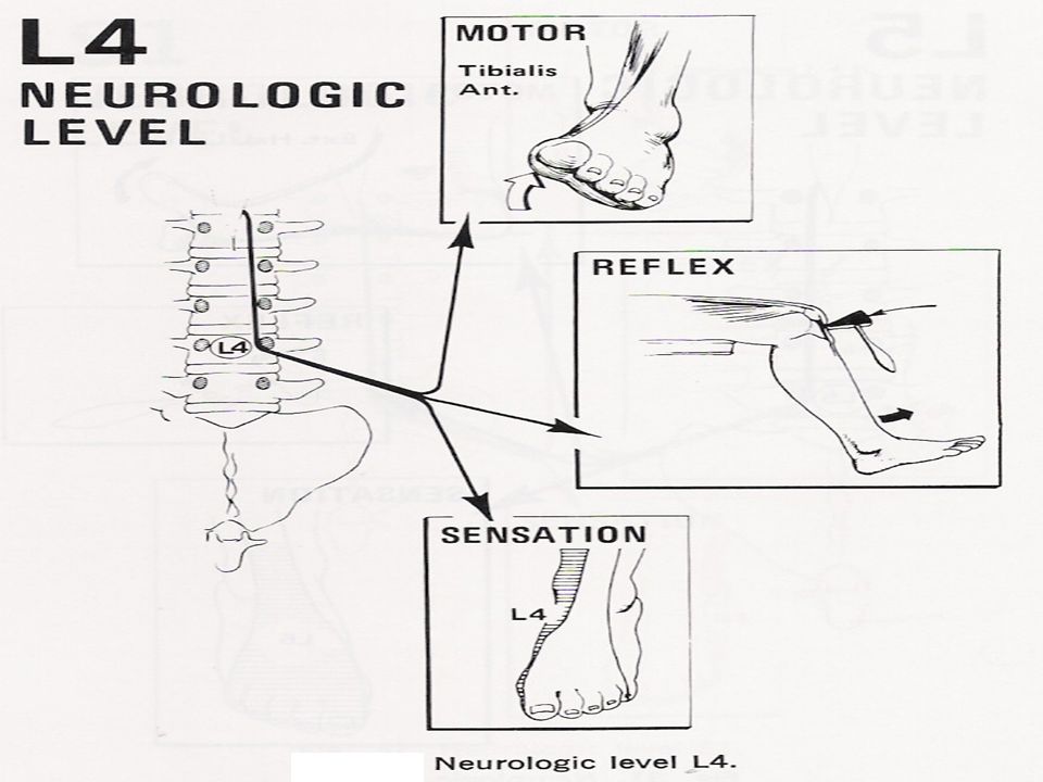
Involvement of the fifth lumbar nerve root can lead to weakness in extension of the great toe and, in a few cases, weakness of the everters and dorsiflexors of the foot. A sensory deficit can appear over the anterior of the leg and the dorsomedial aspect of the foot down to the great toe

46
PHYSICAL EXAMINATION LUMBAR SPINE
Primary reflex changes do not generally occur, but on occasion, a diminution in the posterior tibial reflex can be elicited. There must be asymmetry in obtaining this reflex for it to have any clinical significance.

48
PHYSICAL EXAMINATION LUMBAR SPINE
With compression of the fourth lumbar nerve root, the quadriceps muscle is affected; the patient may note weakness in knee extension, which is often associated with instability. Atrophy of the thigh musculature can be marked. Sensory loss may be apparent over the anteromedial aspect of the thigh, and the patellar tendon reflex can be diminished.

49
PHYSICAL EXAMINATION LUMBAR SPINE
Sensory deficit on physical examination is used to identify the level of spinal nerve root compression. Nitta and co-workers identified the areas of sensory deficit associated with nerve blocks at three lumbar levels, L4, L5, and S1. A total of 71 patients received 86 lumbar spinal nerve blocks.

50
PHYSICAL EXAMINATION LUMBAR SPINE
Characteristic areas of numbness were noted on the medial side of the lower part of the leg in 88% of L4 injection, the dorsum of the first digit in 82% of L5 injections, and the lateral aspect of the fifth digit in 83% of S1 injections.

51
PHYSICAL EXAMINATION LUMBAR SPINE
Nerve root sensitivity can be elicited by any method that creates tension. The straight leg-raising (SLR)test is the one most commonly used. This test is performed with the patient supine.
test is the one most commonly used. This test is performed with the patient supine.")
52
PHYSICAL EXAMINATION CERVICAL SPINE
Physical examination of the neck usually shows some limitation of motion, and on occasion the patient may tilt the head in a cocked-robin position (torticollis) toward the side of the herniated cervical disc.
 toward the side of the herniated cervical disc.")
53
PHYSICAL EXAMINATION CERVICAL SPINE
Neurologic Examination: A neurologic examination that shows abnormalities is the most helpful aspect of the diagnostic work-up, although the examination may remain normal despite a chronic radicular pattern.

54
PHYSICAL EXAMINATION CERVICAL SPINE
The presence of atrophy helps document the location of the lesion, as well as its chronicity. The presence of subjective sensory changes is often difficult to interpret and requires a coherent and cooperative patient to be of clinical value.

55
PHYSICAL EXAMINATION CERVICAL SPINE
When the third cervical root is compressed, no reflex change and motor weakness can be identified. The pain radiates to the back of the neck and toward the mastoid process and pinna of the ear.

56
PHYSICAL EXAMINATION CERVICAL SPINE
Involvement of the fourth cervical nerve root leads to no readily detectable reflex changes or motor weakness. The pain radiates to the back of the neck and superior aspect of the scapula. Occasionally, the pain radiates to the anterior chest wall. The pain is often exacerbated by neck extension.

57
PHYSICAL EXAMINATION CERVICAL SPINE
Unlike the third and the fourth cervical nerve roots, the fifth through eighth cervical nerve roots have motor functions. Compression of the fifth cervical nerve root is characterized by weakness of shoulder abduction, usually above 90 degree, and weakness of shoulder extension.

58
PHYSICAL EXAMINATION CERVICAL SPINE
The biceps reflexes are often depressed and the pain radiates from the side of the neck to the top of the shoulder. Decreased sensation is often noted in the lateral aspect of the deltoid, which represents the autonomous area of the axillary nerve.

60
PHYSICAL EXAMINATION CERVICAL SPINE
Involvement of the sixth cervical nerve root produces biceps muscles weaknes as well as diminished brachioradial reflex. The pain again radiates from the neck down the lateral aspect of the arm and forearm to the radial side of hand (index finger, long finger, and thumb). Numbness occurs occasionally in the tip of the index finger, the autonomous area of the sixth cervical nerve root.
. Numbness occurs occasionally in the tip of the index finger, the autonomous area of the sixth cervical nerve root.")
62
PHYSICAL EXAMINATION CERVICAL SPINE
Compression of the seventh cervical nerve root produces reflex changes in the triceps jerk test with associated loss of strength in the triceps muscles, which extend the elbow. The pain from this lesion radiates from the lateral aspect of the neck down the middle of the area to the middle finger.

63
PHYSICAL EXAMINATION CERVICAL SPINE
Sensory changes occur often in the tip of the middle finger, the autonomous area for the seventh nerve. Patients should also be tested for scapular winging, which may occur with C6 or C7 radiculopathy.

65
PHYSICAL EXAMINATION CERVICAL SPINE
Finally, involvement of the eighth cervical nerve root by a herniated C7-T1 disc produces significant weakness of the intrinsic musculature of the hand. Such involvement can lead to rapid atrophy of the interosseous muscles because of the small size of these muscles.

66
PHYSICAL EXAMINATION CERVICAL SPINE
Loss of the interossei leads to significant loss of fine hand motion. No reflexes are easily found, although the flexor carpi ulnaris reflex may be decreased.

67
PHYSICAL EXAMINATION CERVICAL SPINE
The radicular pain from the eighth cervical nerve root radiates to the ulnar border the hand and the ring and little fingers. The tip of the little finger often demonstrates diminished sensation.

69
PHYSICAL EXAMINATION CERVICAL SPINE
Nerve root sensitivity can be elicited by a method that increases tension on the nerve root. Radicular arm pain is often increased by the Valsalva maneuver of by directly compressing the head.

70
PHYSICAL EXAMINATION CERVICAL SPINE
Radicular pain secondary to a herniated cervical disc may be relieved by abduction of the affected arm. Although these signs are helpful when present, their absence alone does not rule out a nerve root lesion.

71
LABORATORY DATA Medical screening laboratory test (blood counts, chemistry panels erythrocyte sedimentation rate [ESR]) are normal in patients with a herniated disc.
 are normal in patients with a herniated disc.")
72
LABORATORY DATA Electrodiagnostic Testing
Electromyography (EMG) is an electronic extension of the physical examination. The primary use of EMG is to diagnose radiculopathy in cases of questionable neurologic origin. EMG findings may be positive in patients with nerve root impingement.
 is an electronic extension of the physical examination. The primary use of EMG is to diagnose radiculopathy in cases of questionable neurologic origin. EMG findings may be positive in patients with nerve root impingement.")
73
RADIOGRAPHIC EVALUATION LUMBAR SPINE
Roentgenograms Plain roentgenograms may be entirely normal in a patient with symtoms and signs of nerve root impingement.

74
RADIOGRAPHIC EVALUATION LUMBAR SPINE
Computed Tomography Radigraphic evaluation by CT scan may demonstrate disc bulging but may not correlate with the level of nerve damage.

75
RADIOGRAPHIC EVALUATION LUMBAR SPINE
Magnetic Resonance MR imaging also allows visualization of soft tissues, including discs in the lumbar spine. Herniated discs are easily detected with MR evaluation . MR imaging is a sensitive technique for the detection of far lateral and anterior disc herniations.

76
RADIOGRAPHIC EVALUATION CERVICAL SPINE
Roentgenograms Plain roentgenograms may be entirely normal in patients with an acute herniated cervical disc. Conversely, 70% of asymptomatic women and 95% of asymptomatic men between the ages of 60 and 65 years have evidence of degenerative disc disease on plain roentgenograms.

77
RADIOGRAPHIC EVALUATION CERVICAL SPINE
Views to be obtained include anteroposterior, lateral, flexion, and extension.

78
RADIOGRAPHIC EVALUATION CERVICAL SPINE
Computed Tomography CT permits direct visualization of compression of neural structures and is therefore more precise than myelography. Advantages of CT over myelography include better visualization of lateral abnormalities such as foraminal stenosis and abnormalities caudal to the myelographic block, less radiation exposure, and no hospitalization.

79
RADIOGRAPHIC EVALUATION CERVICAL SPINE
From a surgical respective, CT is best at distinguishing soft disc compression from hard bony compression. Disadvantages of CT include the length of time to complete to study and changes in spinal configuration between motion segments. Myelographic dye may be injected and CT images obtained.

80
RADIOGRAPHIC EVALUATION CERVICAL SPINE
Magnetic Resonance MRI allows excellent visualization of soft tissues, including herniated discs in the cervical spine. The test is noninvasive. In a study of 34 patients with cervical lesions, MRI predicted 88% of the surgically proven lesions versus 81% for myelography-CT, 58% for myelography, and 50% for CT alone.

81
DIFFERENTIAL DIAGNOSIS LUMBAR SPINE
The initial diagnosis of a herniated disc is ordinarily made on the basis of the history and physical examination. Plain radiographs of the lumbosacral spine will rarely add to the diagnosis but should be obtained to help rule out other causes of pain such as infection or tumor. Other tests such as MR, CT, and myelography are confirmatory by nature and can be misleading when used as screening tests.

82
DIFFERENTIAL DIAGNOSIS LUMBAR SPINE
Spinal Stenosis Patient with spinal stenosis may also suffer from back pain that radiates to the lower extemities. Patients with spinal stenosis tend to be older that those in whom herniated discs develop.

83
DIFFERENTIAL DIAGNOSIS LUMBAR SPINE
Characteristically, patients with spinal stenosis experience lower exremity pain (pseudoclaudication=neurogenic claudication) after walking for an unspecified distance. They also complain of pain that is exacerbated by standing or extending the spine.
 after walking for an unspecified distance. They also complain of pain that is exacerbated by standing or extending the spine.")
84
DIFFERENTIAL DIAGNOSIS LUMBAR SPINE
Radiographic evaluation is usually helpful in differentiating individuals with disc herniation from those with bony hypertrophy associated with spinal stenosis. In a study of 1293 patients, lateral spinal stenosis and herniated intervertebral discs coexisted in 17.7% of individuals. Radicular pain may becaused by more than one pathologic process in an individual.

85
DIFFERENTIAL DIAGNOSIS LUMBAR SPINE
Facet Syndrome Facet syndrome is another cause of low back pain that may be associated with radiation of pain to structures outside the confines of the lumbosacral spine. Degenetation of articular structures in the facet joint causes pain to develop.

86
DIFFERENTIAL DIAGNOSIS LUMBAR SPINE
In most circumstances, the pain is localized over the area of the affected joint and is aggravated by extension of the spine (standing). A deep , ill-defined, aching discomfort may also be noted in the sacroiliac joint, the buttocks, and the legs. The areas of sclerotome affected show the same embryonic origin as the degenerated facet joint.
. A deep , ill-defined, aching discomfort may also be noted in the sacroiliac joint, the buttocks, and the legs. The areas of sclerotome affected show the same embryonic origin as the degenerated facet joint.")
87
DIFFERENTIAL DIAGNOSIS LUMBAR SPINE
A direct association between facet joint disease and production of pain is questioned by some investigators. Many individuals with arthritic changes in their facet joints visible on radiographic evaluation experience no symptoms.

88
DIFFERENTIAL DIAGNOSIS LUMBAR SPINE
Patients with pain secondary to faced joint disease may have relief of symptoms with apophyseal injection of a long-acting local anesthetic. The true role of facet joint disease in the production of back and leg pain remains to be determined.

89
DIFFERENTIAL DIAGNOSIS LUMBAR SPINE
Other mechanical causes of sciatica include congentenial abnormalites of the lumbar nerve roots, external compression of the sciatic nerve (wallet in a back pants pocket), and muscular compression of the nerve (piriformis syndrome). In rare circumstances,, cervical or thoracic lesion should be considered if the lumbar spine is clear of abnormalities. Medical causes of sciatica (neural tumors or infections, forexample) are usually associated with systemic symptomsin addition to nerve pain in a sciatic distribution.
, and muscular compression of the nerve (piriformis syndrome). In rare circumstances,, cervical or thoracic lesion should be considered if the lumbar spine is clear of abnormalities. Medical causes of sciatica (neural tumors or infections, forexample) are usually associated with systemic symptomsin addition to nerve pain in a sciatic distribution.")
90
DIFFERENTIAL DIAGNOSIS CERVICAL SPINE
No diagnostic criteria exist for the clinical diagnosis of a herniated cervical disc. The provisional diagnosis of a herniated cervical disc is made by the history and physical examination. Individuals who frequently lift heavy objects on the job, smoke, or often dive from a diving board are at increased risk for cervical disc herniation.

91
DIFFERENTIAL DIAGNOSIS CERVICAL SPINE
The plain roentgenogram is usually nondiagnostic, although occasionally disc space narrowing at the suspected interspace or foraminal narrowing on oblique films is seen. The value of roentgenograms is to exclude other causes of neck and arm pain, such as infection and tumor. MR imaging and CT-myelography are the best confirmatory examinations for disc herniation.

92
DIFFERENTIAL DIAGNOSIS CERVICAL SPINE
Cervical disc herniations may affect structures other than nerve roots. Disc herniation may cause vessel compression (vertebral artery) associated with vertebrobasilar artery insufficiency and be manifested as blurred vision and dizziness.
 associated with vertebrobasilar artery insufficiency and be manifested as blurred vision and dizziness.")
93
DIFFERENTIAL DIAGNOSIS CERVICAL SPINE
Other mechanical causes of arm pain should be excluded. The most common is some form of compression on a peripheral nerve. Such compression can occur at the elbow, forearm, or wrist. An example is compression of the median nerve by the carpal ligament leading to carpal tunnel syndrome.

94
DIFFERENTIAL DIAGNOSIS CERVICAL SPINE
The best diagnostic test to rule out these peripheral neuropathies is EMG. Excessive traction onthe arm secondary to heavy weights may cause radicular pain without disc compression of nerve roots. Spinal cord abnormalities must be considered if signs of myelopathy are present in conjunction with radiculopathy.

95
DIFFERENTIAL DIAGNOSIS CERVICAL SPINE
Spinal cord lesions such as syringomyelia are identified by MRI, and motor neuron disease is identified by EMG. Multiple sclerosis should be considered in a patient with radiculopathy if the physical signs indicate lesions above the foramen magnum (optic neuritis). In very rare circumstances, lesions of the parietal lobe corresponding to the arm can mimic the findings of cervical radiculopathy.
. In very rare circumstances, lesions of the parietal lobe corresponding to the arm can mimic the findings of cervical radiculopathy.")
96
TREATMENT NATURAL HISTORY
The location and type of disc herniation played a role in the subsequent resorption. Sequestrated discs exposed to vasculature in the epidural space were more likely to recede in size than subligamentous herniations were. The authors suggest that nonsurgical treatment is appropriate for sequestrated discs even in the setting of motor weakness.

97
TREATMENT Nonoperative Therapy
Treatment for most patients with a herniated disc is nonoperative inasmuch as 80%of them will respond to conservative therapy when monitored over a period of 5 years.

98
TREATMENT The efficacy of nonoperative treatment, however, depends on a healthy relationship between a capable physician and a well-informed patient. If patients have insight into the rationale for the prescribed treatment and follow instructions, the changes for success are greatly increased.

99
TREATMENT Controlled Physical Activity
The primary element in the nonoperative treatment of acute disease is controlled physical activity. For the first several days in the acute situation, bed rest may be necessary and can usually be accomplished at home.

100
TREATMENT The semi-Fowler position with the hips and knees comfortably flexed is ideal because it keeps intradiscal pressure down and reduces nerve root tension. After the first few days, the patient should be gradually mobilized. Walking should be encouraged, whereas sitting is prohibited because it causes excessive pressure on the nerve root.

101
TREATMENT NSAIDs/Corticosteroids
Drug therapy is another important part of the treatment, and three categories of pharmacologic agents are commonly used: anti-inflammatory drugs, analgesics, and muscle relaxants or tranquilizers. Inasmuch as symptoms of low back pain and sciatica result from an inflammatory reaction, as well as mechanical compression, anti-inflammatory drugs are indicated

102
TREATMENT Adequate doses of aspirin have been found to work quite well, although other NSAIDs are frequently used. The patient’s pain will generally be relieved once the inflammation is brought under control. Residual numbness or tingling in the involvement extremity may be present but is usually tolerable.

103
TREATMENT Some patients wh fail to respond to anti-inflammatory medication may get dramatic relief from a short course of systemic steroids adminstered in decreasing dosages over a period of weeks.

104
TREATMENT The initial dose of corticosteroid is 20 mg/day of prednisone. Prednisone is continued with the other nonsteroidal medications. The dose is maintained at 20 mg while the patient is monitored for resolution of radiculopathy.

105
TREATMENT The prednisone is gradually tapered over weeks as the signs of radiculopathy resolve (pain, numbness). If the patients has no response to the prednisone, use of the medication is discontinued at the end of 6 weeks.

106
TREATMENT Dyck and co-workers used intravenous methylprednisolone for lumbosacral radiculopathy, 10 received infusions of intravenous methylprednisolone (1g/week) for 16 weeks and 1 received the equivalent dosage of oral prednisone. This dose of steroids was effective in all 11 patients.
 for 16 weeks and 1 received the equivalent dosage of oral prednisone. This dose of steroids was effective in all 11 patients.")
107
TREATMENT The concern is the long-term effects of such a large dose of corticosteroids. The toxicity is likely to outweigh the benefits with large, prolonged doses of methylprednisolone.

108
TREATMENT Analgesics Analgesic medication is administered to control pain if it is severe. Codeine is recommended for home use. If codeine does not work, hospitalization should be considered so that a stronger analgesic medication such as morphine sulfate can be strictly conrolled. Long-term use of narcotics for these patients should be modified as the radiculopathy resolves.

109
TREATMENT Muscle Relaxants
Muscle relaxants are used in patients with uncontrolled muscle contraction associated with nerve impingement. The mechanism of action these agents is unknown. Most agents, other than diazepam, do not act directly on muscle fibers but act on the central nervous system by diminishing reflex contractions.

110
TREATMENT The beneficial effects of this group of drugs were thought to be related to their tranquilizing properties. In a study reported by Borenstein and Korn, the efficacy of cyclobenzaprine at 5 mg was not associated with the presence or absence of somnolence. In patients with severe muscle spasm, muscle relaxants do appear to be effective.

111
TREATMENT It should be remembered, however, that the use of diazepam for muscle spasm should be discouraged. When used on a chronic basis, diazepam may become a depressant.

112
TREATMENT Diazepam will only add to the psychological problems of patients with chronic pain. If other muscle relaxants without depressant properties are used from the outset, the problems related to depression, tolerance, and addiction can be prevented.

113
TREATMENT Injection Therapy
Epidural corticosteroids injections are useful for patients with radiculopathy. Injection should be considered for patiets with radiculopathy who are not responding to modified activities, NSAID therapy, and muscle relaxants

114
TREATMENT Tumor Necrosis Factor Inhibitors
TNF inhibitors show promise as therapy for patients with radiculopathy secondary to a herniated disc. In animal models of disc herniation and spinal nerve inflammation, TNF is located at the site of nerve damage.

115
TREATMENT TNF is produced by nucles pulposus cell.
Exogenous TNF produces neuropathologic alterations (wallerian degeneration of nerve fibers, macrophage recruitment to phagocytize the debris, and splitting of the myelin sheath) similar to those induced by herniated nucleus pulposus.
 similar to those induced by herniated nucleus pulposus.")
116
TREATMENT Surgical Intervention
Surgical intervention is reserved for patients in whom conservative therapy fails. Patients with radicular pain, abnormal physical findings, and confirmatory radiographic tests are candidates for surgical intervention.

117
TREATMENT Cauda Equina Syndrome
Cauda equina syndrome is associated with compression of the spinal nerve roots that supply neurologic function to the bladder and bowel. Rapid diagnosis and decompression of this abnormality are essential to prevent permanent neurologic dysfunction.

118
TREATMENT In a study of 44 patients with urologic problems of retention, incontinence, or saddle anesthesia, the syndrome developed in 39 in less than 24 hours. MRI in 23 patients or CT in 21 patients identified nerve compression with massive disc herniation.

119
TREATMENT Surgery was performed in 20 patients within 48 hours, whereas 24 patients underwent surgery after 48 hours, with a mean delay of 9 days. Delay in surgery beyond 48 hours was associated with persistent severe motor deficit persistent sciatica , and sexual dysfunction. Cauda equina requires emergency diagnosis and surgical decompression within a 48 hour period to decrease the risk of permanent neurologic sequelae

120
TREATMENT Kennedy and co workers also described a group of patients with cauda equina syndrome. Patients with a poor outcome were those who had perineal sensory loss and urinary dysfunction . The 14 of 19 patients who had a good outcome after surgical decompression had the operation within 14 hours, whereas those with a poor outcome underwent surgery within 30 hours.

121
TREATMENT Cauda equina lesions can cause neural impingement.
Many of these lesions are neoplasms(benign and malignant) such as ependymomas, nerve sheath tumors, meningiomas and lipomas.
 such as ependymomas, nerve sheath tumors, meningiomas and lipomas.")
122
TREATMENT A variety of symptoms were seen , including bilateral lower extremity weakness and tenderness. No specific relationship exists between pathologic diagnosis and symptoms.

123
TREATMENT Cervical Spine
Treatment of most patients with a herniated cervical disc is nonoperative because most respond to conservative treatment for 2 to 3 months. The efficacy of the nonoperative approach depends heavily on the physician patients relationship.

124
TREATMENT If a patient is well informed, insightful, and willing to follow instructions, the changes for a succesful nonoperative outcome are greatly improved. The cornerstone of management of a herniated cervical disc is rest and immobilization. The use of a cervical orthosis greatly increases the likelihood that he patients will rest.

125
TREATMENT Initially, the patient should remain at home resting in bed except for necessary trips, to the bathroom. Controlled physical activity sould be maintained for at least 2 weeks, and the patient should wear the cervical orthosis at all times. The Philadelphia collar is a plastic collar that offers greater support for individuals with cervical radiculopathy.

126
TREATMENT Careful fitting is required in the suboccipital, submental, and sternal areas to maximize inhibition of lower cervical motion. After the acute pains to abate, the patient should gradually increase activity and decrease use of the orthosis. Most people are able to return to work in a month in a light-duty capacity.

127
TREATMENT NSAIDs Drug therapy is an important adjunct to controlled physical activity and immobilization. Anti-inflammatory medications, analgesics, and muscle relaxants have been use in the acute management of these patients.

128
TREATMENT Because it is commonly believed that radicular pain is in part secondary to inflammation of the nerve root, the use of aspirin or other NSAIDs are appropriate. All these medications have gastrointestinal side effects but are generally well tolerated for brief periods.

129
TREATMENT If NSAIDs are required for longer periods, medications such as prostaglandin analogues are available to protect the gastrointestinal tract in individuals who have had a previous history of gastric ulcer. Oral systemic corticosteroids administered in a tapering dosage for 7 day may provide relief in more refractory cases but should not be use routinely.

130
TREATMENT Injection Therapy
A trigger point injection may give dramatic relief of referred muscle pain. Epidural corticosteroid injections have been shown to improve cervical radicular pain. In a study of 16 patients, improvement of pain occured in 12, with improvement in neurologic signs developing in 6 of the same patients.

131
TREATMENT Cervical epidural injections are most beneficial in individuals with radicular pain as opposed to those with axial pain. Epidural corticosteroid injections may also be helpful in decreasing pain in patients with cervical radicular pain lasting 12 months or longer. A lateral approach to injection therapy may likewise be effective for cervical radiculopathy.

132
TREATMENT In radiculopathy patients wha fail other therapies, the lateral percutaneous approach under fluoroscopic guidance is a method to inject periradicular corticosteroids. Improvement can be documented by 14 days and may be sustained for 6 months.

133
TREATMENT Analgesic medication is only rarely needed if the patient is compliant and approaches full bed rest with nearly total immobtility; however, if the pain is severe enough, a brief course of oral codeine may be presciribed. If the patient is resistant to oral narcotic therapy, inpatient hospitalization for intramuscular narcotics may be required in rare circumstances.

134
TREATMENT Muscle relaxants and benzodiazepines have tranquilizing and central nervous system depressant properties. As such, they have at best a limited role in the management of patients with an acute herniated disc.

135
TREATMENT Cervical traction is used to distract the interspace associated with disc herniation. Weights of up to 15 kg are applied for periods of up to 60 seconds with the head flexed. Traction instructions is usually given by a physical therapist, and the traction may be applied by the patient at home.

136
TREATMENT Traction is used for minute session up to three times a day for 4 to 6 weeks. Although the efficacy of traction has not been proved, it is commonly used and thought to be of benefit.

137
PROGNOSIS Lumbar Spine
A prospective study of 11 patients with disc extrusions and radiculopathy monitored the course of symptoms from 8 to 77 months. The extruded portion of the disc is resorbed without any need for surgical removel. All 11 patients had a decrease in neural impingement. Surgical therapy is required for only a very small number of individuals with a herniated disc.

138
PROGNOSIS Cervical Spine
The majority of patients respond to nonoperative treatment. Even patients with MR document herniated cervical discs may have regression of the disc with conservative therapy. Once these patients improve , they should be maintained on a graduated isometric neck exercise program.

Similar presentations


 and pulsating in nature, lasting from 4 to 72 hours; symptoms include.>")


