Download presentation
Presentation is loading. Please wait.

1
Introduction to the Physical Examination

2
Today’s Agenda Overview of course Exam techniques and use of equipment
Vital signs

3
Introduction to the Medical Profession
Not an introduction, but a beginning A new type of learning experience The study of the patient The study of illness as opposed to disease

4
IMP is a two year course IMP I IMP II Primary Care Externship
Communication and Interviewing Physical Examination Clinical Decision Making - EBM IMP II Adv. Communication and Interviewing Physical Diagnosis Radiology, Laboratory and problem-solving Clinical Decision Making-EBM

5
Student Goals: To understand the underlying anatomy and physiology of the normal physical examination To be able to perform a complete screening physical examination in a logical fashion with minimal discomfort to the patient. To be able to recognize normal findings on the physical examination

6
Expectations Attendance Participation Professionalism Honesty Feedback
Attitude

7
Physical Examination Lecture series Small group session CSTAC

8
Assessment Multiple choice examination Practical examination History
Physical examination

9
Basic Clinical Skills 70% of diagnosis can be based on history alone
90% of diagnosis can be made when the physical examination is added Expensive tests often confirm what is found in the H&P

10
“The major effort in becoming a diagnostician consists in acquiring the intellectual background to make his or her perceptions meaningful - in short, he or she must practice and study.” DeGowin and DeGowin

11
Physical Examination: Two Tiers of Investigation
Screening or Comprehensive Examination The foundation of clinical skills Uses Undifferentiated patient New patient Pt wishing a “complete” H&P

12
Physical Examination: Two Tiers of Investigation
Extended or Problem-Focussed Examination Physician follows leads Usually involves an extended assessment of a system or region

13
Physical Examination Knowledge Base Technical Skills Perceptual Skills
Exam skills Use of equipment Perceptual Skills Sensory Interpretation Communication Skills Interpersonal Skills

14
Knowledgebase Normal examination Anatomy Physiology Techniques
Equipment Expected normal findings Normal variations Changes with age Extrapolation to common abnormalities

15
Learning the Physical Examination
A key to a thorough and accurate physical examination is developing a systematic sequence of examination

16
Learning the Physical Examination
An important goal is to minimize the number of times you ask the patient to change positions

17
Learning The Physical Examination
Systems Approach Regional Approach Small group sessions with preceptor Lecture series Reading Bates Practice Review session with SPs

18
Format of Small Group Sessions:
Read material ahead of time Use objectives as a guide Do the practice questions and review with preceptor Practice exam techniques Use checklist as a guideline

19
The Syllabus

20
Lecture Schedule

21
Small Group Sessions: 1.Getting started 2. HEENT, neck, lymph nodes
3. Cardiovascular, peripheral vascular 4. Chest, pulmonary 5 Abdomen 6 Neurological 7. Musculoskeletal 8.&9.Putting it all together 10.Patients

22
Practice Questions

23
Checklist

24
Checklist Explained Applying the cuff Correct setting for patient as above Palpate brachial (or radial) artery Position arm so brachial artery is at heart level 4th intercostal space at its junction with the sternum Center bladder over brachial artery Cuff’s lower boarder 2.5 cm above antecubical crease Secure cuff snugly Slightly flex arm at elbow Estimating Systolic Pressure Inflate cuff until pulse is no longer palpable Read this pressure Deflate cuff Systolic blood pressure should be estimated the first time a patient's blood pressure is taken. This is done by palpating the brachial or radial arteries; after the pulse is palpated, slowly inflate the blood pressure cuff and note the blood pressure at which the pulse is no longer palpable.

25
Learning Resources: Required Textbook: Bates. . A Guide
to Physical Exam and History Taking. 9th ed. Philadelphia: Lippincott, 2005

26
Examination Techniques and Equipment

27
Examination Techniques and Equipment
Objectives for each section: General Appearance Appreciate the importance of observation Exam techniques Inspection List what some examples of what to look for in general observation List a few conditions that are diagnosed from general inspection The type of lighting is best for observing couture Percussion Definition of percussion Types of percussion Uses of percussion The technique of percussion Be able to perform direct and indirect percussion The percussion notes and what they indicate Recognize percussion notes Be able to interpret physical exam findings based on percussion

28
Examination Techniques:
Inspection Percussion Palpation Auscultation

29
Observation (Inspection):
Least mechanical part of the physical examination Hardest to learn Yields the most physical signs More diagnoses are made by inspection than all others combined Depends upon the knowledge of the observer

30
How to Observe Keep your eyes open Keep an open mind Ask questions
Learn what to observe Reflect on what you have observed and look for what you may have missed

31
Finished files are the re-
sult of years of scientif- ic study combined with the experience of years.

32
Observation “Never mind,” said Holmes, laughing; “it is my business to know things. Perhaps I have trained myself to see what others overlook. If not, why should you come to consult me?” “A case of Identity” from Adventures of Sherlock Holmes

33
“The precise and intelligent recognition and appreciation of minor differences is the real essential factor in all successful medical diagnosis” - Joseph Bell, MD (1890) The character of Sherlock Holmes was based on Dr. Bell, an English surgeon who taught Arthur Conan Doyle during medical school.

34
Enhancing Your Powers of Observation
Learning physical examination techniques is all about becoming a better observer A skilled clinician has enhanced powers of observation and the knowledge to use these observations in the care of patients

35
“Don’t touch the patient - state first what you see; cultivate your powers of observation.” Sir William Osler

36
“The student must teach the eye to see, the fingers to feel, and the ear to hear.” Sir William Osler

38
Observation: What you see What you hear (listening)
Know what to look for What you hear (listening) Olfactory diagnosis What you feel emotionally
 Olfactory diagnosis. What you feel emotionally.")
39
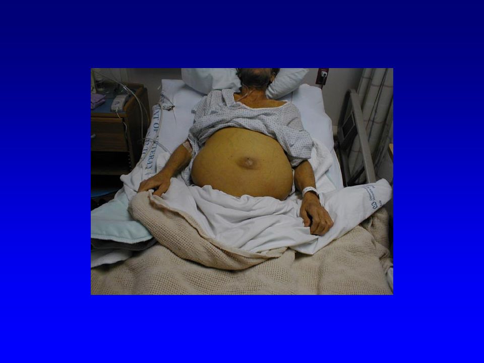
Observation: Inspection
Least mechanical aspect of the physical examination Hardest to learn Yields the most physical signs More diagnosis are made by inspection than all other techniques combined Depends upon the knowledge of the observer

40
Inspection Begins when you first see the patient and ends when they leave Systematic part of each component of the physical examination Part of the mental status examination Subtle observations probably account for “the sixth sense” of astute clinicians

41
Inspection: General Appearance
State of consciousness Signs of distress (sick or not sick?) Apparent state of health Skin:discoloration or obvious lesions Dress, grooming, and personal hygiene Facial expression Gait and posture Motor activity
 Apparent state of health. Skin:discoloration or obvious lesions. Dress, grooming, and personal hygiene. Facial expression. Gait and posture. Motor activity.")
42
Dress, grooming, and personal hygiene

43
Inspection: General Appearance
State of nutrition Body habitus Symmetry Stated age vs. physiologic age Mood, attitude, affect Speech Olfactory diagnosis Bodily excretions (Effuvia)
")
44
Olfactory Diagnosis: “Medical olfaction can often be an important aspect of clinical examination if clinicians approach patient encounters with an “open nose” as well as an open mind.” Hayden, GF: Olfactory diagnosis in medicine, Post Graduate Medicine, 1980

45
Olfactory Diagnosis: “Characteristic patient odors accompany many
diseases and intoxications, and their recognition can provide diagnostic clues, guide the laboratory evaluation, and affect the choice of immediate therapy.” Hayden, GF: Olfactory diagnosis in medicine, Post Graduate Medicine, 1980

46
Inspection: Olfactory Diagnosis:
Detection of ingestions or toxins Alcohol Tobacco Toluene Cyanide Detection of certain infections Anaerobic Necrotic material Diagnosis of certain diseases Fruity; acetone like = Diabetic ketoacidosis Urine-like = Uremia Inborn errors of metabolism

47
Inspection: Bodily Excretions (Effluvia)
Video

48
Inspection: Bodily Excretions (Effluvia)
Urine, stool, sputum, vomitus, exudates, sweat Color, odor, constancy, or smell Examples: Acholic (clay colored) stool of biliary obstruction “Coffee ground” emesis of upper gastrointestinal hemorrhage “Rusty sputum” of pneumococcal pneumonia Melena the black tarry stool from an upper gastrointestinal hemorrhage has a distinct odor “Uremic frost” of severe renal failure
 stool of biliary obstruction. Coffee ground emesis of upper gastrointestinal hemorrhage. Rusty sputum of pneumococcal pneumonia. Melena the black tarry stool from an upper gastrointestinal hemorrhage has a distinct odor. Uremic frost of severe renal failure.")
62
Recording General Observations:
Consider the patient with lung cancer with a superimposed pneumonia: A brief statement at the beginning of the physical examination: “A cachextic cyanotic white male sitting upright on the edge of the bed in moderate reparatory distress” During the vital signs: Respiratory rate 24 and labored with use of accessory muscles During parts of the physical examination: HEENT: Temporal wasting Chest: Barrel chested Skin: Cyanotic and diaphoretic

63
Percussion surface of the body is struck to emit sounds that
“Method of physical examination in which the surface of the body is struck to emit sounds that vary in quality according to the density of the underlying tissues.”

64
Percussion Vibration produced by impact of the finger against underlying tissue Sound waves (resonance) arise from vibrations 4 to 6 cm deep in the body tissue The more dense the material, the quieter the tone
 arise from vibrations 4 to 6 cm deep in the body tissue. The more dense the material, the quieter the tone.")
65
Techniques of Percussion
Direct Striking finger, hand, or lunar aspect of fist directly against the body. Indirect One finger tip (dominate middle finger) used as a hammer (plexar) To strike the PIP joint of the middle finger of the non-dominate hand as the PIP joint is pressed firmly against the area to be percussed (pleximeter)
 used as a hammer (plexar) To strike the PIP joint of the middle finger of the non-dominate hand as the PIP joint is pressed firmly against the area to be percussed (pleximeter)")
66
Percussion Tones Tympany Gastric air bubble
Hyperresonace Emphysemic lung Resonance Healthy lung Dullness Liver Flatness Muscle, thigh

67
Uses of Percussion Sonorous percussion – determine density
Definitive percussion – mapping extent of border of an area Ex: liver It is easier to hear the change from resonance to dullness – so proceed with percussion from areas of resonance to areas of dullness Detection of areas of tenderness Ex: flank percussion in pyleonephritis

68
Palpation Sensitive parts of the hand
Tactile sense – finger pads more sensitive than finger tips Vibratory sense – ulnar aspect of hands, palmer metacarpalphalangeal joints Position and consistency – grasping fingers Temperature – dorsum of hand

69
Qualities Elicited by Palpation:
Texture – skin and hair Moisture – skin Temperature – skin Masses Size, shape, consistency, motility, pulsatile Precordial cardiac thrust Crepitus Tenderness Vocal Fremitus

70
Special Methods of Palpation
Light palpation – up to 1 cm Deep palpation – up to 4 cm Ballottement Fluid wave

71
Auscultation Heart Murmurs, clicks, opening snap, gallops, pericardial friction rubs and knocks Lungs Breath sounds, whispers, voice, crackles (rales), pleural friction rubs Abdomen Bowel sounds, bruits Neck Bruits – carotid, thyroid Head Bruit of AV fistula Joints Crepitus Scrotum Bowel sounds from hernia
, pleural friction rubs. Abdomen. Bowel sounds, bruits. Neck. Bruits – carotid, thyroid. Head. Bruit of AV fistula. Joints. Crepitus. Scrotum. Bowel sounds from hernia.")
72
Instruments Stethoscope Ophthalmoscope Otoscope Near vision chart
Tuning forks Reflex hammer

73
Stethoscope Conveys a vibrating column of air from the body wall to the ears Does not amplify, but sounds may be altered Excludes extraneous noises

74
Stethoscope Heart and lung sounds have a frequency between 60 and 3000 cycles per second Hearing range in a young person is 30 to 20,000 cycles per second, but is dependent upon intensity. At low intensity range is 70 to 150 cycles per second. Therefore some low-pitched sounds may be near the limits of auscultation.

75
Components of the stethoscope
Chest piece Bell piece Transmits all sounds Low pitches are transmitted well Lightly touch test Should have rubber edge Diaphragm Filters out low pitched sounds Isolates high pitched sounds Press firmly Hold between second and third fingers

76
Components of the stethoscope
Rubber tubing Thick walled, stiff, and heavy 30 to 40 cm (12 to 18 inches) Angled Biaurals Point ear pieces towards the nose Ear pieces Snug Comfortable
 Angled Biaurals. Point ear pieces towards the nose. Ear pieces. Snug. Comfortable.")
77
Ophthalmoscope Lenses and mirrors -20 to +40 diopters Light source
Various apertures Small - small pupils Red free filter - green beam, optic disc pallor and minute vessels changes Slit - Anterior eye, elevation of lesions Grid - size of fundal lesions

84
Otoscope Speculum narrows and directs the beam of light
Glass plate magnifying glass Pneumatic attachment - TM mobility May be used for nasal examination

86
Tuning Forks Auditory to 1000 HZ Vibratory to 400 HZ

87
Reflex Hammer Tomahawk Babinski Neurologic hammer

88
Other Safety pins Pen light Tape measure Ruler Q-tips Tongue blades
Near vision chart

89
Near Vision Chart

90
Vital Signs

91
Vital Signs Equipment Needed A Stethoscope A Blood Pressure Cuff
A Watch Displaying Seconds A Thermometer

92
Temperature Temperature can be measured is several different ways:
Oral with a glass, paper, or electronic thermometer (normal 98.6F/37C) Axillary with a glass or electronic thermometer (normal F/36.3C) Rectal or "core" with a glass or electronic thermometer (normal 99.6F/37.7C) Aural (the ear) with an electronic thermometer (normal F/37.7C) Of these, axillary is the least and rectal is the most accurate.
 Axillary with a glass or electronic thermometer (normal 97.6F/36.3C) Rectal or core with a glass or electronic thermometer (normal 99.6F/37.7C) Aural (the ear) with an electronic thermometer (normal 99.6F/37.7C) Of these, axillary is the least and rectal is the most accurate.")
93
Temperature: Fever (pyrexia): elevated body temperature
Hyperpyrexia: extreme fever, > 106F/41.1C Hypothermia: extremely low temperature< 95F/35C

94
False measurements: Patient smoking or drinking hot or cold liquids
Rapid respiratory rate Failure to use thermometer correctly

95
Recording: Temperature in degrees Which scale? Location,
(Type of thermometer) ex: 106F, axillary, (glass)
 ex: 106F, axillary, (glass)")
96
Pulse Sit or stand facing your patient.
Grasp the patient's wrist with your free (non-watch bearing) hand (patient's right with your right or patient's left with your left). There is no reason for the patient's arm to be in an awkward position, just imagine you're shaking hands. Compress the radial artery with your index and middle fingers.
 hand (patient s right with your right or patient s left with your left). There is no reason for the patient s arm to be in an awkward position, just imagine you re shaking hands. Compress the radial artery with your index and middle fingers.")
97
Pulse Note whether the pulse is regular or irregular:
Regular - evenly spaced beats, may vary slightly with respiration Regularly Irregular - regular pattern overall with "skipped" beats Irregularly Irregular - chaotic, no real pattern, very difficult to measure rate accurately Count the pulse for 15 seconds and multiply by 4. Count for a full minute if the pulse is irregular. Record the rate and rhythm.

98
Pulse: Interpretation
A normal adult heart rate is between 50 and beats per minute A pulse greater than 100 beats/minute is defined to be tachycardia. Pulse less than 60 beats/minute is defined to be bradycardia. Tachycardia and bradycardia are not necessarily abnormal. Athletes tend to be bradycardic at rest (superior conditioning). Tachycardia is a normal response to stress or exercise.
. Tachycardia is a normal response to stress or exercise.")
99
Respiration Best done immediately after taking the patient's pulse. Do not announce that you are measuring respirations. Without letting go of the patients wrist begin to observe the patient's breathing. Is it normal or labored?

100
Respiration Count breaths for 15 seconds and multiply this number by 4 to yield the breaths per minute. In adults, normal resting respiratory rate is between breaths/minute. Rapid respiration is called tachypnea.

101
Measurement of Blood Pressure
“Although the arterial blood pressure is measured many time a day by doctors all over the world, few physicians have devoted much thought to the problems and principles involved in measuring blood pressure accurately…From the very beginning, students must learn to record the blood pressure properly. Accurate blood pressure recording will then become a habit that will remain with the physician for a lifetime."

102
Blood Pressure: Systolic = highest BP in the cycle
Diastolic = lowest BP in the cycle Pulse pressure = difference between systolic and diastolic Mean arterial pressure = (1/3)(SBP – DBP) + DBP
(SBP – DBP) + DBP.")
103
Blood Pressure: Hypertension Hypertension is a risk factor
For adults >140/90 Graded by severity Malignant hypertension = acute target organ damage Hypertension is a risk factor

104
Blood Pressure Classification
BP Classification SBP mmHg DBP mmHg Normal <120 and <80 Prehypertension 120–139 or 80–89 Stage 1 Hypertension 140–159 or 90–99 Stage 2 Hypertension >160 or >100

105
Sphygmomanometers Types Mercury-gravity Aneroid Automated Components:
Components: Pressure manometer Inflatable rubber bladder within an inelastic covering Size is important Width - 40% arm circumference Length – 80% arm circumference Most are marked Rubber hand bulb and pressure control valve

106
THE BLOOD PRESSURE CUFF
BLADDER CUFF

107
Technique of Blood Pressure Measurement:
The patient Not smoking, ingesting caffeine, or vigorous activity for 30 min prior Rest sitting comfortably for 5 – 10 min Room quiet and warm Arm rested and free of clothing

108
Technique of Blood Pressure Measurement:
Be aware of conditions which may alter BP Dialysis fistula Lymphedema Atherosclerosis Anxiety (white coat hypertension) Circadian variation
 Circadian variation.")
114
THE AUSCULTATORY GAP THE DISAPPERANCE OF THE PHASE 1 KOROTKOFF SOUNDS IN SYSTOLE WITH REAPPEARANCE ABOVE THE DIASTOLIC PRESSURE. AVOID BY PALPATING THE DISTAL PULSE UNTIL IT DISAPPEARS DURING CUFF INFLATION. MECHANISM UNKNOWN ?ATHEROSCLEROTIC PLAQUE. 20% OF ELDERLY PATIENTS. MAY LEAD TO INACCURATE SYSTOLIC AND DIASTOLIC READING. FALSELY LOW SBP OR FALSELY HIGH DBP. 150/98 200/98 WITH AN AUSCULTATORY GAP BETWEEN CAVALLINI MC ANN INTERN MED 124: ;1996 BATES GUIDE TO THE PHYSICAL EXAMINATION 8TH ED.

115
Phases of the Korotkoff Sounds
Starts with a loud “thud” Recorded at level when 2 beats heard in a row Systolic There may be an auscultatory gap Phase 2 A blowing or swishing sound Phase 3 Softer thud than phase 1 Still crisp Phase 4 Muffing Softer blowing sounds that disappears Phase 5 Silence Diastolic

116
Diastolic Blood Pressure:
Special Considerations: Some controversy if phase 4 or phase 5 is DBP Recorded at phase 5, disappearance of sounds Usually phase 4 and 5 are close, < 5 mm Hg If more than 10 mm Hg apart Record as:160/90/68 In some patients, ex: Aortic regurgitation, sounds never disappear. Record as: 150/70/0

117
Blood Pressure 1. Position the patient's arm so the anticubital fold is level with the heart. Support the patient's arm with your arm or a bedside table. 2. Center the bladder of the cuff over the brachial artery approximately 2.5 cm above the anticubital fold. Proper cuff size is essential to obtain an accurate reading. Be sure the index line falls between the size marks when you apply the cuff. Position the patient's arm so it is slightly flexed at the elbow.

118
Blood Pressure 3. Palpate the radial pulse and inflate the cuff until the pulse disappears. This is a rough estimate of the systolic pressure. 4. Place the stethoscope over the brachial artery.

119
Blood Pressure 5. Inflate the cuff to 30 mmHg above the estimated systolic pressure. Release the pressure slowly, no greater than 5 mmHg per second. The level at which you consistently hear beats is the systolic pressure.

120
Blood Pressure 6. Continue to lower the pressure until the sounds muffle and disappear. This is the diastolic pressure. Record the blood pressure as systolic over diastolic ("120/70" for example).
.")
121
Errors in BP Measurement
Cuff too small Cuff too large Arm held below heart Loose cuff

122
Accurate BP Measurements
Proper patient conditions - Sitting, relaxed, no caffeine or smoking, etc Errors in measurement – Cuff size, technique “White coat” hypertension Pseudohypertension Home BP measurements 24 hour ambulatory measurements

123
CIRCADIAN PATTERNS OF BLOOD PRESSURE
NORMALLY BLOOD PRESSURE FALLS AT NIGHT AND EARLY MORNING. NEJM 347: ;2002

124
Next Week ENT Eye

Similar presentations


 WIPERS Take name & DOB Ask arm preferences, make sure no tight.>")


 Clinical skills for pharmacists.>")


