Download presentation
Presentation is loading. Please wait.

1
Blood disorders

2
What is hematology? Hematology is the study of blood and is concerned mainly with the formed elements in the blood. The formed elements in the blood include: The white blood cells (leukocytes) which include the neutrophils, eosinophils, basophils, monocytes, and lymphocytes (. The red blood cells (erythrocytes) The platelets (thrombocytes) All of the formed elements in the blood are derived from same pluripotential stem cell in the bone marrow
 which include the neutrophils, eosinophils, basophils, monocytes, and lymphocytes (. The red blood cells (erythrocytes) The platelets (thrombocytes) All of the formed elements in the blood are derived from same pluripotential stem cell in the bone marrow.")
3
What is hematology continued
Erythrocytes function in the transport of oxygen to the tissues. Leukocytes function in both specific (immune responses) and non-specific defenses against foreign invasion. Thrombocytes function in hemostasis or blood clotting.
 and non-specific defenses against foreign invasion. Thrombocytes function in hemostasis or blood clotting.")
4
Hemostasis Disorders of bleeding Anemia Blood malignancies

5
Hemostasis

6
definition Maintenance of fluidity of blood while in vessel and formation of hemostatic plug on vascular injury

7
Balance between clot formation and bleeding is maintained

8
Hemostasis involves Clot formation Anti clotting mechanisms

9
At a site of a vascular injury
1.Vasoconstriction 2.Primary hemostatic plug formation 3.Secondary hemostasis due to activation of coagulation cascade by tissue factor and phospholipid via extrinsic pathway- the end result being fibrin which traps the cells in the blood forming a clot

11
Vasoconstriction due to local neural response, and release of endothelin from the endothelium vessels constricted

12
Primary hemostatic plug formation
due to platelet adhesion activation degranulation(ADP, TXA2) recruitment of other platelets
 recruitment of other platelets.")
13
In a site of vessel wall injury platelets in circulation comes in to contact with the ECM
On contact with ECM constituents, platelets undergo 3 reactions: 1) ADHESION and shape change 2) SECRETION (release reaction) 3) AGGREGATION
 ADHESION and shape change. 2) SECRETION (release reaction) 3) AGGREGATION.")
14
PLATELET ADHESION To sub-endothelial ECM constituents Bridged by vWF, a product of endothelial cells

15
PLATELET SECRETION Occurs soon after adhesion Platelets release ADP and calcium ADP activation of platelets is essential for platelet aggregation, further release of ADP

16
Platelet aggregation product of platelet set up a reaction leading to build-up of an enlarging platelet aggregate, the primary hemostatic plug

17
Vascular and platelet responses are important in reducing bleeding but their activity is limited.
To arrest bleeding the proper ‘clot’ should be formed This is brought about by the clotting cascade

18
Coagulation cascade The coagulation cascade is essentially a series of enzymatic conversions, turning inactive proenzymes into activated enzymes and culminating in the formation of thrombin. Thrombin then converts the soluble plasma protein fibrinogen into the insoluble fibrous protein fibrin. This results in formation of the definitive clot

20
Anti clotting mechanism
Once activated the coagulation cascade must be restricted to the local site of vascular injury to prevent clotting of the entire vascular tree. Regulated by natural anticoagulants Anti thrombin III Protein C and Protein S Tissue palsminogen With onset of coagulation cascade, fibrinolytic cascade is also activated to limit the the size of final clot Primarily accomplished by plasmin

22
Disorders of hemostasis
Clot formation inappropriately -thrombosis Bleeding disorders

23
Bleeding disorders

24
Types of skin bleeds –terminology
Petechie - Minute (1- to 2-mm) hemorrhages into skin, mucous membranes, or serosal surfaces
 hemorrhages into skin, mucous membranes, or serosal surfaces.")
25
Types of skin bleeds –terminology
Purpuras - Slightly larger i.e 3- to 5-mm hemorrhages are called purpuras

26
Types of skin bleeds –terminology
Ecchymoses - Larger i.e 1- to 2-cm or more subcutaneous hematomas (bruises)
")
27
Bleeding disorders Vessel wall disorders Platelet disorders
Coagulation disorders

28
Vessel wall disorders Defective collagen due to connective tissue disorders, vitamin C deficiency

29
Platelet disorders Low platelet count (thrombocytopenia )
Platelet function disorders

30
Causes of thrombocytopenia
Decreased platelet production -bone marrow disorders like cancers,aplastic anemia, -drugs, infections Increased destruction -immune thrombocytopenic purpura -DIC -HUS Enlarged spleen

31
Coagulation disorders
Hemophilia A Hemophilia B Vitamin K deficiency Von Willebrand Disease

32
Platelet and vessel wall defects usually present as
skin and mucous membranes-Petechie,Ecchymosis Gum bleeding and epistaxis Menorrhagia Gastrointestinal bleeding Intracranial bleeding

33
Clotting factor disorders may present as
Bleeding Into joints - Haemarthroses Into deep tissues – Hematoma Muscle bleeds

34
Coagulation disorders
Hemophilia A Hemophilia B Vitamin K deficiency Von Willebrand Disease

35
Question why does vitamin K deficiency give rise to bleeding?

36
Hemophilia A & B clinically similar:
occur in approximately 1 in 5,000 male births account for 90% of congenital bleeding disorders Hemophilia A is approximately 5 times more common than B

37
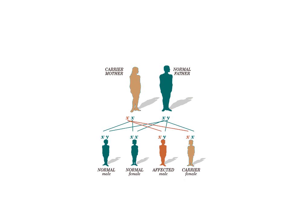
Etiology Inherited as a sex linked recessive trait with bleeding manifestations only in males genes which control factor VIII and IX production are located on the x chromosome; if the gene is defective synthesis of these proteins is defective female carriers transmit the abnormal gene A disease of males

39
Classification % normal factor level Causes of bleeding Severe < 1%
bleeding after trivial injury or spontaneous Moderate 1 - 5% bleeding after minor injury; occasional spontaneous bleeds Mild % following major trauma, surgical or dental procedures

40
Diagnosis Atypical bleeding at circumcision or bruising at neonatal vaccines Toddlers with lip bleeding or unusual bruising when learning to walk Hx of affected males on mother’s side Elevated PTT Factor assays

41
Clinical Features – Joint Bleeds
Joints (Hemarthrosis) Knees, ankles and elbows most common sites begin as the child begins to crawl and walk Single joint bleed: stiffness, swelling, pain With repeated bleeding into same jt---arthropathy-> stiffness and contractures
 Knees, ankles and elbows most common sites. begin as the child begins to crawl and walk. Single joint bleed: stiffness, swelling, pain. With repeated bleeding into same jt---arthropathy-> stiffness and contractures.")
43
Clinical Features – Muscle Bleeds
Bleeding into muscle or soft tissue Sites: calf Symptoms: pain, swelling, muscle spasm Complications: nerve compression, contracture

44
Other Sites of Hemorrhage
Abdomen GI tract Intracranial bleeds Around vital structures in the neck Can cause death…

45
They have high risk of HIV,Hep B and Hep C due to repeated transfusion of blood products

46
Management Specific Hemophillia A Fac viii preparations Cryo DDAVP
Hemophillia B Fac ix CPP

47
General Avoid NSAIDs Avoid contact sports Avoid IM injections
Good dental care Education – life long management Acute and long term management of musculoskeletal problems

48
Von Willabrand disease
Read…..

49
Investigations in bleeding disorders
Bleeding time-vessel wall and paltelet defects detected Prothrombin time (PT)-prolonged in disorders of the extrinsic pathway Activated partial thromboplastin time(APTT) –prolonged in intrinsic pathway disorders
-prolonged in disorders of the extrinsic pathway. Activated partial thromboplastin time(APTT) –prolonged in intrinsic pathway disorders.")
51
Thank you…..

Similar presentations


 Thrombocytes. PLATELETS (PLT) Thrombocytes.>")


