Download presentation
Presentation is loading. Please wait.

1
Chronic hind limb lameness in a 10 year old Oldenburg gelding Part 2

2
MRI and CT Examinations Following discussion of the results of the referral nuclear medicine and subsequent radiographic examinations, the horse was referred to our hospital for MRI and CT examinations.

3
MRI Right Hind Proximal Suspensory MRI Proton density (PD) images shown Distal to proximal
 images shown Distal to proximal")
4
Lateral

34
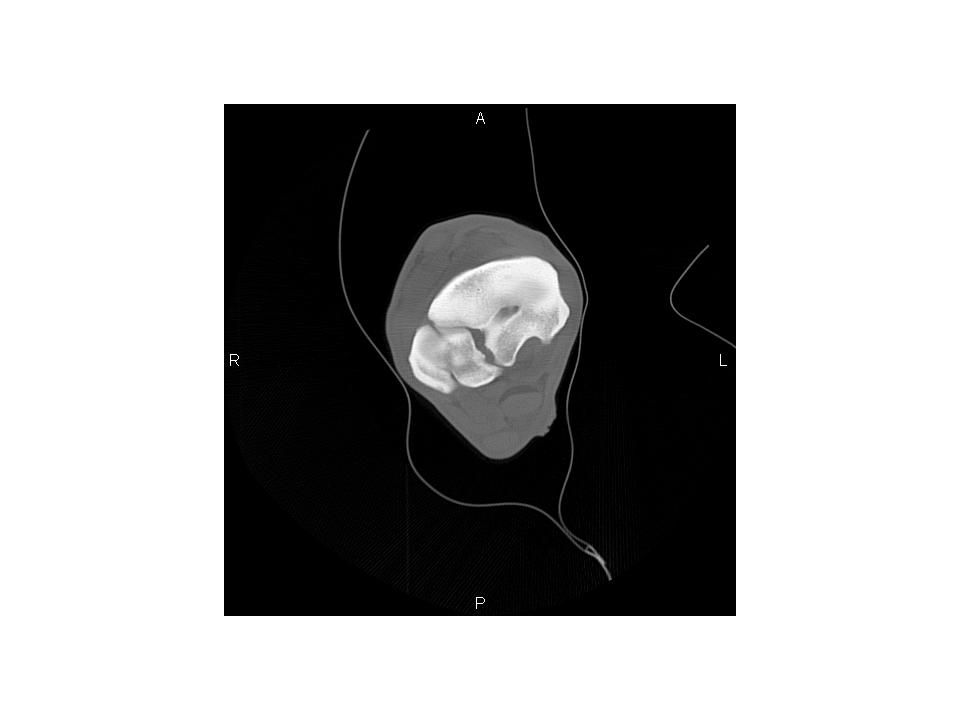
CT – Transverse Plane Bone window and level only shown Distal to proximal

35
Lateral

65
CT Sagittal Reconstruct

66
MRI Findings The proximal suspensory ligament is diffusely enlarged. PD (and T2, STIR, not shown) hyperintensity of the dorsal aspect of the ligament. Marked sclerosis of the proximoplantar aspect of MT3. (STIR hyperintensity of the cortical bone was present - not shown). Large enthesophyte at the origin of the suspensory ligament.
 hyperintensity of the dorsal aspect of the ligament. Marked sclerosis of the proximoplantar aspect of MT3. (STIR hyperintensity of the cortical bone was present - not shown). Large enthesophyte at the origin of the suspensory ligament..")
67
MRI Diagnosis Severe desmitis of the proximal right hind suspensory ligament with associated sclerosis and enthesopathy of the MT3.

68
CT Findings Sclerosis of the plantar aspect of proximal MT3 with irregulary marginated, multifocal, sharp enthesophytes. One particulary large enthesophyte exends along the dorsomedial of the suspensory ligament. Moderate proliferation and synostosis between MT2 and MT3 is present. Bridging periosteal proliferation is present abaxially between MT4 and MT3, approximately 3 cm distal to the head of MT4. The proximal suspensory ligament is enlarged proximally (with hypoattenuation in its dorsal aspect, soft tissue window/level, not shown).
..")
69
CT Diagnosis Severe enthesophytosis of the origin of the right hind suspensory ligament with associated severe desmitis.

70
Case Outcome Suspensory ligament fasciotomy and regional neurectomy with autologous PRP suspensory ligament injection were performed. Discharged with instructions for restricted exercise with incremental increases of defined activity over several months. The horse’s rehabilitation is in progress.

71
Commentary The imaging portion of this case began with a referral nuclear medicine examination in which a severe bony response of the plantar aspect of MT3 was identified. The apparent response of adjacent MT2 and MT4 was a bit unusual for a typical case of suspensory ligament origin desmitis and radiographs were subsequently obtained by the referral hospital.

72
Commentary The radiographic examination showed severe bony response of the plantar aspect of MT3, in addition to exuberant reaction of proximal MT2 and MT4. Initial images also suggested a fracture of the medial plantar surface of MT3, shown to be an artifact created by the degenerative joint disease between MT3 and MT2.

73
Commentary Further imaging at this point was directed primarily at assessing the damage to the proximal suspensory ligament and thus the horse was referred to our hospital for a MRI examination. MRI underestimated the degree of bony changes seen radiographically, so CT was performed immediately afterward (the CT suite is in close proximity to MRI) to better understand the degree of bony changes.
 to better understand the degree of bony changes..")
74
Commentary This case was chosen to illustrate several points. First, the severity of bony changes associated with chronic suspensory ligament origin desmitis was felt unusual. Secondly, MRI underrepresented the bony changes that were evident radiographically, subsequently fully appreciated using CT.

Similar presentations