Download presentation
Presentation is loading. Please wait.

1
William S. Klug Michael R. Cummings Charlotte A
William S. Klug Michael R. Cummings Charlotte A. Spencer Concepts of Genetics Eighth Edition Chapter 7 Sex Determination and Sex Chromosomes Copyright © 2006 Pearson Prentice Hall, Inc.

2
Sexual Differentiation and Life Cycles
Chlamydomonas

3
Figure 7-1 Copyright © 2006 Pearson Prentice Hall, Inc.
Figure 7-1 The life cycle of Chlamydomonas. Unfavorable conditions stimulate the formation of isogametes of opposite mating type that may fuse in fertilization. The resulting zygote undergoes meiosis, producing two haploid cells of each mating type. The photograph shows vegetative cells of this green alga. Figure Copyright © 2006 Pearson Prentice Hall, Inc.

4
Figure 7-2 Copyright © 2006 Pearson Prentice Hall, Inc.
Figure 7-2 llustration of mating types during fertilization in Chlamydomonas. Mating will occur only when plus and minus cells are together. Figure Copyright © 2006 Pearson Prentice Hall, Inc.

5
Sexual Differentiation and Life Cycles
Caenorhabditis elegans

6
Figure 7- 4 Copyright © 2006 Pearson Prentice Hall, Inc.
Figure 7-4 (a) Photomicrograph of a hermaphroditic nematode, C. elegans; (b) The outcomes of self-fertilization in a hermaphrodite and a mating of a hermaphrodite and a male worm. Figure Copyright © 2006 Pearson Prentice Hall, Inc.
 Photomicrograph of a hermaphroditic nematode, C. elegans; (b) The outcomes of self-fertilization in a hermaphrodite and a mating of a hermaphrodite and a male worm. Figure 7- 4 Copyright © 2006 Pearson Prentice Hall, Inc.")
7
X and Y Chromosomes Were First Linked to Sex Determination Early in the 20th Century

8
Figure 7- 5a Copyright © 2006 Pearson Prentice Hall, Inc.
Figure 7-5a (a) The Protenor mode of sex determination where the heterogametic sex (the male in this example) is XO and produces gametes with or without the X chromosome; (b) The Lygaeus mode of sex determination, where the heterogametic sex (again, the male in this example) is XY and produces gametes with either an X or a Y chromosome. In both cases, the chromosome composition of the offspring determines its sex. Figure 7- 5a Copyright © 2006 Pearson Prentice Hall, Inc.
 The Protenor mode of sex determination where the heterogametic sex (the male in this example) is XO and produces gametes with or without the X chromosome; (b) The Lygaeus mode of sex determination, where the heterogametic sex (again, the male in this example) is XY and produces gametes with either an X or a Y chromosome. In both cases, the chromosome composition of the offspring determines its sex. Figure 7- 5a Copyright © 2006 Pearson Prentice Hall, Inc.")
9
Figure 7- 5b Copyright © 2006 Pearson Prentice Hall, Inc.
Figure 7-5b (a) The Protenor mode of sex determination where the heterogametic sex (the male in this example) is XO and produces gametes with or without the X chromosome; (b) The Lygaeus mode of sex determination, where the heterogametic sex (again, the male in this example) is XY and produces gametes with either an X or a Y chromosome. In both cases, the chromosome composition of the offspring determines its sex. Figure 7- 5b Copyright © 2006 Pearson Prentice Hall, Inc.
 The Protenor mode of sex determination where the heterogametic sex (the male in this example) is XO and produces gametes with or without the X chromosome; (b) The Lygaeus mode of sex determination, where the heterogametic sex (again, the male in this example) is XY and produces gametes with either an X or a Y chromosome. In both cases, the chromosome composition of the offspring determines its sex. Figure 7- 5b Copyright © 2006 Pearson Prentice Hall, Inc.")
10
The Y Chromosome Determines Maleness in Humans

11
Figure 7-6 Copyright © 2006 Pearson Prentice Hall, Inc.
Figure 7-6 The traditional human karyotypes derived from a normal female and a normal male. Each contains 22 pairs of autosomes and two sex chromosomes. The female (a) contains two X chromosomes, while the male (b) contains one X and one Y chromosome (see arrows). Figure Copyright © 2006 Pearson Prentice Hall, Inc.
 contains two X chromosomes, while the male (b) contains one X and one Y chromosome (see arrows). Figure 7-6 Copyright © 2006 Pearson Prentice Hall, Inc.")
12
Figure 7-6a Copyright © 2006 Pearson Prentice Hall, Inc.
Figure 7-6a The traditional human karyotypes derived from a normal female and a normal male. Each contains 22 pairs of autosomes and two sex chromosomes. The female (a) contains two X chromosomes, while the male (b) contains one X and one Y chromosome (see arrows). Figure 7-6a Copyright © 2006 Pearson Prentice Hall, Inc.
 contains two X chromosomes, while the male (b) contains one X and one Y chromosome (see arrows). Figure 7-6a Copyright © 2006 Pearson Prentice Hall, Inc.")
13
Figure 7-6b Copyright © 2006 Pearson Prentice Hall, Inc.
Figure 7-6b The traditional human karyotypes derived from a normal female and a normal male. Each contains 22 pairs of autosomes and two sex chromosomes. The female (a) contains two X chromosomes, while the male (b) contains one X and one Y chromosome (see arrows). Figure 7-6b Copyright © 2006 Pearson Prentice Hall, Inc.
 contains two X chromosomes, while the male (b) contains one X and one Y chromosome (see arrows). Figure 7-6b Copyright © 2006 Pearson Prentice Hall, Inc.")
14
The Y Chromosome Determines Maleness in Humans
Klinefelter and Turner Syndromes

15
Figure 7-7a Copyright © 2006 Pearson Prentice Hall, Inc.
Klinefelter syndrome Figure 7-7a The karyotypes and phenotypic depictions of individuals with (a) Klinefelter syndrome (47,XXY) and (b) Turner syndrome (45,X). Figure 7-7a Copyright © 2006 Pearson Prentice Hall, Inc.
 Klinefelter syndrome (47,XXY) and (b) Turner syndrome (45,X). Figure 7-7a Copyright © 2006 Pearson Prentice Hall, Inc.")
16
Figure 7-7b Copyright © 2006 Pearson Prentice Hall, Inc.
Turner syndrome Figure 7-7b The karyotypes and phenotypic depictions of individuals with (a) Klinefelter syndrome (47,XXY) and (b) Turner syndrome (45,X). Figure 7-7b Copyright © 2006 Pearson Prentice Hall, Inc.
 Klinefelter syndrome (47,XXY) and (b) Turner syndrome (45,X). Figure 7-7b Copyright © 2006 Pearson Prentice Hall, Inc.")
17
The Y Chromosome Determines Maleness in Humans
47,XXX Syndrome 47,XYY Condition The Y Chromosome and Male Development

18
Table 7-1 Copyright © 2006 Pearson Prentice Hall, Inc.
Table 7.1 Frequency of XYY Individuals in Various Settings Table Copyright © 2006 Pearson Prentice Hall, Inc.

19
Figure 7-8 Copyright © 2006 Pearson Prentice Hall, Inc.
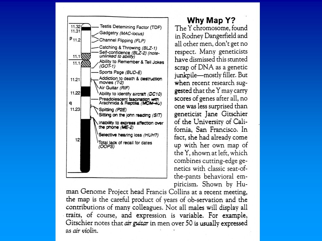
Figure 7-8 The various regions of the human Y chromosome. Figure Copyright © 2006 Pearson Prentice Hall, Inc.

21
The Ratio of Males to Females in Humans Is Not 1.0

22
Dosage Compensation Prevents Excessive Expression of X-Linked Genes in Humans and Other Mammals
Barr Bodies

23
Figure 7-9 Copyright © 2006 Pearson Prentice Hall, Inc.
Figure 7-9 Photomicrographs comparing cheek epithelial cell nuclei from a male that fails to reveal Barr bodies (bottom) with a female that demonstrates Barr bodies (indicated by an arrow in the top image). This structure, also called a sex chromatin body, represents an inactivated X chromosome. Figure Copyright © 2006 Pearson Prentice Hall, Inc.
 with a female that demonstrates Barr bodies (indicated by an arrow in the top image). This structure, also called a sex chromatin body, represents an inactivated X chromosome. Figure 7-9 Copyright © 2006 Pearson Prentice Hall, Inc.")
24
Figure 7-10 Copyright © 2006 Pearson Prentice Hall, Inc.
Figure 7-10 Barr body occurrence in various human karyotypes, where all X chromosomes except one are inactivated. Figure Copyright © 2006 Pearson Prentice Hall, Inc.

25
Dosage Compensation Prevents Excessive Expression of X-Linked Genes in Humans and Other Mammals
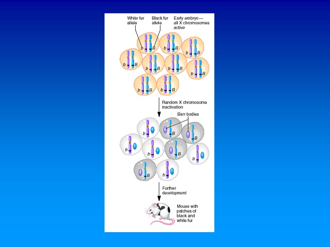
The Lyon Hypothesis

27
Figure 7-11a Copyright © 2006 Pearson Prentice Hall, Inc.
Figure 7-11a (a) A calico cat, where the random distribution of orange and black patches illustrates the Lyon hypothesis. The white patches are due to another gene; (b) A tortoiseshell cat, which lacks the white patches characterizing calicos. Figure 7-11a Copyright © 2006 Pearson Prentice Hall, Inc.
 A calico cat, where the random distribution of orange and black patches illustrates the Lyon hypothesis. The white patches are due to another gene; (b) A tortoiseshell cat, which lacks the white patches characterizing calicos. Figure 7-11a Copyright © 2006 Pearson Prentice Hall, Inc.")
28
Figure 7-11b Copyright © 2006 Pearson Prentice Hall, Inc.
Figure 7-11b (a) A calico cat, where the random distribution of orange and black patches illustrates the Lyon hypothesis. The white patches are due to another gene; (b) A tortoiseshell cat, which lacks the white patches characterizing calicos. Figure 7-11b Copyright © 2006 Pearson Prentice Hall, Inc.
 A calico cat, where the random distribution of orange and black patches illustrates the Lyon hypothesis. The white patches are due to another gene; (b) A tortoiseshell cat, which lacks the white patches characterizing calicos. Figure 7-11b Copyright © 2006 Pearson Prentice Hall, Inc.")
29
Figure 7-12 Copyright © 2006 Pearson Prentice Hall, Inc.
anhidrotic ectodermal dysplasia Figure 7-12 Depiction of the absence of sweat glands (shaded regions) in a female heterozygous for the X-linked condition anhidrotic ectodermal dysplasia. The locations vary from female to female, based on the random pattern of X chromosome inactivation during early development, resulting in unique mosaic distributions of sweat glands in heterozygotes. Figure Copyright © 2006 Pearson Prentice Hall, Inc.
 in a female heterozygous for the X-linked condition anhidrotic ectodermal dysplasia. The locations vary from female to female, based on the random pattern of X chromosome inactivation during early development, resulting in unique mosaic distributions of sweat glands in heterozygotes. Figure 7-12 Copyright © 2006 Pearson Prentice Hall, Inc.")
30
Dosage Compensation Prevents Excessive Expression of X-Linked Genes in Humans and Other Mammals
The Mechanism of Inactivation Xist

31
FYI--This figure is not from your book

32
The Ratio of X Chromosomes to Sets of Autosomes Determines Sex in Drosophila

33
Figure 7-13 Copyright © 2006 Pearson Prentice Hall, Inc.
Figure 7-13 Chromosome compositions, the ratios of X chromosomes to sets of autosomes, and the resultant sexual morphology in Drosophila melanogaster. The normal diploid male chromosome composition is shown as a reference on the left (XY2A). Figure Copyright © 2006 Pearson Prentice Hall, Inc.
. Figure 7-13 Copyright © 2006 Pearson Prentice Hall, Inc.")
34
The Ratio of X Chromosomes to Sets of Autosomes Determines Sex in Drosophila
Dosage Compensation in Drosophila Drosophila Mosaics

35
Figure 7-14 Copyright © 2006 Pearson Prentice Hall, Inc.
bilateral gynandromorph w+ m+ w+ m+ w m Figure 7-14 A bilateral gynandromorph of Drosophila melanogaster formed following the loss of one X chromosome in one of the two cells during the first mitotic division. The left side of the fly, composed of male cells containing a single X, expresses the mutant white-eye and miniature-wing alleles. The right side is composed of female cells containing two X chromosomes heterozygous for the two recessive alleles. Figure Copyright © 2006 Pearson Prentice Hall, Inc.

36
Temperature Variation Controls Sex Determination in Reptiles

37
Figure 7-15 Copyright © 2006 Pearson Prentice Hall, Inc.
Figure 7-15 Three different patterns of temperature-dependent sex determination (TSD) in reptiles, as described in the text. The relative pivotal temperature is crucial to sex determination during a critical point during embryonic development (FTfemale-determining temperature; MTmale-determining temperature). Figure Copyright © 2006 Pearson Prentice Hall, Inc.
 in reptiles, as described in the text. The relative pivotal temperature is crucial to sex determination during a critical point during embryonic development (FTfemale-determining temperature; MTmale-determining temperature). Figure 7-15 Copyright © 2006 Pearson Prentice Hall, Inc.")
Similar presentations