Download presentation
Presentation is loading. Please wait.

1
Cartilage Cartilage belongs to the skeletal tissues and is a specialized form of connective tissue Cartilage is composed of cells, chondrocytes (2-5% of the tissue volume only) located in lacunae surrounded by an intercellular matrix.
 located in lacunae surrounded by an intercellular matrix.")
2
Cartilage is an avascular tissue Cartilage is a tissue of very low metabolic activity and cell turnover (except in the embryo) Cartilage receives its nutrients from blood vessels from a surrounding dense connective tissue, the perichondrium Nerves are not present in cartilage, but nerves and nerve ending are present in the perichondrium.
 Cartilage receives its nutrients from blood vessels from a surrounding dense connective tissue, the perichondrium Nerves are not present in cartilage, but nerves and nerve ending are present in the perichondrium.")
3
Cartilage is classified as: –Hyaline cartilage –Elastic cartilage –Fibrocartilage

4
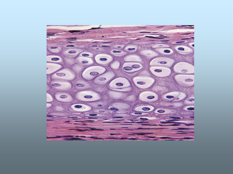
HYALINE CARTILAGE Hyaline cartilage is the most common form of cartilage. Fresh hyaline cartilage is a semi-transparent (translucent), milky-white tissue, that is both flexible and resilient to mechanical forces. In adults hyaline cartilage is found in the respiratory tract (nose, larynx, trachea, bronchi), the ventral part of ribs, and on articulating surfaces of long bones and joints (articular cartilage). Hyaline cartilage is much more common in the embryo, where it plays an important role in long bone development.
, milky-white tissue, that is both flexible and resilient to mechanical forces. In adults hyaline cartilage is found in the respiratory tract (nose, larynx, trachea, bronchi), the ventral part of ribs, and on articulating surfaces of long bones and joints (articular cartilage). Hyaline cartilage is much more common in the embryo, where it plays an important role in long bone development..")
6
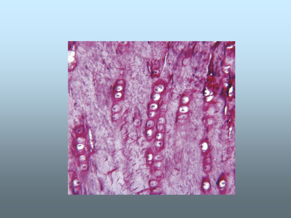
Chondrogenesis Like all connective tissue, cartilage is derived in the embryo from mesenchyme Mesenchyme cells grow and differentiate into young cartilage cells or chondroblasts, that are very active in secreting the surrounding matrix. The chondroblasts grow and develop in lacunae. These chondroblasts further differentiate into mature cartilage cells or chondrocytes. There are two different types of chondrogenesis: appositional growth interstitial growth

8
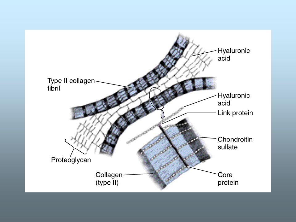
Type II collagen fibers are embedded in the matrix and provide structural support & its fibers constitute about 40% of the dry weight of cartilage The proteoglycans represent a complex of protein and sulfated glycosaminoglycans (GAGs), and in particular: –chondroitin-4-sulfate –chondroitin-6-sulfate –keratan sulfate These sulfated GAGs provide the basophilic staining characteristics There are molecules of a non-sulfated GAG: hyaluronic acid The hyaluronic acid is a long thread-like molecule, to which are attached link proteins periodically along its length. Proteoglycan monomers (which resemble a test-tube brush) are attached to the link-proteins and consist of core proteins to which are connected the sulfated GAGs.
 are attached to the link-proteins and consist of core proteins to which are connected the sulfated GAGs..")
10
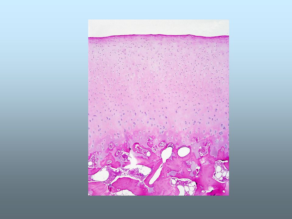
The biomechanical properties of the cartilage allow it to function as a biomechanical spring or shock-absorber, to spread the load at joints and prevent too great pressures on bones In histological sections the matrix surrounding the lacunae (nearest to the cells) is stained more intensely and is more basophilic (capsular or territorial matrix) because the relative concentration of GAGs is greater than in the mass of the matrix, where the staining is less intense (inter-territorial matrix).
 is stained more intensely and is more basophilic (capsular or territorial matrix) because the relative concentration of GAGs is greater than in the mass of the matrix, where the staining is less intense (inter-territorial matrix).")
11
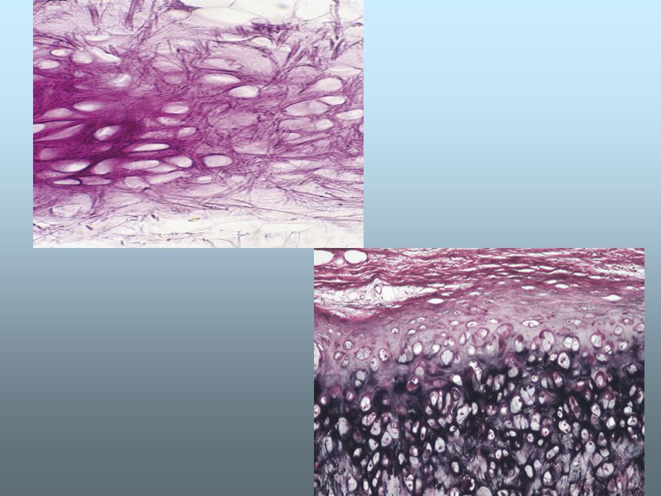
ELASTIC CARTILAGE Elastic cartilage is characterized by its great flexibility and elasticity owing to the large quantities of elastic fibers in the matrix. These elastic fibers provide the yellowish color in the fresh tissue. The elastic fibers in the matrix near the perichondrium are less-densely packed (and easier to see) than those deeper in the tissue. Elastic cartilage is found in the external ear, in the walls of the external auditory meatus and Eustachian tube and also in the epiglottis.
 than those deeper in the tissue. Elastic cartilage is found in the external ear, in the walls of the external auditory meatus and Eustachian tube and also in the epiglottis..")
13
FIBROCARTILAGE Fibrocartilage is found in areas of the body subject to high mechanical stress or weightbearing. It lacks the flexibility of the other cartilage types. Fibrocartilage is present in: –intervertebral disks –pubic symphysis –temporo-mandibular joints –at sites of connection of many ligaments to bones (e.g. Ligamentum teres femoris) – tendon insertions.
 – tendon insertions..")
15
Fibrocartilage is characterized by large numbers and concentrations of collagen fibers in the matrix These collagen fibers are the dominant feature of the matrix and with relatively little amorphous matrix The large amounts of collagen fibers result in the matrix appearing acidophilic in histological sections after H&E staining Fibrocartilage is not surrounded by perichondrium

16
The intervertebral disks consist of fibrocartilage plates between the vertebrae and act as mechanical shock absorbers. In sections they are seen to be formed of two components: – annulus fibrosus, which is the outer region consisting of orderly concentric arrangements of cells and matrix dominated by type I collagen (as in tendons) – nucleus pulposus (large vacuolated cells, that are vestiges of the embryonic notochord.
 – nucleus pulposus (large vacuolated cells, that are vestiges of the embryonic notochord..")
17
nucleus pulposus

18
Secondary cartilage This refers to cartilage that develops in association with specific bones formed by intramembranous ossification after the bones are already formed This is the opposite of cartilage associated with endochondral ossification, where the cartilage precedes the bone formation The cartilage of the temporomandibular joint is an example of a secondary cartilage

19
FUNCTIONS OF CARTILAGE Cartilage is important for: –skeletal support in the embryo prior to the development of the bony skeleton. –elongation of developing long bones (endochondral ossification). –articulating joints (articular cartilage). –flexible support in the ear and eartubes, and in the larger tubes of the respiratory tract (trachea, bronchi).
. –articulating joints (articular cartilage). –flexible support in the ear and eartubes, and in the larger tubes of the respiratory tract (trachea, bronchi)..")
20
Regeneration and repair Despite the fact that cartilage is found in relatively few sites in the adult body, its functions are important for our well-being Cartilage in adults has very little regenerative ability if damaged and is subject to tear and wear with aging This is due to the dearth of cartilage cells, minimal mitosis, absence of an integral blood supply and overall low metabolic activity of the tissue The clinical problems of damage or aging of the tissue (osteoarthritis) are substantial. With aging the cartilage matrix may develop calcified deposits (calcified cartilage).
..")
Similar presentations


 RESISTS COMPRESSION AVASCULAR – nutrients diffuse through matrix.>")


