Download presentation
Presentation is loading. Please wait.

1
Sickle Cell Anemia An example of why: a change in protein can lead to disease a change in DNA can lead to a change in protein

2
Ground Rules for Class Discussions and Workshops Be on time. Speak so that everyone from front to back can hear you. Listen when others are speaking. If it’s review for you, use you intellect to hear it in a new way. Write down your answers or consolidate to print.

3
Central Dogma DNA RNA Protein

5
What do you already know about hemoglobin? What is the function of hemoglobin? What class of biomolecules does hemoglobin belong to? What are the symptoms of sickle cell anemia? Is sickle cell anemia hereditary? What does that tell us?

6
Symptoms of Sickle Cell Anemia pain episodes strokes increased infections leg ulcers bone damage yellow eyes or jaundice early gallstones lung blockage kidney damage and loss of body water in urine painful erections in men (priapism) blood blockage in the spleen or liver (sequestration) eye damage low red blood cell counts (anemia) delayed growth
 blood blockage in the spleen or liver (sequestration) eye damage low red blood cell counts (anemia) delayed growth")
8
Proteins synthesized from amino acids

9
Circle; Triangle; Square; Bond; Amino terminal; Carboxy terminal

10
* 4 classes of structure.

11
Website for Amino acid interactive Workshop Amino acids – everyone open to this page http://www.biomed.curtin.edu.au/biochem/tutori als/AAs/AA.html http://www.biomed.curtin.edu.au/biochem/tutori als/AAs/AA.html

12
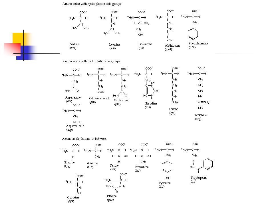
Power of the R Groups Note the one letter and 3 letter abbreviations for your amino acid(s). Identify the atoms in red, blue, white, gray, and other colors Find the carboxy group, amino group, beta carbon, R group Categorize the amino acids – and be able to say why – some fit in more than one category! Aromatic Aliphatic, unbranched Aliphatic, branched Polar Positively charged (basic) Negatively charged (acidic) Small Has a sulfur atom in the R group Hmm? Which are hydrophobic vs. hydrophilic? Which would attract each other if brought together? Which would repel? Which would likely fold to the interior in an aqueous environment? Which would likely fold to the exterior in a lipid environment?
 Negatively charged (acidic) Small Has a sulfur atom in the R group Hmm. Which are hydrophobic vs. hydrophilic. Which would attract each other if brought together. Which would repel. Which would likely fold to the interior in an aqueous environment. Which would likely fold to the exterior in a lipid environment .")
14
Amino acid characteristics

15
Alpha helix is the “default” Ala, Glu, Leu Beta sheet Branched R groups Val, Thr, Ile Helix disrupters with close H bond participants: Ser, Asp, Asn Turns (not shown): Gly, Asp, Pro
: Gly, Asp, Pro")
17
The Nucleic Acids: DNA and RNA DNA synthesized from deoxynucleotide triphosphates (dNTPs) RNA synthesized from nucleotide triphosphates (NTPs)
 RNA synthesized from nucleotide triphosphates (NTPs)")
19
OH dNTP NTP 5’ and 3’

21
DNA sequence Write the primary sequence of the DNA displayed in 3B from the 5’ to the 3’ end of both strands Website for interactive workshop for DNA analysis

22
DNA RNA PROTEIN Replication

23
Central Dogma DNA RNA Protein Transcription Translation

24
DNA RNA PROTEIN Transcription

25
Mature mRNA RNA, but NOT mRNA DNA RNA PROTEIN RNA processing

26
Central Dogma DNA RNA Protein Transcription Translation

28
DNA RNA PROTEIN Translation Etc. 5’ UTR

29
DNA RNA (with ribosomes)
")
30
Translation exercise Translate the following sequence using the codon table: ATG GTG CAC CTG ACT CCT GAG GAG AAG TCT GCC GTT ACT Perform same procedure on the sequence below using a software program: ATG GTG CAC CTG ACT CCT GTG GAG AAG TCT GCC GTT ACT http://us.expasy.org/tools/dna.html http://us.expasy.org/tools/dna.html How many nucleotides have changed in the codon in boldface? What is the amino acid difference in the two sequences? What is the quality of that difference with respect to R groups?

32
*

33
The early evidence that sickle cell anemia is caused by an amino acid change in hemoglobin. Tryptic digest: the protease trypsin cleaves C terminal to lysine and arginine.

34
Summary DNA (mutated = changed) RNA (mutated) Protein (possibly mutated) Remember: Mutation is not always “bad”! For example: Mutation → Evolution → An additional normal genome

35
Multiple sequence alignment for cytochrome C – mutation and conservation Human protein accession number AAA35732 (see next slide) Dog protein accession number XP_532493 Yeast protein number from structure database 1YCC CLUSTAL W PROGRAM
 Dog protein accession number XP_ Yeast protein number from structure database 1YCC CLUSTAL W PROGRAM")
36
Hemoglobin HBB1 >gi|4504349|ref|NP_000509.1| beta globin [Homo sapiens] MVHLTPEEKSAVTALWGKVNVDEVGGEALGRLLVVYPWTQRFFESFGDLSTPDA VMGNPKVKAHGKKVLGAFSDGLAHLDNLKGTFATLSELHCDKLHVDPENFRLLG NVLVCVLAHHFGKEFTPPVQAAYQKVVAGVAN ALAHKYH FASTA format for a protein sequence in single letter code
![Hemoglobin HBB1 >gi| |ref|NP_ | beta globin [Homo sapiens] MVHLTPEEKSAVTALWGKVNVDEVGGEALGRLLVVYPWTQRFFESFGDLSTPDA VMGNPKVKAHGKKVLGAFSDGLAHLDNLKGTFATLSELHCDKLHVDPENFRLLG NVLVCVLAHHFGKEFTPPVQAAYQKVVAGVAN ALAHKYH FASTA format for a protein sequence in single letter code](http://images.slideplayer.com/17/5323511/slides/slide_36.jpg "Hemoglobin HBB1 >gi| |ref|NP_ | beta globin [Homo sapiens] MVHLTPEEKSAVTALWGKVNVDEVGGEALGRLLVVYPWTQRFFESFGDLSTPDA VMGNPKVKAHGKKVLGAFSDGLAHLDNLKGTFATLSELHCDKLHVDPENFRLLG NVLVCVLAHHFGKEFTPPVQAAYQKVVAGVAN ALAHKYH FASTA format for a protein sequence in single letter code")
37
How to prepare the sequences for the MSA on ClustalW For Human and Dog Go to NCBINCBI Select to search the protein database from the dropdown menu Enter the Accession Number (previous slide) and GO Click on the link Change the display to a FASTA file Copy the FASTA output for both species into a single text file. Make sure the header is separate from the sequence. For Yeast Clink on the link, find the FASTA format and copy into the same file Copy or upload the file into ClustalW

38
Workshop due as email to me by 9AM Wednesday, 6/24. 1.Give the answers to questions/challenges from slides within today’s PowerPoint. 2.Print out your ClustalW results and add a short paragraph discussing how Clustal W gives you a clue as to which part(s) of the Cytochrome C protein you would hypothesize are most important to its function (which is/are the same in all 3 organisms). Start your paragraph as a hypothesis as to which parts are most important, and write your discussion as a defense of your hypothesis. 3.What is the chromosomal location of the gene that causes sickle cell anemia? 4.What is the name of the gene? 5.State the nucleotide change and amino acid change that leads to sickle cell anemia (there may be more than one change that gives rise to the disease) 6.If sickle cell anemia is so devastating, why has it lasted in the population for such a long time? Give a molecular, mechanistic, evolutionary explanation (you may have to do a little research to get this). What does the sickled molecule do that the normal molecule can’t?
 of the Cytochrome C protein you would hypothesize are most important to its function (which is/are the same in all 3 organisms). Start your paragraph as a hypothesis as to which parts are most important, and write your discussion as a defense of your hypothesis. 3.What is the chromosomal location of the gene that causes sickle cell anemia. 4.What is the name of the gene. 5.State the nucleotide change and amino acid change that leads to sickle cell anemia (there may be more than one change that gives rise to the disease) 6.If sickle cell anemia is so devastating, why has it lasted in the population for such a long time. Give a molecular, mechanistic, evolutionary explanation (you may have to do a little research to get this). What does the sickled molecule do that the normal molecule can’t .")
Similar presentations


 2006 After running 16.>")


 (Translation) DNA (genetic information stored in genes) RNA (working copies of genes) Proteins (functional.>")

