Download presentation
Presentation is loading. Please wait.

1
The Periodontal Pocket
DR.HINA ADNAN

2
DEFINITION Pathologically deepened gingiva sulcus.
Deepening of gingival sulcus my occur by coronal movement of the gingiva margin, apical displacement of gingiva attachment or combination of both.

3
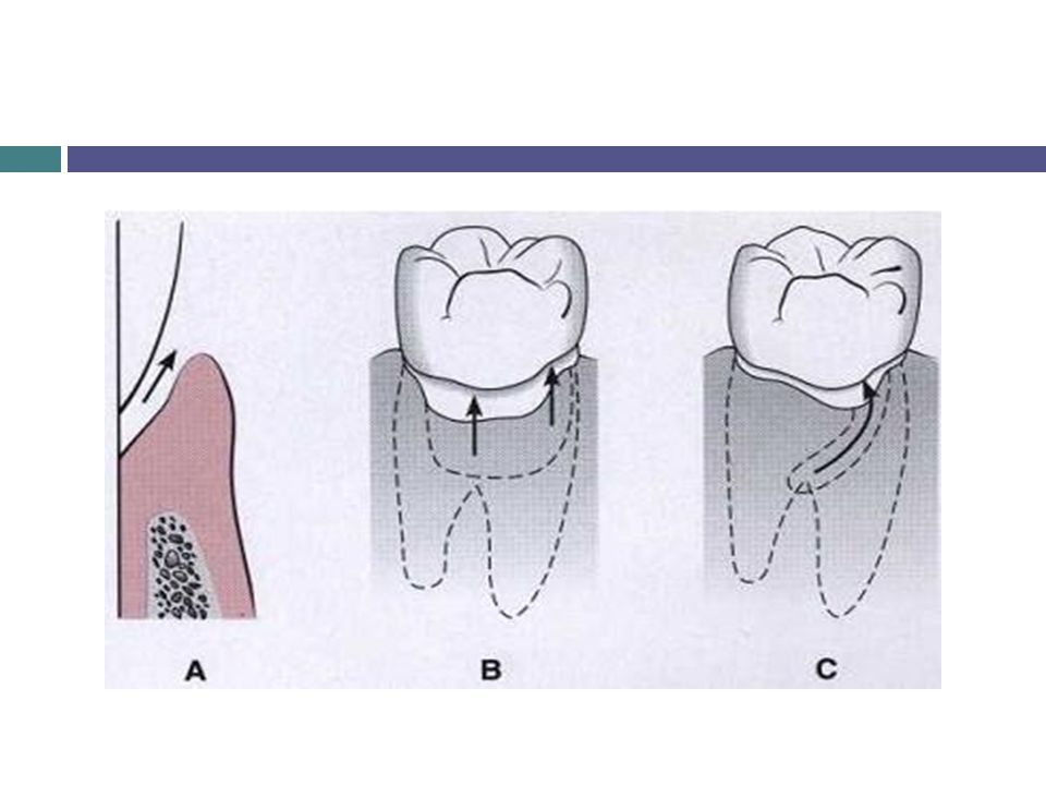
Classification 1(Deepening of the gingival sulcus)
Pocket gingival pocket periodontal pocket suprabony infrabony pocket pocket

6
also called pseudo pocket or relative pocket or false pocket.
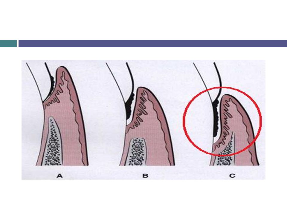
GINGIVAL POCKET PERIODONTAL POCKET also called pseudo pocket or relative pocket or false pocket. Seen in the gingivitis. Formed by the gingiva enlargement without destruction of the underlying periodontal tissue. The sulcus is deep because of increased bulk of gingiva. Also called absolute or true pocket. Seen in periodontitis. Occurs with destruction of supporting periodontal tissues and loosing and exfoliation of the teeth.

7
types of periodontal pockets
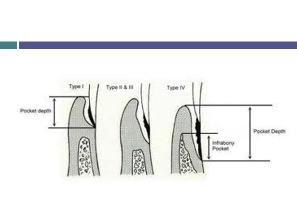
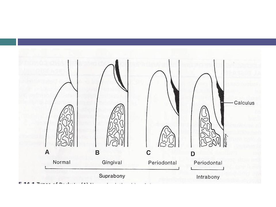
Gingival pocket: There is no destruction of the supporting periodontal tissues. Suprabony pocket: The base of the pocket is coronal to the level of the underlying bone. Bone loss is horizontal. Infrabony pocket: The base of the pocket is apical to the level of the adjacent bone. Bone loss is vertical.

8
Supra bony pocket Infra bony pocket
Also called supra crestal or supra alveolar pocket. Also known as subcrestal or intraalveolar pocket. Bottom of the pocket is coronal to underlying alveolar bone. Bottom of the pocket is apical to the crest of the alveolar bone. Lateral wall consist of soft tissue alone. Lateral wall consist of tissue and bone. Pattern of destruction of bone is horizontal. Pattern of destruction of bone is vertical. Interproximally, transeptal fibers arranged horizontally ( between the base of the pocket and the alveolar bone). Interproximally , transeptal fibers are oblique ( extent from the cementum beneath the base of the pocket along and over the crest of the cementum of the adjacent tooth). On the facial and lingual surfaces , periodontal ligaments fibers , follow the horizontal-oblique course. On the facial and lingual surfaces , periodontal ligaments fibers follow the angular pattern.
. Interproximally , transeptal fibers are oblique ( extent from the cementum beneath the base of the pocket along and over the crest of the cementum of the adjacent tooth). On the facial and lingual surfaces , periodontal ligaments fibers , follow the horizontal-oblique course. On the facial and lingual surfaces , periodontal ligaments fibers follow the angular pattern.")
11
Classification 2 According to the involved tooth surface: Pocket
Simple compound complex or spiral Involved one surface involved more than originate on one surface one surface twisting around the tooth to involve one or more additional surfaces but deep into oral cavity on the surface of its origin

13
Pathogenesis of pocket fotmation
Presence of bacterial plaque on tooth surface. Margin gingiva become inflamed. Gingiva sulcus deepens due to edematous enlargement of gingiva. Gingiva pocket. Anaerobic organisms tend to colonies the sub gingival plaque ( spirochetes and motile rods due to an aerobic environment created in the pocket). Large number of PMN , leukocytes and macrophages migrates to the gingiva tissue in response to bacterial challenge.
. Large number of PMN , leukocytes and macrophages migrates to the gingiva tissue in response to bacterial challenge.")
14
Two mechanism of collagen loss :
I )lysosomal enzymes ( collegenase ) released by PMN , leukocytes destruction of collagen fibers in gingival C.T II) fibroblast phagocytes collagen fibers by cytoplasmic process to the ligaments cementum interface. When the collagen fibers apical to junction epithelial get destroyed , the epithelial cells proliferate along the root surface in an apical direction until they become in contact with healthy collagen fibers. At the same time coronal portion of the junction epithelium get detached from the tooth surface.
lysosomal enzymes ( collegenase ) released by PMN , leukocytes destruction of collagen fibers in gingival C.T. II) fibroblast phagocytes collagen fibers by cytoplasmic process to the ligaments cementum interface. When the collagen fibers apical to junction epithelial get destroyed , the epithelial cells proliferate along the root surface in an apical direction until they become in contact with healthy collagen fibers. At the same time coronal portion of the junction epithelium get detached from the tooth surface.")
15
PMN cells migrate towards the coronal portion of junction epithelium.
When volume of PMN leukocytes at the coronal portion of junctional epithelium exceed 60% the epithelium cells separate from the tooth surface. Pocket information . Plaque removal is difficult or impossible from deep pocket. Favoring growth of pathogenic organism in that protected environment. Further attachment loss.

16
Horizontal bone loss. If I.F.O present then vertical bone loss occurs ( angular bone loss).
.")
17
The initial lesion in the development of periodontitis is the inflammation of the gingiva in response to a bacterial challenge.

18
Clinical Features Gingival pocket wall presents various degrees of bluish red discoloration , a smooth shiny surface and pitting on pressure. Less frequently , gingival wall may be pink and firm. Bleeding is presenting by gently probing soft tissue wall of pocket . Painful during probing. Pus may be present: It is not an indication of the depth of the pocket or the severity of the destruction of the supporting tissues. is a common feature of periodontal disease loss of stippling. Tooth mobility and diastema formation.

20
Pockets content Debris of microorganism and their products.
Gingival fluid. Salivary mucin. Food remnants. Desquamated epithelial cells. Leukocytes.

21
Root surface Wall The root surface wall of periodontal pockets often undergoes changes that are significant because they may perpetuate the periodontal infection cause pain and complicate periodontal treatment. The root surface that gets expose to the oral environment as a result of periodontal attachments loss , undergoes following changes: structure, chemical and cytotoxic.

22
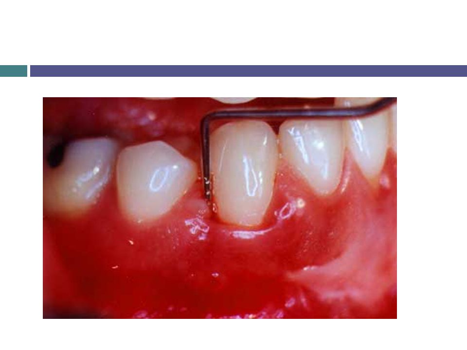
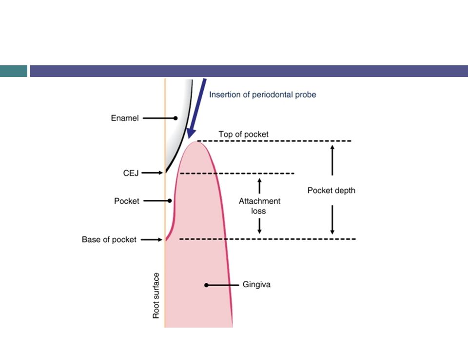
Diagnosis and Detection
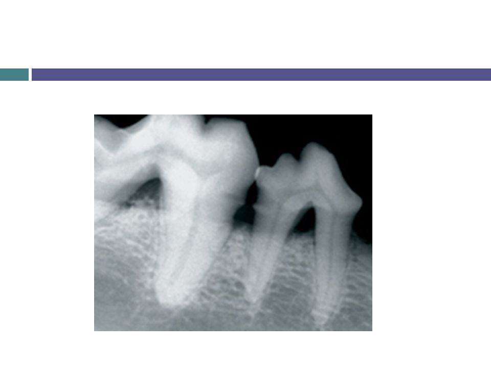
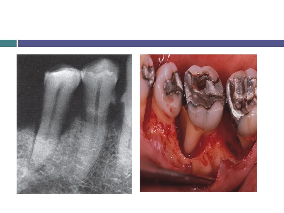
Careful exploration with a periodontal probe. Radiographic: pocket are not detected by radiographic examination because pocket is a soft tissue change. Radiograph indicates area of bone loss where pocket may be suspected they do not show pocket presence or depth.

23
Note: Gutta Percha points or calibrated silver points can be used with radiograph to assist in determining the level of attachment of periodontal pocket.

Similar presentations