Download presentation
Presentation is loading. Please wait.

1
RHEUMATOID ARTHRITIS

2
Chronic multisystem disease of unknown cause Characteristic features: Persistent inflammatory synovitis Involves peripheral joints Symmetric distribution Hallmark of the disease: Synovial inflammation causes cartilage damage and bone erosions and subsequent changes in bone integrity Source: Harrison’s Principles of Internal Medicine, 17 th edition

3
PATHOLOGY AND PATHOGENESIS Earliest lesions in rheumatoid synovitis: - Microvascular injury - Increase in the # of synovial lining cells o Seen along with perivascular infiltration w/ mononuclear cells Source: Harrison’s Principles of Internal Medicine, 17 th edition

4
PATHOLOGY AND PATHOGENESIS Characteristic constellation of features (light microscopy): - Hyperplasia and hypertrophy of synovial lining cells - Focal or segmental vascular changes o Microvascular injury o Thrombosis o Neovascularisation - Edema - Infiltration w/ mononuclear cells Source: Harrison’s Principles of Internal Medicine, 17 th edition
: - Hyperplasia and hypertrophy of synovial lining cells - Focal or segmental vascular changes o Microvascular injury o Thrombosis o Neovascularisation - Edema - Infiltration w/ mononuclear cells Source: Harrison’s Principles of Internal Medicine, 17 th edition")
5
PATHOLOGY AND PATHOGENESIS Source: http://www.bio.davidson.edu/Courses/Immunology/Students/Spring2003/Super/jointandcells.gif

6
PATHOLOGY AND PATHOGENESIS Source: Harrison’s Principles of Internal Medicine, 17 th edition

7
PATHOLOGY AND PATHOGENESIS Source: Harrison’s Principles of Internal Medicine, 17 th edition Collagenase and cathepsins - Degrade components of the articular matrix Osteoclasts - Prominent at sites of bone erosion

8
DIAGNOSIS OF RA Source: Harrison’s Principles of Internal Medicine, 17 th edition 1. Morning stiffness: stiffness in and around the joints lasting 1 hr before maximal improvement

9
DIAGNOSIS OF RA Source: Harrison’s Principles of Internal Medicine, 17 th edition 2. Arthritis of 3 or more joint areas: At least three joint areas have soft tissue swelling or joint effusions, not just bony overgrowth. The 14 possible joint areas involved are right or left PIP, MCP, wrist, elbow, knee, ankle, and MTP joints.

10
DIAGNOSIS OF RA Source: Harrison’s Principles of Internal Medicine, 17 th edition 3. Arthritis of hand joints: Arthritis of wrist, MCP joint, or PIP joint

11
DIAGNOSIS OF RA Source: Harrison’s Principles of Internal Medicine, 17 th edition 4. Symmetric arthritis: Simultaneous involvement of the same joint areas on both sides of the body.

12
DIAGNOSIS OF RA Source: Harrison’s Principles of Internal Medicine, 17 th edition 5. Rheumatoid nodules: Subcutaneous nodules over bony prominences, extensor surfaces, or juxtaarticular regions.

13
DIAGNOSIS OF RA Source: Harrison’s Principles of Internal Medicine, 17 th edition 6. Serum rheumatoid factor: Demonstration of abnormal amounts of serum rheumatoid factor by any method for which the result has been positive in less than 5% of normal control subjects. 7. Radiographic changes: Typical changes of RA on posteroanterior hand and wrist radiographs that must include erosions or unequivocal bony decalcification localized in or most marked adjacent to the involved joints.

14
STAGES OF RHEUMATOID ARTHRITIS Source: http://nihseniorhealth.gov/rheumatoidarthritis/faq/faq1c_popup.html

15
STAGE I: - Represents synovitis - Synovial membrane becomes hyperemic and edematous with foci of infiltrating small lymphocytes - Joint effusions w/ high cell count (5,000 to 60,000 per mm3) - X-rays will not yet show destructive changes, but soft tissue swelling or osteoporosis may be seen STAGE II: - Inflamed synovial tissue now proliferates & begins to grow into joint cavity across articular cartilage, which it gradually destroys - Narrowing of joint due to loss of articular cartilage STAGES OF RHEUMATOID ARTHRITIS Source: Wheeless' Textbook of Orthopaedics from http://www.wheelessonline.com/ortho/stages_of_rheumatoid_arthritis
 - X-rays will not yet show destructive changes, but soft tissue swelling or osteoporosis may be seen STAGE II: - Inflamed synovial tissue now proliferates & begins to grow into joint cavity across articular cartilage, which it gradually destroys - Narrowing of joint due to loss of articular cartilage STAGES OF RHEUMATOID ARTHRITIS Source: Wheeless Textbook of Orthopaedics from")
16
STAGE III: - Pannus of synovium - Eroded articular cartilage & exposed sub-chondral bone - X-rays will show extensive cartilage loss, erosions around the margins of joint, and deformities may have become apparent STAGE IV: - End stage disease - Inflammatory process is subsiding - Fibrous or bony ankylosing of joint will end its functional life - Subcutaneous nodules associated w/ severe disease STAGES OF RHEUMATOID ARTHRITIS Source: Wheeless' Textbook of Orthopaedics from http://www.wheelessonline.com/ortho/stages_of_rheumatoid_arthritis

18
Class I Completely able to perform usual activities of daily living (self-care, vocational, and avocational) Class II Able to perform usual self-care and vocational activities, but limited in avocational activities Class III Able to perform usual self-care activities, but limited in vocational and avocational activities Class IV Limited in ability to perform usual self-care, vocational, and avocational activities CLASSIFICATION OF GLOBAL FUNCTIONAL STATUS IN RHEUMATOID ARTHRITIS Source: American College of Rheumatology from http://www.rheumatology.org/publications/classification/ra/raclass.asp
 Class II Able to perform usual self-care and vocational activities, but limited in avocational activities Class III Able to perform usual self-care activities, but limited in vocational and avocational activities Class IV Limited in ability to perform usual self-care, vocational, and avocational activities CLASSIFICATION OF GLOBAL FUNCTIONAL STATUS IN RHEUMATOID ARTHRITIS Source: American College of Rheumatology from")
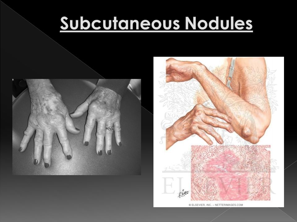
19
EXTRAARTICULAR COMPLICATIONS OF RA Source: Harrison’s Principles of Internal Medicine, 17 th edition Occur in those with high titers of autoantibodies to the fc component of immunoglobulin G or with antibodies to CCP Rheumatoid nodules - Found on periarticular structures, extensor surfaces, or other areas subjected to mechanical pressure - Common locations: olecranon bursa, proximal ulna, achilles tendon, and occiput - Has a central zone of necrotic material including collagen fibrils, noncollagenous filaments and cellular debris

20
EXTRAARTICULAR COMPLICATIONS OF RA Source: Harrison’s Principles of Internal Medicine, 17 th edition Clinical weakness and skeletal muscle atrophy -May be evident within weeks of onset of RA - Most apparent in musculature approximating affected joint Type II fiber atrophy and muscle fiber necrosis with or without mononuclear cell infiltrate Cutaneous vasculitis - Crops of small brown spots in the nail beds, nail folds, and digital pulp

21
EXTRAARTICULAR COMPLICATIONS OF RA Source: Harrison’s Principles of Internal Medicine, 17 th edition Pleuropulmonary manifestations - More common in men - Found during autopsy (symptomatic disease during lifetime is infrequent) - Include: pleural disease, interstitial fibrosis, pleuropulmonary nodules, pneumonitis, arteritis Pericarditis - Pericardial fluid has a low glucose level and is frequently associated with the occurrence of pleural effusion - Increased incidence of congestive heart failure and death from CV disease
 - Include: pleural disease, interstitial fibrosis, pleuropulmonary nodules, pneumonitis, arteritis Pericarditis - Pericardial fluid has a low glucose level and is frequently associated with the occurrence of pleural effusion - Increased incidence of congestive heart failure and death from CV disease")
22
EXTRAARTICULAR COMPLICATIONS OF RA Source: Harrison’s Principles of Internal Medicine, 17 th edition Neurologic manifestations -RA tends to spare the CNS directly, although vasculitis can cause peripheral neuropathy - May result from atlantoaxial or midcervical spine subluxation - Nerve entrapment secondary to proliferative synovitis or joint deformities may produce neuropathies of median, ulnar, radial (interosseous branch) or anterior tibial nerves
 or anterior tibial nerves")
23
EXTRAARTICULAR COMPLICATIONS OF RA Source: Harrison’s Principles of Internal Medicine, 17 th edition Eye manifestations - Episcleritis Usually mild and transient - Scleritis Involves deeper layers of the eye Histologically, the lesion is similar to a rheumatoid nodule and may result in thinning and perforation of the globe (scleromalacia perforans)
")
24
EXTRAARTICULAR COMPLICATIONS OF RA Source: Harrison’s Principles of Internal Medicine, 17 th edition Felty’s syndrome - Consists of: - Chronic RA - Splenomegaly - Neutropenia - Anemia - Patients have high titers of rheumatoid factor, subcutaneous nodules, and other manifestations of systemic rheumatoid disease - May develop after joint inflammation has regressed

25
EXTRAARTICULAR COMPLICATIONS OF RA Source: Harrison’s Principles of Internal Medicine, 17 th edition Osteoporosis - May be aggravated by glucocorticoid therapy may cause significant loss of bone mass, especially early in the course of therapy -Osteopenia in RA involves both juxtaarticular bone and long bones distant from involved joints - Modest decrease in mean bone mass and moderate increase in the risk of fracture

Similar presentations

 CBC with differential.>")


 Gergely Péter dr RHEUMATOID ARTHRITIS (RA) Gergely Péter dr Definition: Chronic destructive diseases characterized by joint inflammation.>")


 M:F 1:3 Age range:>")


>")


