Download presentation
Presentation is loading. Please wait.

1
Copyright © 2004 Pearson Education, Inc., publishing as Benjamin Cummings Fundamentals of Anatomy & Physiology Frederic H. Martini Lecture 5:Chapter 5 The Integumentary System Pages: 153 - 178 Lecturer: Dr. Barjis Room P313 Phone: (718)2605285 E-Mail: ibarjis@citytech.cuny.edu
")
2
Learning Objectives List the components of the integumentary system, including their physical relationships. Specify the functions of the integumentary system. Describe the main features and functions of the epidermis and dermis. Discuss individual and racial differences in skin. Discuss the effects of UV light on the epidermis. Explain the structure and function of the various accessory organs of the skin. Explain how the skin responds to injury and aging.

3
Cutaneous membrane Epidermis Dermis Accessory structures Subcutaneous layer The Integumentary System: An Overview The integumentary system consists of

4
Protection Excretion Temperature maintenance Nutrient storage Vitamin D3 synthesis Sensory detection Integumentary system functions:

5
The Components of the Integumentary System

6
The epidermis is composed of layers of keratinocytes Thin skin = four layers (strata) Thick skin = five layers The Epidermis Thin Skin and Thick Skin
 Thick skin = five layers The Epidermis Thin Skin and Thick Skin")
7
Provides mechanical protection Prevents fluid loss Keeps microorganisms from invading the body The epidermis

8
Stratum germinativum Stratum spinosum Stratum granulosum Stratum lucidum Stratum corneum Layers of the epidermis:

9
The Epidermal Ridges of Thick Skin

10
Cells accumulate keratin and eventually are shed Epidermal ridges are interlocked with dermal papillae Fingerprints Improve gripping ability Langerhans cells (immunity) in s. spinosum Merkel cells (sensitivity) in s. germinativum Epidermal characteristics:
 in s. germinativum Epidermal characteristics:.")
11
The Structure of the Epidermis

12
Blood supply Carotene and melanin Melanocytes produce melanin and protect from UV radiation Epidermal pigmentation Interrupted blood supply leads to cyanosis Skin color depends on

13
Melanocytes

14
Synthesize vitamin D3 (cholecalciferol) when exposed to UV Respond to epidermal growth factor Growth Division Repair Secretion Epidermal cells
 when exposed to UV Respond to epidermal growth factor Growth Division Repair Secretion Epidermal cells")
15
Papillary layer Contains blood vessels, lymphatics, sensory nerves of epidermis Reticular layer Contains network of collagen and elastic fibers to resist tension The Dermis Dermal Organization

16
Dermal Circulation

17
Caused by excessive stretching of the dermis Patterns of collagen and elastic fibers form lines of cleavage Stretch marks

18
Lines of Cleavage of the Skin

19
Cutaneous plexus arteries found in subcutaneous layer/ papillary dermis Cutaneous sensory receptors (light touch, pressure) Dermal Circulation and innervation
 Dermal Circulation and innervation")
20
Stabilizes skins position against underlying organs and tissues The Subcutaneous Layer Hypodermis

21
Originate in hair follicle Composed of root and shaft Root base (hair papilla) surrounded by hair bulb and root hair plexus Hairs have soft medulla and hard cortex Cuticle = superficial dead protective layer Accessory Structures Hairs
 surrounded by hair bulb and root hair plexus Hairs have soft medulla and hard cortex Cuticle = superficial dead protective layer Accessory Structures Hairs")
22
the Anatomy of a Single Hair

23
Vellus hairs (peach fuzz) Terminal hairs ( heavy) Club hair (cessation of growth) Shed and grow according to hair growth cycle Arrector pili muscle attaches to hair Hair types
 Terminal hairs ( heavy) Club hair (cessation of growth) Shed and grow according to hair growth cycle Arrector pili muscle attaches to hair Hair types")
24
Hair Follicles

25
Sebaceous Suderiferous Mammary Ceruminous Glands in the skin

26
Discharge waxy sebum onto hair shaft when associated with hairs Sebaceous follicles discharge onto epidermal surface Sebaceous glands

27
Sebaceous Glands and Follicles

28
Apocrine sweat glands Produce odorous secretion Merocrine (eccrine) sweat gland Sensible perspiration Suderiferous glands
 sweat gland Sensible perspiration Suderiferous glands")
29
Sweat Glands

30
Mammary glands Structurally similar to apocrine sweat glands Ceruminous glands In ear, produce waxy cerumen Other glands

31
Nail body covers the nail bed Nail production occurs at the nail root Eponychium (cuticle) overlies root Free edge of nail extends over hyponychium Nails
 overlies root Free edge of nail extends over hyponychium Nails")
32
The Structure of a Nail

33
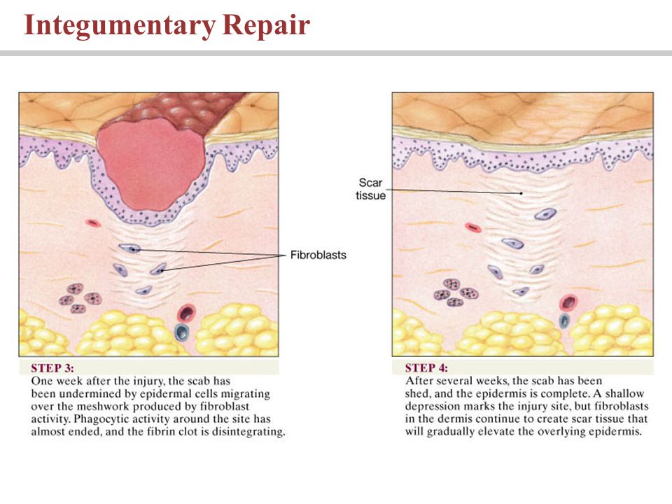
Regenerates easily Regeneration process includes formation of Scab Granulation tissue Scar tissue Local Control of Integumentary Function Injury and repair

34
Integumentary Repair

36
Integument thins Blood flow decreases Cellular activity decreases Repairs occur more slowly Aging and the Integumentary System With age

37
The components of the integumentary system, including their physical relationships. The functions of the integumentary system. The main features and functions of the epidermis and dermis. Individual and racial differences in skin. The effects of UV light on the epidermis. The structure and function of the various accessory organs of the skin. How the skin responds to aging. You should now be familiar with:

Similar presentations