Download presentation
Presentation is loading. Please wait.

1
Central Nervous System Dr. Mohammad Alzoghiabi

2
Organization of the Nervous System Central nervous system 1.Brain 2.Spinal cord Peripheral nervous system 1.Sensory receptors 2.Sensory nerves 3.Ganglia

4
Central Nervous System Functions: Has information about environment Organizes reflexes Plans & execute voluntary movement Memories, thinking & learning

5
Central Nervous System Types of nerves 1.Sensory or afferent division 2.Motor or efferent division

6
Spinal cord TTTThe most caudal portion of CNS FFFFrom the base of the skull to 1st lumbar vertebra CCCContain 31 pairs of spinal nerves

7
Spinal cord and nerves SSSSensory verves: carry information to spinal cord from different organs via dorsal root and cranial nerve ganglia MMMMotor nerves: carry information from spinal cord to periphery, include both: Somatic Autonomic

8
Brain Stem Medulla breathing, blood pressure, swallowing, coughing and vomiting reflexes Pons participates with medulla in regulating breathing gets information from cerebral hemispheres to cerebellum Midbrain Controls eye movements contains neucli of auditory and visual system

9
Cerebellum Functions: i.Coordination of movements ii.Planning and execution of movements iii.Maintenance of posture iv.Coordination of head and eye movements

10
Thalamus and Hypothalamus Thalamus processes all sensory and motor informations Hypothalamus contains centers to regulate 1. Body temp 2. Water balance 3. Pituitary gland 4. ADH and oxytocin Known as Diencephalon “between brain”

11
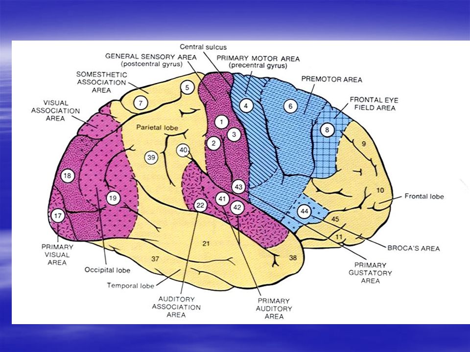
Motor Cortex AAAAnterior to the central cortical sulcus divided into: 1- primary motor cortex 2- premotor area 3- supplementary motor area

12
Primary Motor Area (MI) Right front of the central sulcus Laterally in the sylvian fissure Superiorly to the top of the brain Inferiorly to the longitudinal fissure Topographical representations of the body areas in an inverted manner
 Right front of the central sulcus Laterally in the sylvian fissure Superiorly to the top of the brain Inferiorly to the longitudinal fissure Topographical representations of the body areas in an inverted manner")
16
Primary Motor Area (MI) Main origin of the pyramidal tract More than half of motor area concerned with the hands and muscle of speech Excitation of a single motor neuron causes the excitation of a specific pattern of movement but not a single muscle Contains upper motorneurons which project directly to spinal cord Lesion in MI leads to flaccid paralysis
 Main origin of the pyramidal tract More than half of motor area concerned with the hands and muscle of speech Excitation of a single motor neuron causes the excitation of a specific pattern of movement but not a single muscle Contains upper motorneurons which project directly to spinal cord Lesion in MI leads to flaccid paralysis")
17
Premotor Area Anterior to the primary motor cortex Superiorly to the longitudinal fissure Inferiorly to the sylvian fissure Same topographical organization as the motor cortex Responsible for more complex patterns of movement

18
Supplementary Motor Area Has another topographic organization Contraction often bilateral - bilateral grasping movements (climbing) - attitudinal movements - fixation movements - positional movements of head and eyes
 - attitudinal movements - fixation movements - positional movements of head and eyes")
19
Other Motor Regions Bronca’s Area: responsible for vocalization Voluntary eye movement field: controls the eye movement to certain objects and blinking Head rotation area Area of hand skills destruction motor apraxia

20
White Matter of Spinal Cord Posterior faniculus Anterior faniculus Lateral faniculus Tract: Tract: Composed of nerve fibers sharing a common origin, destination and functions

21
Posterior funiculus Contains one ascending fibers concerned with two modalities - kinesthesia - discriminative touch Lesion of this area causes loss of: - vibration sense - position - two point discrimination - fine touch (tested by piece of cotton) - weight perception
 - weight perception")
23
Lateral & Anterior funiculi Ascending Tracts: Dorsal spinocerebral tract Ventral spinocerebellar tract Spinocervical thalamic tract Lateral spionothalamic tract Anterior spinothalamic tract

24
Lateral & Anterior funiculi Desending Tracts Corticospinal tract Rubrospinal tract Lateral vestibulospinal tract Medial vestibularspinal tract Reticulospinal tract Descending autonomic pathway

25
Ascending Tracts Dorsal spinocerebellar tract: conveys impulses from muscle spindle, golgi organ via (DRG) to cerebellum Ventral spinocerebellar tract: conveys impulses to cerebellum from golgi organ
 to cerebellum Ventral spinocerebellar tract: conveys impulses to cerebellum from golgi organ")
26
Ascending Tracts Spinocervical thalamic tract: conveys impulses to thalamus; carries the kinesthesia & discriminative touch Laeral spinothalamic tract: transmits pain & temperature sensations to thalamus Anterior spinothalamic tract: transmits light touch sensations to brain stem & thalamus

28
Descending Tracts 1. Corticospinal tract (pyramidal) From motor cortex to medulla as follow: 30% from the motor cortex to the muscles 30% from the premotor and supplementary cortex to the muscles 40% from somatosensory areas divided into: 1.Crossed (lateral corticospinal tract) 2.Uncrossed fibers (anterior corticospinal tract)
 From motor cortex to medulla as follow: 30% from the motor cortex to the muscles 30% from the premotor and supplementary cortex to the muscles 40% from somatosensory areas divided into: 1.Crossed (lateral corticospinal tract) 2.Uncrossed fibers (anterior corticospinal tract).")
29
Corticospinal Pathway (pyramidal) Corticospinal Pathway (pyramidal) From the cortex internal capsule thru brain stem via medulla where majority of the fibers cross in the lower medulla lateral coticospinal tract of the cord interneurons (few in dorsal horn and very few in anterior motor neurons) N.B. also a few of fibers do not cross but pass ipsilaterally in ventral corticospinal tract

32
Corticospinal tract (pyramidal) Corticospinal tract (pyramidal) 3% of pyramidal fibers with 16 m myelinated Originate from betz cells in the motor area Impulses conveyed are facilitatory to flexor motor neurons Terminates in internurons laminae IV-VII Lesion: Homolateral paralysis Contralateral paralysis In addition to: spasticity, hyperactive myotatic reflexes and clonus
 Corticospinal tract (pyramidal) 3% of pyramidal fibers with 16 m myelinated Originate from betz cells in the motor area Impulses conveyed are facilitatory to flexor motor neurons Terminates in internurons laminae IV-VII Lesion: Homolateral paralysis Contralateral paralysis In addition to: spasticity, hyperactive myotatic reflexes and clonus")
33
Descending Tracts 2. Rubrospinal tract Neuronal origin is in red nucleus (midbrain) Terminates similarly as pyramidal tract Indirect corticospinal tract Facilitates flexor motor neurons 3. Lateral vestibulospinal tract Neuronal origin lie in pons (lateral vestibular nucleus) Terminates at laminae VII & VIII Facilitates extensor motor neurons
 Terminates similarly as pyramidal tract Indirect corticospinal tract Facilitates flexor motor neurons 3. Lateral vestibulospinal tract Neuronal origin lie in pons (lateral vestibular nucleus) Terminates at laminae VII & VIII Facilitates extensor motor neurons.")
35
Descending Tracts 4. Medial vestibularspinal tract: From media vestibular nucleus in pons Terminates as latera tract Facilitates flexor motor neurons 5. Reticulospinal tract: a. pontine reticulospinal tract from pons terminates at laminae VII & VIII facilitates extensor motor neurons b. medullary reticulospinal tract terminates at VII & IX facilitates flexor motor neurons

36
Descending Tracts 6. Descending autonomic pathway: Neuronal origin at hypothalamus Small caliber fibers Project into intermediolateral of column

Similar presentations


>")
.>")


