Download presentation
Presentation is loading. Please wait.

1
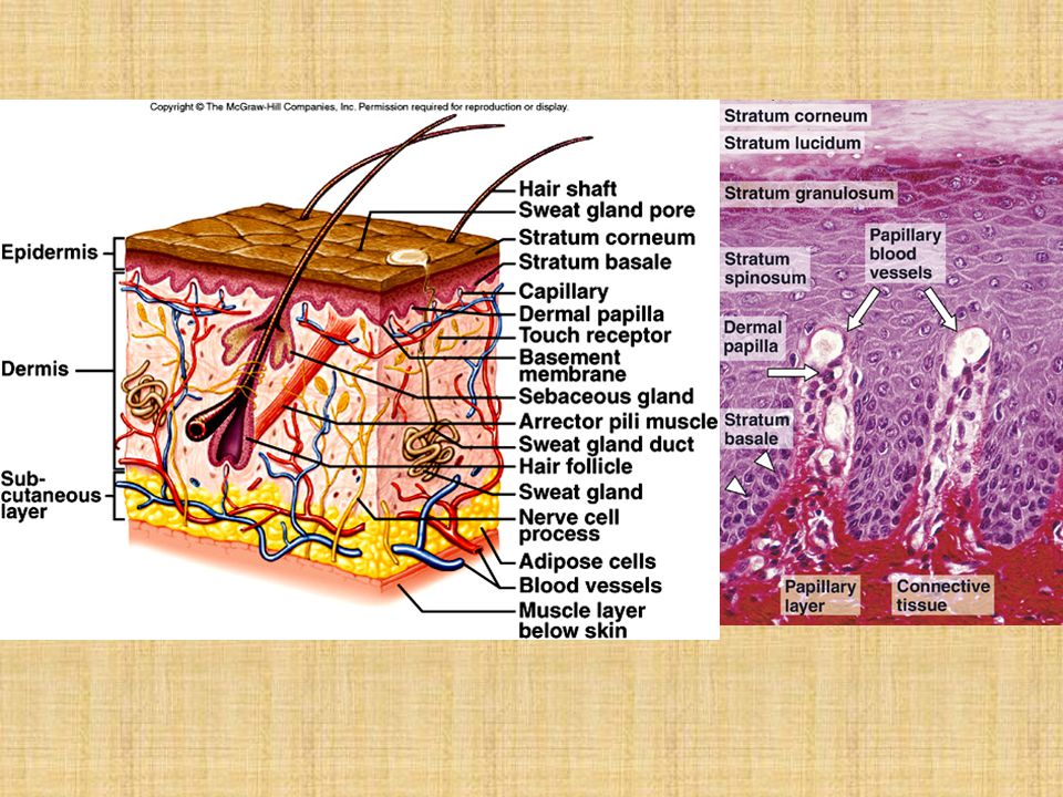
The skin Overview-The Skin 16% of the total body weight.
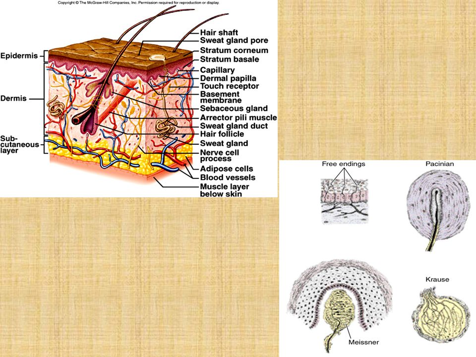
It is composed of the epidermis and dermis. A deeper superficial fascia layer, the hypodermis, lies under the skin. The skin contains several appendages (sweat glands, hair follicles, sebaceous glands, and nails). The skin and its appendages are called the integument. Function. The skin protects the body against injury, infection; regulates body temperature; absorbs ultraviolet (UV) radiation, which is necessary for synthesis of vitamin D; and contains receptors for touch, temperature, and pain stimuli from the external environment.
. The skin and its appendages are called the integument. Function. The skin protects the body against injury, infection; regulates body temperature; absorbs ultraviolet (UV) radiation, which is necessary for synthesis of vitamin D; and contains receptors for touch, temperature, and pain stimuli from the external environment.")
3
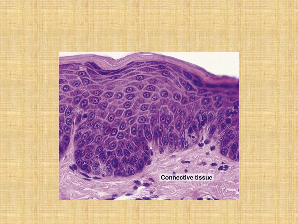
Epidermis superficial layer of the skin ectodermal origin
stratified squamous keratinized epithelium composed predominantly of keratinocytes and three different types of nonkeratinocytes: melanocytes, Langerhans cells, and Merkel cells. constantly being regenerated every days, is carried out by the mitotic activity of keratinocytes It has deep invaginations (interpapillary ridges) that interdigitate with projections of the dermis (dermal papillae) resulting in a highly irregular interface. On the fingertips, these surface ridges are visible as fingerprints, whose configuration is genetically determined and thus unique to each individual.
 that interdigitate with projections of the dermis (dermal papillae) resulting in a highly irregular interface. On the fingertips, these surface ridges are visible as fingerprints, whose configuration is genetically determined and thus unique to each individual.")
5
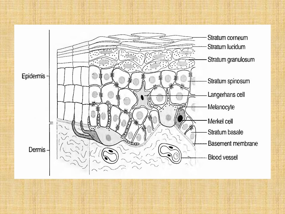
Layers of the epidermis
The stratum basale (germinativum) It is the deepest layer of the epidermis It is composed of keratinocytes that are cuboidal to columnar in shape. These mitotically active cells are attached directly to the basal lamina by hemidesmosomes and to each other by desmosomes. This layer also contains melanocytes and Merkel cells.
 It is the deepest layer of the epidermis. It is composed of keratinocytes that are cuboidal to columnar in shape. These mitotically active cells are attached directly to the basal lamina by hemidesmosomes and to each other by desmosomes. This layer also contains melanocytes and Merkel cells.")
6
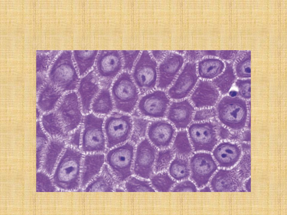
The stratum spinosum consists of a few layers of polyhedral keratinocytes (prickle cells). Their extensions, termed "intercellular bridges" by early histologists, are now known to terminate in desmosomes This layer also contains Langerhans cells. Keratinocytes in the deeper aspects of the stratum spinosum are also mitotically active. The malpighian layer consists of the stratum spinosum and stratum basale. (mitotic activity) In the superficial regions of the stratum spinosum are keratinocytes that contain membrane-coating granules. The contents of these granules are released into the intercellular spaces in the form of lipid-containing sheets that are impermeable to water and many foreign substances.
 In the superficial regions of the stratum spinosum are keratinocytes that contain membrane-coating granules. The contents of these granules are released into the intercellular spaces in the form of lipid-containing sheets that are impermeable to water and many foreign substances.")
8
The stratum granulosum
It is the most superficial layer in which nuclei are still present. It comprises three to five layers of flattened keratinocytes that contain keratohyalin granules, bundles of keratin filaments (tonofilaments), and occasional membrane-coating granules. Keratohyalin granules (not membrane-bound) contain histidine and cystine-rich proteins, which bind the keratin filaments together.
, and occasional membrane-coating granules. Keratohyalin granules (not membrane-bound) contain histidine and cystine-rich proteins, which bind the keratin filaments together.")
9
The stratum lucidum It is a clear, homogeneous layer just superficial to the stratum granulosum It is often difficult to distinguish in histologic sections. It is found only in palmar and plantar skin. This layer consists of keratinocytes that have neither nuclei nor organelles but contain keratin filaments and eleidin, a transformation product of keratohyalin.

10
The stratum corneum It is the most superficial layer of the epidermis.
It may consist of as many as 15 to 20 layers of flattened, nonnucleated, dead "cells" filled with keratin. These nonviable, scale-like structures are called squames (or horny cells) The outermost layer of squames is continuously shed by desquamation.
 The outermost layer of squames is continuously shed by desquamation.")
12
Nonkeratinocytes in the epidermis
Melanocytes are present in the stratum basale and originate from the neural crest. These cells synthesize a dark brown pigment (melanin) in oval-shaped organelles (melanosomes). Melanosomes contain tyrosinase, a UV-sensitive enzyme directly involved in melanin synthesis. Melanosome content, size, rate of transfer, and aggregation pattern in keratinocytes vary with race. Melanin protects against tissue damage caused by UV radiation. Long, melanosome-containing processes extend between the cells of the stratum spinosum. Melanin is transferred, via a unique mechanism known as cytocrine secretion, from these melanosome filled tips into keratinocytes of the stratum spinosum.
 in oval-shaped organelles (melanosomes). Melanosomes contain tyrosinase, a UV-sensitive enzyme directly involved in melanin synthesis. Melanosome content, size, rate of transfer, and aggregation pattern in keratinocytes vary with race. Melanin protects against tissue damage caused by UV radiation. Long, melanosome-containing processes extend between the cells of the stratum spinosum. Melanin is transferred, via a unique mechanism known as cytocrine secretion, from these melanosome filled tips into keratinocytes of the stratum spinosum.")
14
Langerhans cells They are dendritic cells (so named because of their long processes) originate in the bone marrow. They are located primarily in the stratum spinosum, contain characteristic paddle-shaped Birbeck granules function as antigen-presenting cells in immune responses to contact antigens (contact allergies)
")
15
Merkel cells They are present in small numbers in the stratum basale, near areas of well-vascularized, richly innervated connective tissue. They possess desmosomes and keratin filaments, suggesting an epithelial origin. Their pale cytoplasm contains small, dense-cored granules that are similar in appearance to those in some cells of the diffuse neuroendocrine system (DNES). They receive afferent nerve terminals and are believed to function as sensory mechanoreceptors.
. They receive afferent nerve terminals and are believed to function as sensory mechanoreceptors.")
16
Thick and thin skin They are distinguished on the basis of the thickness of the epidermis Thick skin It has an epidermis that is micrometers thick. It is characterized by a prominent stratum corneum, a well-developed stratum granulosum, and a distinct stratum lucidum. It lines the palms of the hands and the soles of the feet. Thick skin lacks hair follicles, sebaceous glands, and arrector pili muscles.

17
Thin skin It has an epidermis that is 75-150 um thick
It has a less prominent stratum corneum than thick skin and generally lacks a stratum granulosum and stratum lucidum Thin skin is present over most of the body and contains hair follicles sebaceous glands, and arrector pili muscles

18
Dermis The dermis is the layer of the skin underlying the epidermis.
It is of mesodermal origin It is composed of dense irregular connective tissue that contains many type I collagen fibers and networks of thick elastic fibers. It is divided into a superficial papillary layer and a deeper, more extensive reticular layer No distinct boundary exists between these layers

19
The dermal papillary layer
It is uneven and forms dermal papillae, which interdigitate with the epidermal downgrowths (interpapillary ridges) It is composed of thin, loosely arranged fibers and cells, and contains capillary loops and Meissner corpuscles, which are fine-touch receptors. The dermal reticular layer It constitutes the major portion ofthe dermis. It is composed of dense bundles of collagen fibers and thick elastic fibers It contains pacinian corpuscles (pressure receptors) and Krause end bulbs (cold receptors) in its deeper aspects.
 It is composed of thin, loosely arranged fibers and cells, and contains capillary loops and Meissner corpuscles, which are fine-touch receptors. The dermal reticular layer. It constitutes the major portion ofthe dermis. It is composed of dense bundles of collagen fibers and thick elastic fibers. It contains pacinian corpuscles (pressure receptors) and Krause end bulbs (cold receptors) in its deeper aspects.")
21
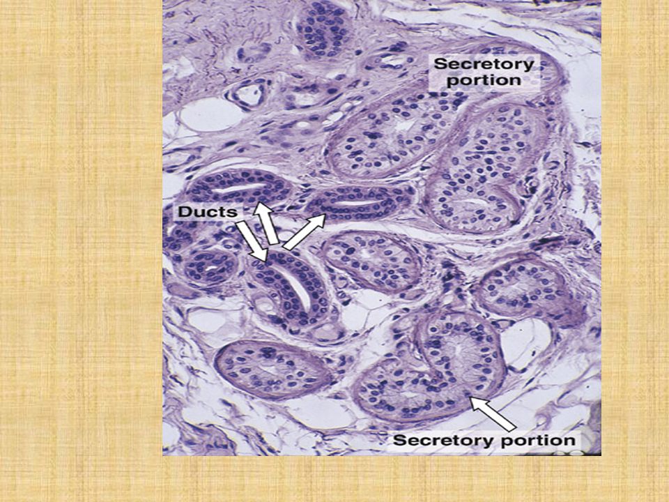
Glands in the Skin Sweat glands
They are simple coiled tubular glands consisting of a secretory unit and a single duct. These glands are present in skin throughout the body. The secretory unit of eccrine sweat glands is embedded in the dermis and is composed of three cell types. Dark cells Clear cells Myoepithelial cells The duct of eccrine sweat glands is narrow and lined by stratified cuboidal epithelial cells, which contain many keratin filaments and have a prominent terminal web. As the secreted material passes through the duct, its cells reabsorb some electrolytes and excrete other substances (such as urea, lactic acid, ions, and certain drugs).
.")
23
Apocrine sweat glands They include the large, specialized sweat glands located in various areas of the body (e.g., axilla, areola of the nipple, perianal region) and the ceruminous (wax) glands of the external auditory canal. These glands do not begin to function until puberty and are responsive to hormonal influences. Their large coiled secretory units are enveloped by scattered myoepithelial cells. These glands empty their viscous secretions into hair follicles at a location superficial to the entry of sebaceous gland ducts.
 and the ceruminous (wax) glands of the external auditory canal. These glands do not begin to function until puberty and are responsive to hormonal influences. Their large coiled secretory units are enveloped by scattered myoepithelial cells. These glands empty their viscous secretions into hair follicles at a location superficial to the entry of sebaceous gland ducts.")
24
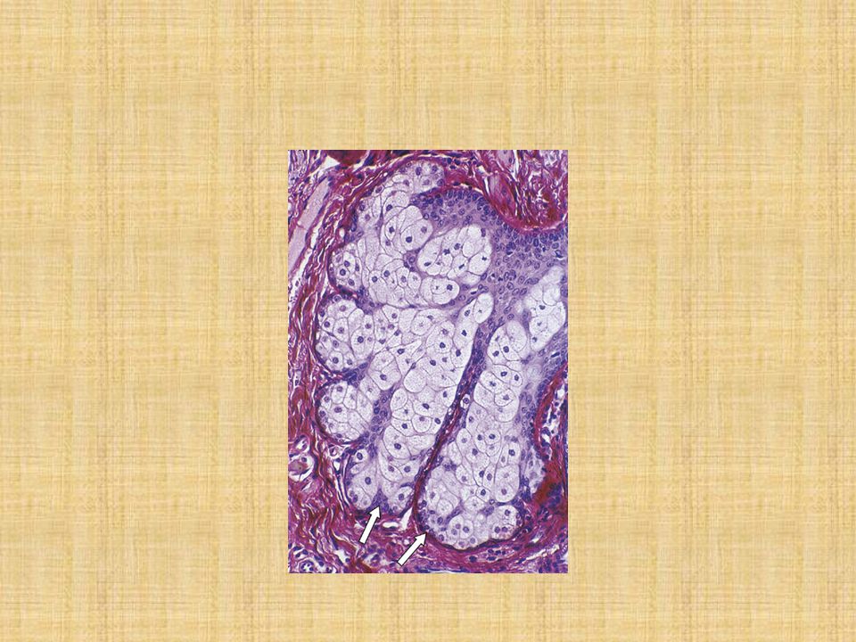
Sebaceous glands They are branched acinar glands having a lobular appearance. The clustered acini of one sebaceous gland empty into a single short duct. The duct empties into the neck of a hair follicle. Sebaceous glands are embedded in the dermis over most of the body's surface but are absent from the palms and soles. They are most abundant on the face, forehead, and scalp. These holocrine glands release sebum (composed of an oily secretion and degenerating epithelial cells).
.")
26
Hair Follicle and Arrector Pili Muscle
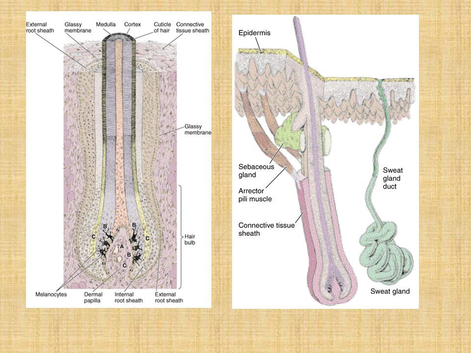
A hair follicle is an invagination of the epidermis extending deep into the dermis. The hair shaft is a long, slender filament that is located in the center of the follicle and extends above the surface of the epidermis. It consists of an inner medulla, cortex, and outer cuticle of the hair. At its deep end it is continuous with the hair root. The hair bulb is the terminal expanded region of the hair follicle in which the hair is rooted. It is deeply indented by a dermal papilla which contains a capillary network necessary for sustaining the follicle. The hair bulb contains cells that form the internal root sheath and medulla of the hair shaft. The internal root sheath lies deep to the entrance of the sebaceous gland.

27
The external root sheath is a direct continuation of the stratum malpighii of the epidermis.
The glassy membrane is a noncellular layer and represents a thickening of the basement membrane, separates the hair follicle from the surrounding dermal sheath. The arrector pili muscle attaches at an oblique angle to the dermal sheath surrounding a hair follicle. It extends superficially to underlie sebaceous glands, passing through the reticular layer of the dermis and inserting into the papillary layer of the dermis. The contraction of this smooth muscle elevates the hair and is responsible for formation of "goose bumps," caused by depressions of the skin where the muscle attaches to the papillary layer of the dermis.

29
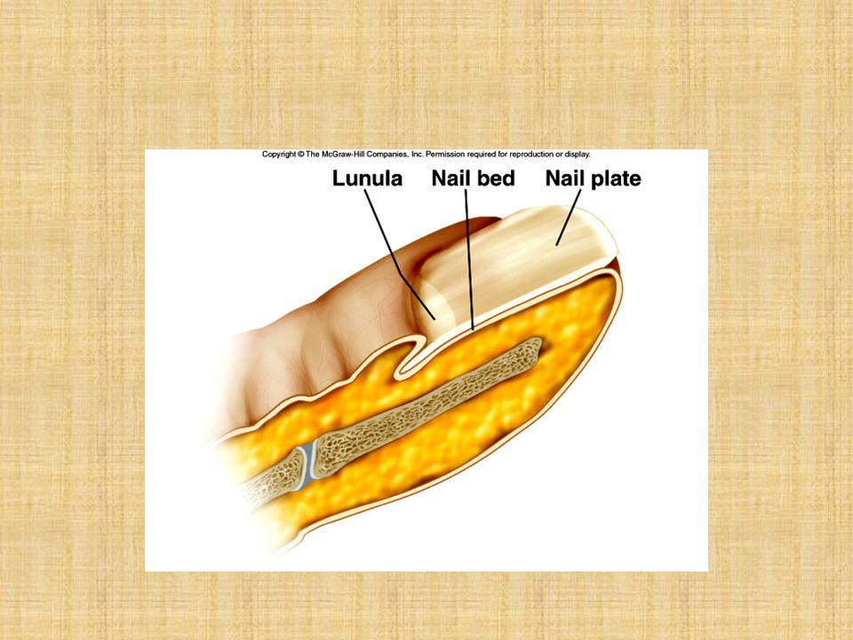
Nails Nails are located on the distal phalanx of each finger and toe.
Nails are hard keratinized plates that rest on a bed of epidermis. At the proximal end, each is covered by a fold of epidermis, called the cuticle or eponychium, which corresponds to the stratum corneum. The cuticle overlies the crescent shaped, whitish lunula. Nails grow as the result of mitoses of cells in the matrix of the nail root.

31
Clinical consideration
Tumors basal cell (stratum basale) squamous cell melanoma
 squamous cell. melanoma.")
Similar presentations


 Roles: protection maintenance of normal body.>")


>")


