Download presentation
Presentation is loading. Please wait.

1
Dr Mahmood Fauzi ASSIST PROF OPHTHALMOLOGY AL MAAREFA COLLEGE
Leocokoria Dr Mahmood Fauzi ASSIST PROF OPHTHALMOLOGY AL MAAREFA COLLEGE

2
Objectives Define leucokoria Explain presenting sign of leucokoria
Enlist differential diagnoses of leucokoria Summarize clinical presentation ,diagnoses and management of some common causes of leucokoria- Retinoblastoma,retinopathy of prematurity ,congenital cataracts,coats disease ,toxoplasmoses,retinal detachment dysplasia, coloboma. Outline diagnostic Workup in a child with leucokoria

3
Leukocoria The term leukocoria means "white pupil" and is the name given to the clinical finding of a white pupillary reflex. While occasionally the reflection of a normal optic disc, leukocoria can also be caused by abnormalities in the lens, vitreous, or retina. It can be the initial manifestation of a wide spectrum of intraocular and systemic disease processes, and the presenting sign of abusive head trauma.

4
Causes of Leukocoria DIFFERENTIAL DIAGNOSIS OF LEUKOCORIA Cataract
Retinoblastoma Retinal detachment ROP PHPV Coat´s disease Toxocariasis Coloboma Retinal dysplasia Norrie´s disease

5
Retinoblastoma Retinoblastoma is the most common and rapidly developing intraocular tumor of childhood, accounting for 1% of childhood cancer deaths and 5% of blindness in children. Develops in the cells of the retina in childhood(1-4) year. Approximately 1in 20,000 birth The disease is bilateral in approximately 30% of cases. Overall mortality from retinoblastoma is now decreased. With modern diagnostic and therapeutic advances, the mortality rate from metastatic or recurrent retinoblastoma has been as low as 5%.
 year. Approximately 1in 20,000 birth. The disease is bilateral in approximately 30% of cases. Overall mortality from retinoblastoma is now decreased. With modern diagnostic and therapeutic advances, the mortality rate from metastatic or recurrent retinoblastoma has been as low as 5%.")
6
RETINOBLASTOMA Leukocoria (60%) Strabismus (20%) OTHER - Uveitis,
CLINICAL MANIFESTATIONS Leukocoria (60%) Strabismus (20%) OTHER - Uveitis, Orbital cellulitis, Hyphaema, Heterochromia, Glaucoma, Bupthalmos
 Strabismus (20%) OTHER - Uveitis, Orbital cellulitis, Hyphaema, Heterochromia, Glaucoma, Bupthalmos.")
7
Presentations of Retinoblastoma
Leukocoria - 60% Strabismus - 20% Secondary glaucoma Anterior segment invasion Orbital inflammation Orbital invasion

8
Rb- GENETICS Less than 10% of retinoblastoma suffers have a family history of the disorder(Rb gene), 90% of cases are sporadic. Of the sporadic cases, the responsible mutation is in a germ cell in 25% of cases and in a somatic cell in 75% of cases

9
RETINOBLASTOMA

10
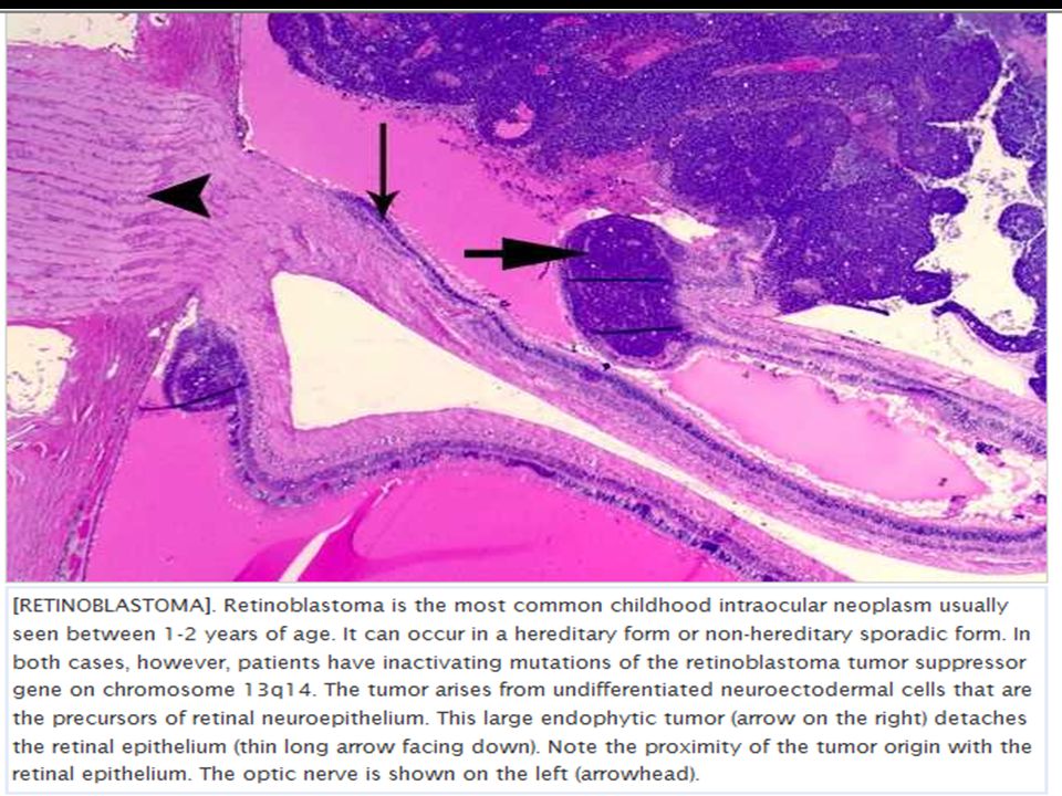
Advanced Endophytic Retinoblastoma
Friable white mass Cottage cheese appearance Fine surface blood vessels Vitreous seedings

11
Exophytic Retinoblastoma
Multilobulated white mass with overlying retinal detachment May be difficult to visualize through deep detachment

12
Diagnosis Eye exam. For a more thorough exam, anesthetics may be used to keep the child still. Imaging tests. Imaging tests include ultrasound, computerized tomography (CT) scan and magnetic resonance imaging (MRI), among others. Consulting with other doctors. oncologist, genetic counselor or a surgeon Biopsies are not usually done to diagnose retinoblastoma because It can be recognized with great accuracy just by examination A biopsy cannot be done easily without harming the eye Biopsy may risk spreading cancer cells
 scan and magnetic resonance imaging (MRI), among others. Consulting with other doctors. oncologist, genetic counselor or a surgeon. Biopsies are not usually done to diagnose retinoblastoma because. It can be recognized with great accuracy just by examination. A biopsy cannot be done easily without harming the eye. Biopsy may risk spreading cancer cells.")
13
Ultrasound

14
CT diagnosis of retinoblastoma
Calcification Optic nerve involvement Orbital and CNS extension Pinealoblastoma

15
Characteristic histo pathology
Arise in primitive photoreceptor cells. poorly differentiated neuroblastic cells with scanty cytoplasm and prominent basophilic nuclei. Necrotic tumour , Cuboidal tumour cells Characteristic Flexner-Wintersteiner rosettes represent an attempt at retinal differentiation. Calcification, usually occurring in necrotic areas. Calcium stains with H&E. It is worth identifying calcium in suspect eyes by ultrasound, or CT scan to differentiate retino-blastomas from other tumors.

17
Management-retinoblastoma
Genetic Counseling Treatment of small (3 mm diameter) tumours Photocoagulation Cryotherapy Chemotherapy Medium sized (upto 12 mm) tumours External beam radiation Large tumours Enucleation Extraocular extension Radiotherapy Metastatic Disease High dose chemotherapy Intra-thecal chemotherapy Total body radiotherapy
 tumours. Photocoagulation. Cryotherapy. Chemotherapy. Medium sized (upto 12 mm) tumours. External beam radiation. Large tumours. Enucleation. Extraocular extension. Radiotherapy. Metastatic Disease. High dose chemotherapy. Intra-thecal chemotherapy. Total body radiotherapy.")
18
Management of retinoblastoma
Advantages Disadvantages Photocoagulation (Laser Therapy) The laser beam focuses on the cancerous tumor, cuts off blood supply to the tumor and shrinks it. Depending on the size of the tumor, chemotherapy may be needed for larger tumors that cannot be shrunk just by laser. Cryotherapy (Freezing Treatment) The tumor is frozen and thawed several times by a cold gas and this causes the tumor to shrink. The tumor will leave a pigmented scar and the eye lid will swell for a couple of days. Chemotherapy After the extensive cycles of chemo, the cancer cells are reduced, thereby, shrinking of the tumor. There are several cycles, and there is an IV port necessary to draw blood, and inject the drugs. Enucleation This is removal of the eyeball and the tumor is extracted when no other option is possible due to the size of the tumor. The whole eyeball is removed with the attendant problems of anophthalmic socket.
 The laser beam focuses on the cancerous tumor, cuts off blood supply to the tumor and shrinks it. Depending on the size of the tumor, chemotherapy may be needed for larger tumors that cannot be shrunk just by laser. Cryotherapy (Freezing Treatment) The tumor is frozen and thawed several times by a cold gas and this causes the tumor to shrink. The tumor will leave a pigmented scar and the eye lid will swell for a couple of days. Chemotherapy. After the extensive cycles of chemo, the cancer cells are reduced, thereby, shrinking of the tumor. There are several cycles, and there is an IV port necessary to draw blood, and inject the drugs. Enucleation. This is removal of the eyeball and the tumor is extracted when no other option is possible due to the size of the tumor. The whole eyeball is removed with the attendant problems of anophthalmic socket.")
19
Follow-up Heritable Retinoblastoma patients can develop recurrences and need to be followed up regularly Examine the patients every 6 months till the age of 5 years and then annually till the age of 10 years.

20
Poor Prognostic Factors
Optic nerve involvement Choroidal invasion Large tumour Bilateral involvement Anterior location Poor cellular differentiation Older children

21
RETINOPATHY OF PREMATURITY (ROP)
Vasoproliferative retinopathy affecting premature infants exposed to high oxygen INCIDENCE Prematurity (<32/40) Birth weight (30% < 1000gm affected) Oxygen duration 90% ROP regresses spontaneously, 5% blindness Retinopathy of prematurity (ROP) is a serious vaso-proliferative disorder that affects extremely premature infants. Retinopathy of prematurity often regresses or heals but can lead to severe visual impairment or blindness. Significant retinopathy of prematurity can lead to lifelong disabilities for the smallest survivors of neonatal ICUs (NICUs). It remains a serious problem despite striking advances in neonatology.
 Birth weight (30% < 1000gm affected) Oxygen duration. 90% ROP regresses spontaneously, 5% blindness. Retinopathy of prematurity (ROP) is a serious vaso-proliferative disorder that affects extremely premature infants. Retinopathy of prematurity often regresses or heals but can lead to severe visual impairment or blindness. Significant retinopathy of prematurity can lead to lifelong disabilities for the smallest survivors of neonatal ICUs (NICUs). It remains a serious problem despite striking advances in neonatology.")
22
Patho-physiology-RETINOPATHY OF PREMATURITY (ROP)
Increased oxygen increases VGEF (vascular endothelial growth factor) which promotes angiogenesis. The proliferating vessels invade the vitreous, inciting fibrosis and contraction. In the later cicatricial stages of ROP, the retina is folded on itself, forming a fibroneural mass that drags the macula and optic disc temporally. The end stage of the disease is marked by total retinal detachment, leukocoria, blindness, and phthisis bulbi.
 which promotes angiogenesis. The proliferating vessels invade the vitreous, inciting fibrosis and contraction. In the later cicatricial stages of ROP, the retina is folded on itself, forming a fibroneural mass that drags the macula and optic disc temporally. The end stage of the disease is marked by total retinal detachment, leukocoria, blindness, and phthisis bulbi.")
23
RETINOPATHY OF PREMATURITY (ROP) Stages Ophthalmological Findings Distinct line between vascularized and avascularized region of retina The line noted in stage 1 gains both depth and hight Vessels extend beyond the retina into the viterious Partial retinal detachment Complete retinal detacchment
 Stages Ophthalmological Findings 1 Distinct line between vascularized and avascularized region of retina 2 The line noted in stage 1 gains both depth and hight 3 Vessels extend beyond the retina into the viterious 4 Partial retinal detachment 5 Complete retinal detacchment")
24
RETINOPATHY OF PREMATURITY (ROP)
LOCATION zone 1 - centred on disc, 2x disc to fovea distance zone 2 - outer limit equator temporally, ora nasally zone 3 - temporal peripheral crescent –

25
extraretinal fibrovascular proliferation
Regressing ROP Marked “plus” disease. retinal detachment. Stage 4 ROP extraretinal fibrovascular proliferation

26
Developmental Cataracts
Non-traumatic unilateral cataracts first detected after 6 months of age. A history of recent-onset strabismus or leukocoria, preservation of good alignment with central steady fixation (even on a light), family photographs documenting symmetrical red fundus reflexes, or paediatrician's records of red reflex observation can help to establish a good visual prognosis.
, family photographs documenting symmetrical red fundus reflexes, or paediatrician s records of red reflex observation can help to establish a good visual prognosis.")
27
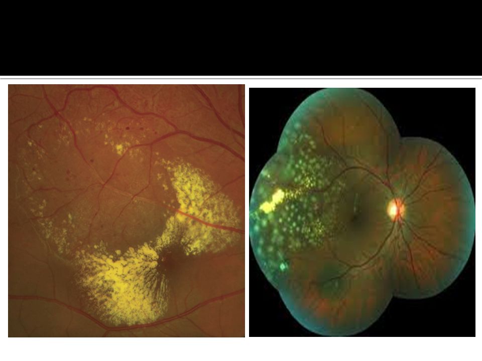
Congenital retinal telangiectasis (Coats' disease)
Idiopathic Retinal vascular disorder Usually affects young male patients unilaterally in their first or second decade of life. Up to 1/3rd of patients are >30 years at time of presentation. No defined familial inheritance. Presentation- patients may present with decreased vision, as well as strabismus or leukocoria in children. The hallmark feature of congenital retinal telangiectasis is localized fusiform aneurysmal dilations of the retinal vessels reminiscent of tiny light bulbs …….

30
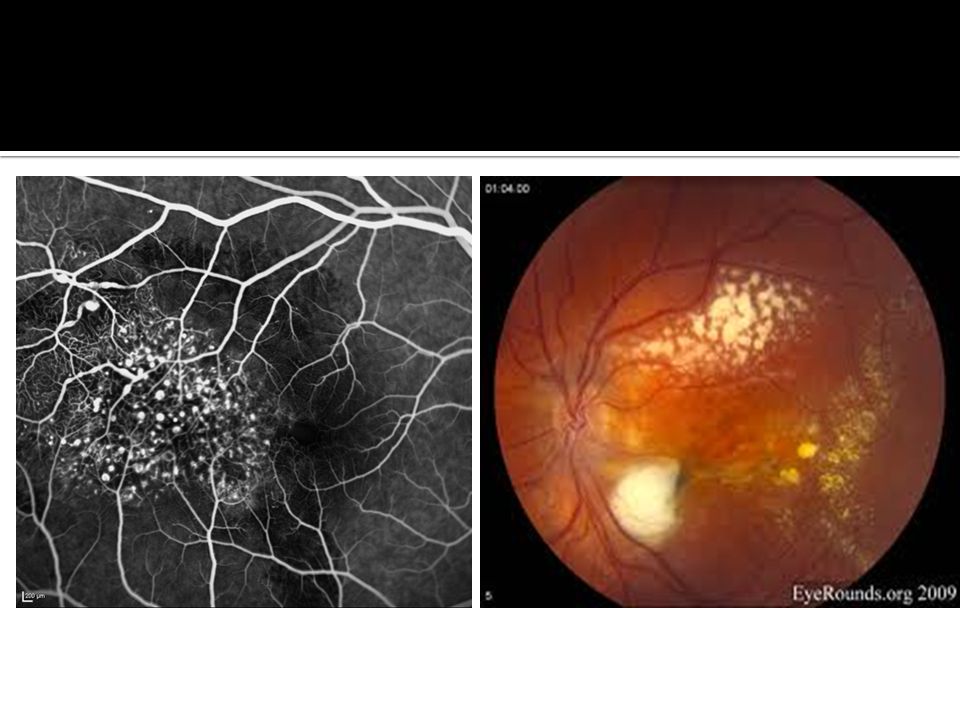
Pathogenesis and Treatment
Pathogenesis: Coats' disease result from breakdown of t blood-retinal barrier in the endothelial cell, resulting in leakage of blood products containing cholesterol crystals and lipid-laden macrophages into the retina and subretinal space. Accumulation of this proteinaceous exudate thickens the retina, leading to massive, exudative retinal detachment. Treatment: Laser surgery or cryotherapy(freezing) can be used to destroy the abnormal blood vessels, thus halting progression of the disease. if leaking blood vessels are clustered around the optic nerve, this treatment is not recommended as accidental damage to the nerve itself can result in permanent blindness.
 can be used to destroy the abnormal blood vessels, thus halting progression of the disease. if leaking blood vessels are clustered around the optic nerve, this treatment is not recommended as accidental damage to the nerve itself can result in permanent blindness.")
31
Persistent hyperplastic primary vitreous (PHPV)
Congenital anomaly in which the primary vitreous fails to regress in utero. Highly vascular mesenchymal tissue forms a mass behind the lens. A grey-yellow retro-lental membrane may produce leukocoria. The globe is white and slightly micro-ophthalmic. Patients have no history of prematurity or oxygen administration. PHPV are mostly unilateral and non-hereditary. When bilateral, PHPV may follow autosomalrecessive or autosomaldominant inheritance pattern.

32
Toxoplasmosis Toxoplasmosis gondi -protozoa causing up to 50% cases of posterior uveitis. Ocular infection is characterized by focal necrotizing retinochoroiditis with vitritis. In congenital infection (Highest transmission occurs in the IIIrd trimester) The most important signs in the diagnosis of congenital toxoplasmosis are the three Cs. (Convulsions, calcification (intracranial), Chorio retinitis) The eye may also be affected by cataract, microphthalmos, and optic atrophy
 The most important signs in the diagnosis of congenital toxoplasmosis are the three Cs. (Convulsions, calcification (intracranial), Chorio retinitis) The eye may also be affected by cataract, microphthalmos, and optic atrophy.")
33
Congenital Toxoplasmosis and Chorioretinitis
Chorioretinitis is present in 80% of children with congenital toxoplasmosis and is most often bilateral toxoplasmosis is considered one of the most common causes of chorioretinitis.

34
Investigation of Toxoplasmosis
ELISA IgM in neonates, rising IgG in adults (although not that helpful in adults). Fluorescein angiography (hypofluorescence in the early stages and then progressive leakage). Indocyanine angiography - multiple small dark spots seen around the visible lesions. This sign may be useful in assessing the effect of treatment.
. Fluorescein angiography (hypofluorescence in the early stages and then progressive leakage). Indocyanine angiography - multiple small dark spots seen around the visible lesions. This sign may be useful in assessing the effect of treatment.")
35
Indications For Active Treatment of Toxoplasmosis
Lesions that involve macula or optic disc Large, active lesions. Immuno compromised patients. Treatment with antiparasitic drugs is effective for active infections but not for the encysted form. The classic treatment includes triple drug therapy with pyrimethamine (0.5-1 mg/kg/d), sulfadiazine ( mg/kg/d), and prednisone. Concurrent folinic acid helps to minimize bone marrow toxicity produced by the pyrimethamine. Prevention of fetal infection in maternal Toxoplasma during pregnancy is spiramycin. A 60% decrease has been reported in the congenital infection rate in patients who received this treatment;
, sulfadiazine ( mg/kg/d), and prednisone. Concurrent folinic acid helps to minimize bone marrow toxicity produced by the pyrimethamine. Prevention of fetal infection in maternal Toxoplasma during pregnancy is spiramycin. A 60% decrease has been reported in the congenital infection rate in patients who received this treatment;")
36
Ocular toxocariasis infection nematode presents as strabismus,
leukocoria, severe posterior uveitis, subretinal granuloma present in the posterior pole decreased vision. Retinal damage as a result of inflammatory response

37
Retinal detachment in childhood
Retinal detachment in childhood can be confused with retinoblastoma, and vice versa. The possibility of an underlying retinoblastoma should always be considered when a child presents with retinal detachment and vitreous haemorrhage, even when a history of trauma is obtained.

38
Causes of Retinal Detachment in Children
■ Retinopathy of prematurity ■ Systemic diseases Incontinentia pigmenti Familial exudative vitreoretinopathy ■ Trauma ■ Toxocara ■ Coat’s disease

39
Norrie disease X-linked recessive heritable disorder characterized by bilateral leukocoria caused by retinal detachment. Affected boys classically have a triad of blindness, deafness, and mental retardation. usually progress to phthisis bulbi.

40
Retinal dysplasia Characteristic Ocular Findings in Trisomy 13
May present with bilateral Leukocoria. Rarely, retinal dysplasia occurs unilaterally in the congenitally malformed eyes of otherwise healthy persons.

41
COLOBOMA Greek word koloboma meaning mutilated or curtailed.
Occurs due to failure of closure of choroidal fissure Coloboma of optic disc: Coloboma of choroid and retina: Coloboma of macula: Associations CHARGE Syndrome,Trisomy 13 (Patau syndrome) Trisomy 18 (Edwards syndrome),Cat-eye syndrome Posteriorly located coloboma can involve the optic nerve, retina, and choroid. If the retina is involved, it appears as an area of whitening often with pigment deposition at the junction of the coloboma and normal retina. patients with coloboma have increased risk for retinal detachment.
 Trisomy 18 (Edwards syndrome),Cat-eye syndrome. Posteriorly located coloboma can involve the optic nerve, retina, and choroid. If the retina is involved, it appears as an area of whitening often with pigment deposition at the junction of the coloboma and normal retina. patients with coloboma have increased risk for retinal detachment.")
42
All children with newly discovered leukocoria should be referred promptly to an ophthalmologist to exclude retinoblastoma and other life- or sight-threatening conditions

43
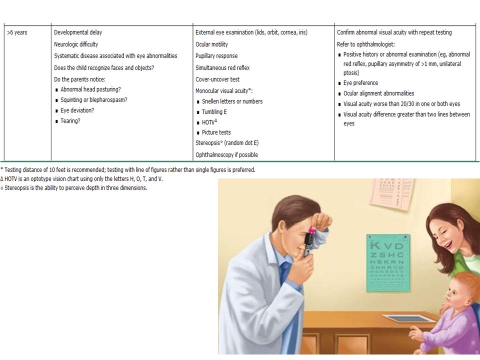
Eye examination for infants and children

45
Leucokoria-Key points
■The term leukocoria means "white pupil" or clinical finding of a white pupillary reflex. ■All children with newly discovered leukocoria should promptly be referred to an ophthalmologist to exclude retinoblastoma and other life- or sight-threatening conditions. ■leukocoria can also be caused by abnormalities in the lens, occasionally the reflection of a normal optic disc, vitreous, or retina. It can be the initial manifestation of a wide spectrum of intraocular and systemic disease processes, and the presenting sign of abusive head trauma. ■The history for the child with leukocoria should include prenatal exposures (toxins, infection, medications) and complications, birth history, postnatal course (infection, oxygen exposure, medications), medical history, recent exposures (puppies, kittens) family history (particularly for retinoblastoma or other eye tumors, eye loss, osteogenic sarcoma, and fetal loss or miscarriage), growth pattern, development, and review of systems. ■Ophthalmic ultrasound is sometimes used to determine the presence or absence of intraocular calcium (indicative of retinoblastoma). ■Laboratory evaluation of the child with leukocoria should be directed by the history and physical examination findings, may include serology or other testing for congenital infections, metabolic studies (eg. for galactosemia), and genetic studies for various syndromes (e.g., Turner syndrome).
 and complications, birth history, postnatal course (infection, oxygen exposure, medications), medical history, recent exposures (puppies, kittens) family history (particularly for retinoblastoma or other eye tumors, eye loss, osteogenic sarcoma, and fetal loss or miscarriage), growth pattern, development, and review of systems. ■Ophthalmic ultrasound is sometimes used to determine the presence or absence of intraocular calcium (indicative of retinoblastoma). ■Laboratory evaluation of the child with leukocoria should be directed by the history and physical examination findings, may include serology or other testing for congenital infections, metabolic studies (eg. for galactosemia), and genetic studies for various syndromes (e.g., Turner syndrome).")
46
Resources http://www.aapos.org/terms/conditions/67

Similar presentations


1 RETINOBLASTOMA. 2 RETINOBLASTOMA It is the most common primary ocular malignancy of childhood. It formed 15% of all childhood cancers.>")


 Waxman MD PhD>")


