Download presentation
Presentation is loading. Please wait.

1
ZLY 303 Phylum Porifera

2
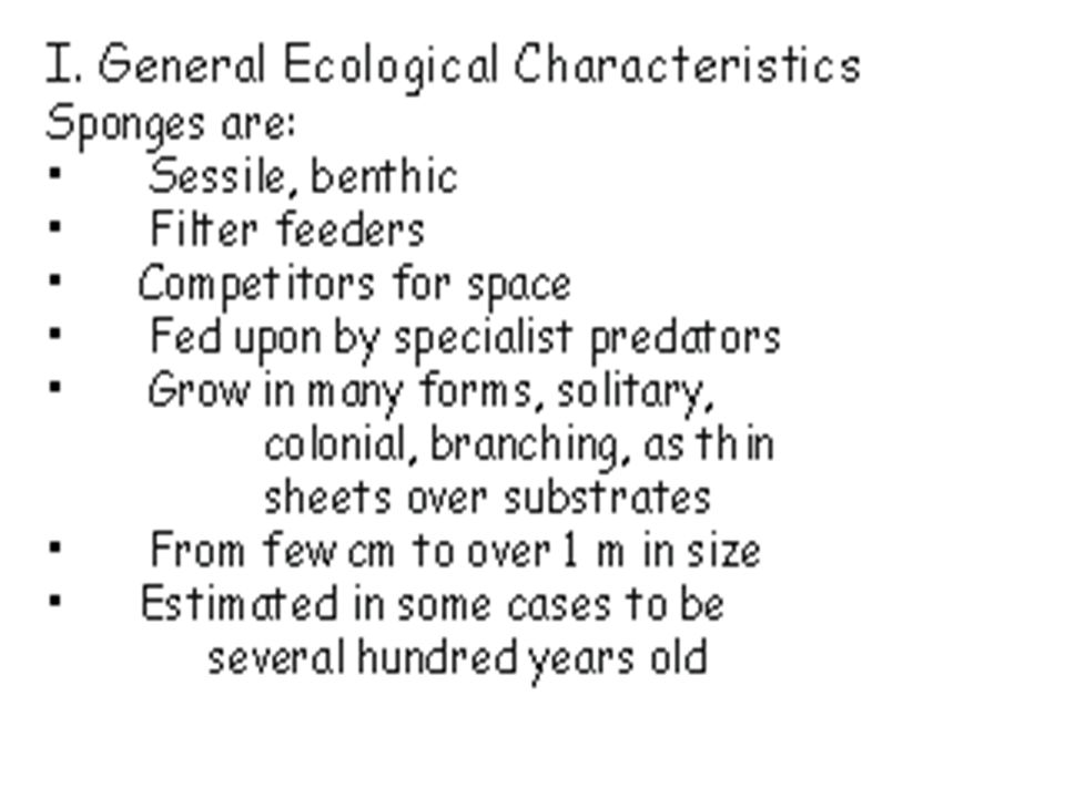
Phylum Porifera (Sponges)
spp. mostly marine some freshwater none terrestrial 3 classes, most important distinctions are skeletal Most primitive animal group

3
Distribution There are approximately 9,000 living species and above 2200 fossil forms. All are aquatic most are marine; found at all depths from intertidal to the abyssal zone, a few occur in freshwater (~150 spp.) most range from <1/2 inch to over 6 ft. tall (loggerhead sponges), some are round, flat, grow as crusts or vase-like, some are radially symmetrical; most are, Assymetrical, often brightly colored: yellows, reds, greens, oranges, lavenders
 most range from <1/2 inch to over 6 ft. tall (loggerhead sponges), some are round, flat, grow as crusts or vase-like, some are radially symmetrical; most are, Assymetrical, often brightly colored: yellows, reds, greens, oranges, lavenders.")
4
Simplicity They are closely related to the group of protozoan protists, called the choanoflagellates, The protozoan protists cells closely resemble the collar cells of sponges, Approximately, 1/4th of Sponges genes are shared by all other animals, All sponges are sessile, but multicellular their structure is unlike any other animal group.

6
Ecology Growth is entirely dependent on the following:- shape of the substratum to which they attach, the direction or speed of water currents, Space availability Therefore environment plays significance in growth patterns.

7
Sponge Biological Associations

8
Excoriations by sponges into shells and Carapace of Bivalves and Brachipods

9
Abundance of fossil records
In 2010, newly discovered fossils were found in old rocks that were million years old. About 400 million years ago, sponges dominated the oceans as reef builders, Some fossil sponge reefs are much larger than the great barrier reef, Sponges covered an arc across most of Northern Europe some 200 million years ago. They were fossilized into hard rock for building castles in the Middle age.

10
Spindle diagram showing Originating Era

11
Stromatoporoids A B Stromatoporoids Showing calcareous Layers

12
Phylogenetics of Porifera
Diagnostic feature of the Porifera was the presence of spicules, Groups with a solid calcareous skeleton such as the Archaeocyatha, chaetetids, sphinctozoans, stromatoporoids, and receptaculids were problematic. 15 extant species of sponges having a solid calcareous skeleton were recently added, With living sponges in hand, histological, cytological, and larval characteristics were observed. Calcarea and the Demospongia are more closely related to each other than they were to the Hexactinellida.

13
Phylogenetics of Porifera
chaetetids, stromatoporoids, and sphinctozoans were living with a fourth class was erected and called sclero-spongia, Sclero-spongia is not a natural monophyletic group and thus has being abandoned, The Archaeocyatha pose a special case because no living representative of this group has been discovered but their organization is consistent with that of living sponges. Phylogenetic analysis included archaeocyaths with other sponges and grouped them as sisters to the demosponges due to the presence of choanoflagellates. On the contrary, sponges only achieve collar cells after embryological development.

14
Adaptive Radiation in Poriferans
The enormous diversification centres on a unique ability of members to use a perfect water-current system to channel food, oxygen and eliminate waste via the same system. The proliferation of the flagellated chamber observed in the leuconoid sponges favoured them because of the largeness in size of this group as compared to the asconoid and synconoid.

15
Morphology Arrows shows water flow directions

17
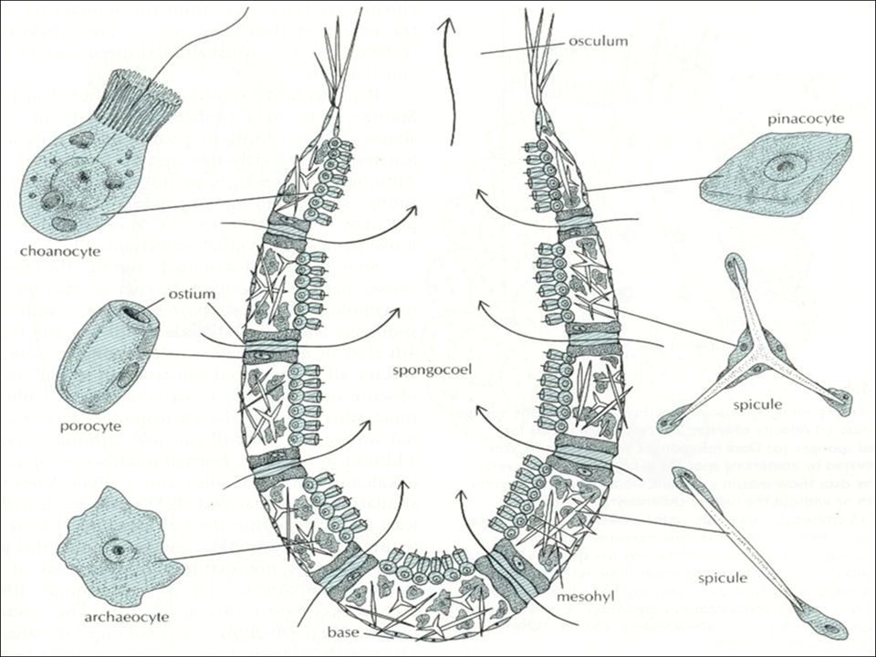
Microscopic view of Porifera cells

18
Choanocytes (collar cells)
They line the flagellated canals and chambers They are ovoid with one end embedded in the mesohyl Adjacent Microvilli are connected to each other by microfibrils They act as a pump to bring water into the sponge Food engulfed is passed to archeocyte for digestion flagellum Collar microvilli Food Vacuole Nucleus

19
Collagen - Support Collagen is found between the inner canals and chambers Mesohyl Amoeboid cells located in the mesohyl, have different roles:- Archeocytes: motile in the mesohyl: Phagocytize particles in the pinacoderm, Recieves particles for digestion Sclerocytes: Specialized, secrete spicules Spongocytes: Secrete spongin fibres for skeleton Collencytes: Secrete fibrillar collagen Lophocytes: Secrete large quantities of Collagen distinguishable from collencytes

20
Body wall: Support Small Section through Sponge wall

21
Types of Spicules Demospongiae & Sclerospongiae secrete siliceous spongin silicon dioxide (SiO2). Calcareous sponges secrete crystalline calcium carbonate (CaCO3) spicules with one to four rays Collagen is stiffened by addition of microscopic mineral accretions or additional protein fibers (spongin) or both
 spicules with one to four rays. Collagen is stiffened by addition of microscopic mineral accretions or additional protein fibers (spongin) or both.")
22
Types of Spicules

23
Types of Canal Systems Osculum Osculum Choanocytes Porocytes
Spongocoel Choanocytes Spongocoel Ostium Ostium Ascon Sycon Leucon

24
Anatomy of Ascon & Sycon

25
Leuconoid Anatomy The most common network of water channels

26
Asconoids Flagellated spongocoels, Simplest organization, Small, branched and tube shaped, Spongocoels allow water into the cells lined with choanocytes, Present only in Calcarea E.g. Leucosolenia, Clathrina canariensis,

27
Syconoids Flagellated canals larger editions of asconoids,
Tubular body but single osculum, Thicker & complex body wall containing radial canals lined with choanocytes Spongocoels lined with epithelia & not flagella Water enters through dermal ostia and filters through Prosophyles, Food is forced through internal pores called apopyles, No branched colonies E.g. Calcarea, hexactinellida

28
In flux & Out flux of Water in Syconoid

29
Leuconoids Flagellated chambers, Complex organization
Suitably adapted for increasing size, Forms large masses with numerous oscula, Flagellated cluster chambers filled from incurrent canals, and opens into excurrent canals, Most are leuconoids, Clear adaptive value, E.g. Calcarea, Leuconoids

30
Classification Phylum Porifera Class Calcarea, Class Demospongiae, Class Hexactinellida, Sclerospongiae (no longer considered a class)
 .")
31
Classification Class Calcarea (calcareous sponges) small, vase shaped, primitive group, mostly drab coloured; a few are yellow, red, green, lavender, all marine, especially shallow waters, show all 3 types of canal systems; mostly asconoid canals, spicules of CaCO3, needle shaped or 3-4 rayed, monaxons, Grows in colonies Straight spicules around the osculum that discourages small animals from entering

32
Class Demospongiae (Most sponges)
Possess spicules made of silicon dioxide (SiO2) or spongin or a combination of both Most sponges belong to this class (90%) Nearly all are leuconoid body type, 95% are living, encrusting plants Mostly found on the continental shelf in well oxygenated habitats Spongilla spp. (Bath sponge), present in midsummer and later form gemmules after disintegration
 or spongin or a combination of both. Most sponges belong to this class (90%) Nearly all are leuconoid body type, 95% are living, encrusting plants. Mostly found on the continental shelf in well oxygenated habitats. Spongilla spp. (Bath sponge), present in midsummer and later form gemmules after disintegration.")
33
Class Hexactinellida (Glass sponges)
Spicules are made of silica, Usually found in deep water on soft substrates in the tropics 200-1,000m. Spicules are six- rayed, pointed and have a lattice-like structure, Cup, vase or urn shape, Radially symmetrical, Formed as one by trabecular net of living tissues from fusion of pseudopodia of archaeocytes, E.g. Euplectella (Venus flower)
")
34
Reproduction in Sponges
ASEXUAL Marine sponges Budding Fragmentation Regeneration Freshwater sponges Gemmules SEXUAL Male & female gametes are formed (monoecious). Archeocytes become eggs Choanocytes filter sperm out of the water Fertilization is involved. Planktonic larvae or mini flagellated colonies are released to colonize new areas. Most are viviparous
. Archeocytes become eggs. Choanocytes filter sperm out of the water. Fertilization is involved. Planktonic larvae or mini flagellated colonies are released to colonize new areas. Most are viviparous.")
35
Development during Reproduction (Demosponges)
Parenchymula larva is free – swimming, Outward flagellated cells invaginates and becomes the choanocytes,

36
Development during Reproduction (Calcarea)
A hollow blastula (Amphiblastula) develops with flagellated cells interiorly, Blastula turns inside-out, Flagellated end appears outward (micromeres), Larger nonflagellated end (macromeres) turns into pinacoderm & Sclerocytes, Flagellated cells become the choanocytes, archeocytes & collencytes
 develops with flagellated cells interiorly, Blastula turns inside-out, Flagellated end appears outward (micromeres), Larger nonflagellated end (macromeres) turns into pinacoderm & Sclerocytes, Flagellated cells become the choanocytes, archeocytes & collencytes.")
37
Larvae of Sponges A Gemmule of Spongillidae Asexual Asexual budding
Formation of internal buds/Gemmules by freshwater sponges Regeneration: can regenerate from broken pieces Allorecognition Sexual Sexual usually hermaphroditic with male and female cells scattered throughout the connective tissue. A Gemmule of Spongillidae

38
Human Impacts of Sponges
Bath sponges In use since bronze age (4000 yrs.) Holds up to X 35s its weight in water Takes 5 yrs. to reach marketable size Creates job opportunities for harvesters & collectors Sponges were challenged by red tides and a fungal disease that wiped out the sponge beds Synthetic sponges were introduced to the market
 Holds up to X 35s its weight in water. Takes 5 yrs. to reach marketable size. Creates job opportunities for harvesters & collectors. Sponges were challenged by red tides and a fungal disease that wiped out the sponge beds. Synthetic sponges were introduced to the market.")
39
Human Impacts of Sponges
Production of a wide variety of bioactive compounds Pharmaceuticals: Antibiotics, Asthma, Arthritis, Anticancer drugs, Chemicals that promote wound healing, Anti-inflammatories Examples antibiotics against bacteria such as E. coli and Staph aureus, e.g. Acyclovir from Caribbean sponge 1st antiviral compound approved for human use fights herpes infections (in use since 1982), e.g. Vidabarine. may attack AIDS virus, e.g. a species of S/Pacific sponge produces chemicals that is bactericidal against Candida albicans (thrush and vaginal infections)
, e.g. Vidabarine. may attack AIDS virus, e.g. a species of S/Pacific sponge produces chemicals that is bactericidal against Candida albicans (thrush and vaginal infections)")
40
Human Impacts of Sponges
In 2009, a new chemical derived from sponge has the ability to re-sensitize bacterial pathogens to antibiotics (Ensures loss of resistance to all antibiotics and die) Material Science The strongest glass structure derived from Euplectella (Venus flower) is a source for study due to its strength Aquarium Trade
 Material Science. The strongest glass structure derived from Euplectella (Venus flower) is a source for study due to its strength. Aquarium Trade.")
41
End of Presentation Thanks for Listening

Similar presentations


 1. Mesozoa 2. Parazoa 3. Eumetazoa.>")
 Composed of a network of cells; no true.>")


