Download presentation
Presentation is loading. Please wait.

1
Maintaining the Internal Environment
Chapter 32 Maintaining the Internal Environment Copyright © The McGraw-Hill Companies, Inc. Permission required for reproduction or display.

2
How the Animal Body Maintains Homeostasis
Homeostasis is defined as the dynamic constancy of the internal environment conditions fluctuate continuously within narrow limits most of the regulatory mechanisms of the vertebrate body that are not devoted to reproduction are concerned with maintaining homeostasis

3
How the Animal Body Maintains Homeostasis
To maintain internal constancy, the vertebrate body needs sensors that are able to measure each condition of the internal environment an integrating center that contains the set point, or proper value for a particular internal condition effectors are generally muscles or glands that can change the value of the condition back toward the set point

4
How the Animal Body Maintains Homeostasis
The integrating center is often a particular region of the brain or spinal cord, but could also be cells of endocrine glands it receives messages from several sensors and then determines if the condition is deviating from the set point it sends a message to the certain effectors to either decrease or increase their activity the activity of the effectors is influenced by the effects they produce in a negative feedback loop

5
A generalized diagram of a negative feedback loop

6
How the Animal Body Maintains Homeostasis
Examples of negative feedback loops in homeostasis regulating body temperature humans, as well as mammals and birds, are endothermic this means that they can maintain relatively constant body temperature other vertebrates are ectothermic, meaning their body temperatures depend more or less on the environmental temperature but they can modify their behavior to affect body temperature regulating blood glucose

7
Control of blood glucose levels

8
Regulating the Body’s Water Content
Animals use various mechanisms for osmoregulation, the regulation of the body’s osmotic composition this refers to how much water and salt the body contains the proper operation of many vertebrate organ systems of the body requires that the osmotic concentration of the blood be kept within narrow bounds

9
Regulating the Body’s Water Content
In many animals, the removal of water and salts from the body is coupled with the removal of metabolic wastes through the excretory system for example, protists, like Paramecium, employ contractile vacuoles water and metabolic wastes are collected by endoplasmic reticula that connect to feeder canals, which lead to the vacuole the water and wastes are then expelled through a pore

10
The Vertebrate Kidney The kidney is a complex organ made up of many, many units called nephrons blood pressure forces the fluid in the blood through a capillary bed at the top of each nephron, called a glomerulus the glomerulus excludes blood cells, proteins, and other large molecules from the filtrate the remainder of the nephron tube reabsorbs anything else useful from the filtrate

11
Basic organization of the vertebrate nephron

12
Vertebrate Kidney Function
Because the original glomerular filtrate is isotonic blood, all vertebrates can produce a urine that is isotonic to blood by reabsorbing ions hypotonic to blood by making the urine more dilute Only birds and mammals can reabsorb water from the glomerular filtrate to produce a urine that is hypertonic (more concentrated than) blood
 blood.")
13
Evolution of the Vertebrate Kidney
Kidneys are thought to have evolved first among the freshwater fish Body fluids of a freshwater fish have a greater osmotic concentration than the surround water, these animals face two serious problems water tends to enter the body from the environment solutes tend to leave the body and enter the environment

14
The Vertebrate Kidney - Fish
Freshwater fish address these problems by not drinking water excreting a large volume of dilute urine reabsorbing ions (mainly NaCl) across the nephron tubule from the glomerular filtrate actively transporting NaCl across the gills from the surrounding water into the blood
 across the nephron tubule from the glomerular filtrate. actively transporting NaCl across the gills from the surrounding water into the blood.")
15
Fish Kidneys Marine fish probably evolved from freshwater ancestors
because their bodies are hypotonic to the surrounding seawater, they faced problems in that water tends to leave their bodies through osmosis across the gills they lose water in their urine to compensate, marine fish drink lots of seawater they excrete isotonic urine

16
Freshwater and marine teleosts (bony fish) face different osmotic problems
 face different osmotic problems")
17
Sharks’ (and their realtives’)Kidneys
Elasmobranchs are the most common subclass of cartilaginous fish they solve their osmotic problem posed by their seawater environment by reabsorbing urea from the nephron tubules this elevates the osmotic concentration in the blood so that they do not have to continually drink seawater the blood is approximately isotonic to the surrounding sea

18
Figure 32.9 Osmoregulation in elasmobranchs

19
32.3 Evolution of the Vertebrate Kidney
The amphibian kidney is identical to that of freshwater fish amphibians produce a very dilute urine and actively transport Na+ across their skin The kidneys of terrestrial reptiles absorb much salt and water in the nephron tubules their urine is still hypotonic but they can absorb additional water in the cloaca

20
Other Vertebrate Kidneys
Because mammals and birds can produce hypertonic urine, they can excrete their waste products in a small volume of water the production of the hypertonic urine is possible due to a looped portion of the nephron, called the Loop of Henle marine birds additionally drink sea water and excrete excess salt through salt glands

21
Osmoregulation by some vertebrates

22
Marine birds drink seawater and then excrete the salt through the salt glands

23
Functions of the Mammalian Urinary System
Removal of metabolic wastes (especially nitrogenous wastes e.g. urea & uric acid). Water balance (and therefore blood pressure). Control of electrolyte balance. Control of pH. Removal of toxins.
. Water balance (and therefore blood pressure). Control of electrolyte balance. Control of pH. Removal of toxins.")
24
The Mammalian Kidney Each kidney receives blood from a renal artery, and it is from this blood that urine is produced urine drains from each kidney through a ureter the ureters carry urine to a urinary bladder urine passes out of the body through the urethra

25
The mammalian urinary system contains two kidneys, each of which contain about a million nephrons within the renal cortex and renal medulla

26
The Mammalian Kidney Within the kidney, the mouth of the ureter flares open to form a funnel-like renal pelvis the renal pelvis has cup-like extensions that receive urine from the renal tissue the renal tissue is divided into an outer renal cortex an inner renal medulla

27
About 25% of your cardiac output flows through your kidneys each minute!

28
The Nephron: functional unit of the kidney
Interlobular artery Afferent Arteriole Glomeruli

29
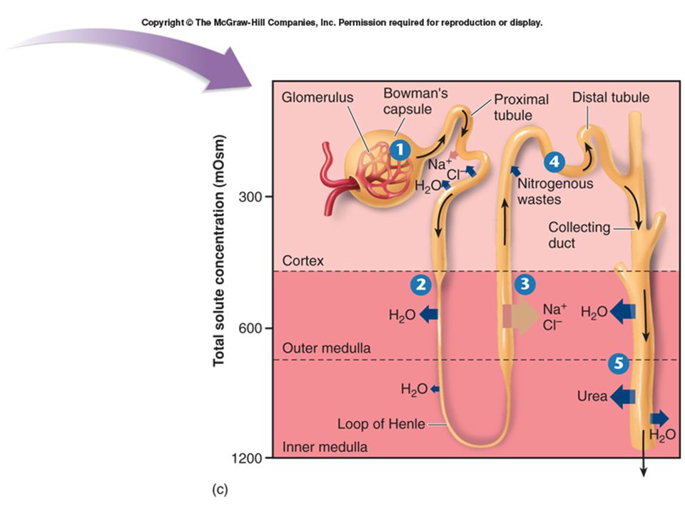
Urine formation The mammalian nephron is composed of three regions. Each region has specific roles. Filtration the filtration device at the top of each nephron is called the Bowman’s capsule which receives filtrate from the glomerular capillaries Tubular reabsorption and secretion the Bowman’s capsule is connected to a long renal tubule, which includes the Loop of Henle, that acts as a reabsorption device Water and pH balance the renal tubule empties into a collecting duct that operates as a water conservation device

30
Formation of urine in the kidney
Filtration (glomerulus) Tubular reabsorption (kidney tubules) Tubular secretion (kidney tubules) Water conservation (collecting system)
 Tubular reabsorption (kidney tubules) Tubular secretion (kidney tubules) Water conservation (collecting system)")
31
Filtration occurs when, driven by blood pressure, small molecules are pushed across the thin walls of the glomerulus into the Bowman’s capsule large particles, such as blood cells and proteins, are excluded from the filtrate the filtrate contains water, nitrogenous wastes (mostly urea), nutrients (principally glucose and amino acids), and a variety of ions
, nutrients (principally glucose and amino acids), and a variety of ions.")
32
Filtration pressures: NFP must be positive for U2P

33
The filtration membrane

34
Tubular reabsorption Reabsorption of filtered solutes occurs in the Proximal Convoluted Tubules. Reabsorbed substances: glucose amino acids water vitamins fatty acids urea electrolytes (Na, K, HCO3- etc.)
")
35
Reabsorption of water The “thin” segment of the descending arm of the Loop of Henle is permeable to the passage of water, but impermeable to salts and urea this leaves a more concentrated filtrate to go to the ascending arm of the Loop of Henle

36
32.4 The Mammalian Kidney NaCl (salt) reabsorption
as the filtrate passes up the ascending arm of the Loop of Henle, the walls become thicker and permeable to salts (but not to water) which pass out into the surrounding tissues and enter the blood in the upper regions of the ascending arm of the Loop of Henle, active transport channels pump out more salt.
 which pass out into the surrounding tissues and enter the blood. in the upper regions of the ascending arm of the Loop of Henle, active transport channels pump out more salt.")
37
32.4 The Mammalian Kidney Tubular secretion
Mostly occurs in the “distal convoluted tubule” and the collecting duct. This involves active transport of other nitrogenous wastes, such as uric acid and ammonia, as well as excess hydrogen ions

38
32.4 The Mammalian Kidney Further reabsorption of water
in the collecting duct, the lower reaches are permeable to urea, which exits to the surrounding tissues this makes water even more likely to leave

40
Eliminating Nitrogenous Wastes
Amino acids and nucleic acids are nitrogen-containing molecules When animals metabolize these substances, they produce nitrogen-containing by-products, called nitrogenous wastes, that must be eliminated by the body

41
The first step in the metabolism of amino acids and nucleic acids is the removal of the amino (--NH2 group) this group is then combined with H+ to form ammonia (NH3) this takes place in the liver
 this takes place in the liver.")
42
Ammonia is toxic Ammonia is quite toxic and is safe only in very dilute concentrations for fish and tadpoles, ammonia can be directly eliminated across the gills or excreted in dilute urine in sharks, adult amphibians, and mammals, the nitrogenous waste is eliminated as urea, which is less toxic reptiles, birds, and insects excrete nitrogenous wastes in the form of uric acid, which can be excreted with very little water

43
Figure 32.14 Nitrogenous wastes

44
How mammals do it! Mammals also make some uric acid but it is a waste product of the breakdown of nucleotides most mammals have an enzyme, uricase, that converts the uric acid into a more soluble form called allantoin humans, apes, and dalmation dogs lack this enzyme and must excrete the uric acid in humans, an excessive accumulation of uric acid is called gout

45
Clinical Diseases that are harmful to the urinary system: Hypertension
Diabetes mellitus Liver dysfunction Congestive heart failure Hormone imbalances These can all lead to renal failure! Some can also result in …

46
Kidney stones Renal Calculi

47
“Well Mr. Osborne, I don’t think that it’s kidney stone after all”

Similar presentations


. Other excretory.>")
